



ACVR1 Protein, Canine (HEK293, His)

Based on 1 Customer Validation
Activin A receptor, type I (ACVR1), also known as ALK-2, is a bone morphogenetic protein (BMP) type I receptor. ACVR1 is involved in a wide variety of biological processes, including bone, heart, cartilage, nervous, and reproductive system development and regulation. ACVR1 Protein, Canine (HEK293, His) is produced in HEK293 cells with a C-Terminal His-tag.
- Species: Canine
- Source: HEK293
-
Storage:Stored at -20°C for 2 years from date of receipt. After reconstitution, it is stable at 4°C for 1 week or -20°C for longer (with carrier protein). It is recommended to freeze aliquots at -20°C or -80°C for extended storage.


To place orders, for customer services and technical support, please contact: MedChemExpress USA
Tel: 609-228-6898 E-mail: [email protected] [email protected]


-
 Biological Activity
-
Technical Parameters
-
Product Properties
- Documentation
- References
- Help & FAQs




Biological Activity
Description
Activin A receptor, type I (ACVR1), also known as ALK-2, is a bone morphogenetic protein (BMP) type I receptor. ACVR1 is involved in a wide variety of biological processes, including bone, heart, cartilage, nervous, and reproductive system development and regulation[1]. ACVR1 Protein, Canine (HEK293, His) is produced in HEK293 cells with a C-Terminal His-tag.
Background
Activin A receptor type I (ACVR1) gene, also known as ALK-2, is located in chromosome 2q23-q24 and encodes for the 509 amino acid protein. The ACVR1 protein product is initially described as an activin type I receptor, and it is found to be expressed in several tissues and different human cell lines[1].
The sequence of amino acids in ACVR1 proteins from different species is very stable, which leads to the conclusion that in the process of evolution, ACVR1 has been only slightly altered, and that both in humans and in animals, its function is similar.
As a member of the BMP/TGFβ receptor family, the ACVR1 protein contains an extracellular N-terminal ligand-binding domain, a transmembrane (TM) domain, an intracellular glycine-serine-rich (GS) domain, and a protein kinase (PK) domain. The loop positioned in the helix-loop-helix of the GS domain contains the key residues responsible for ACVR1 activation upon phosphorylation. As a type I receptor, ACVR1 forms heterotetrameric receptor complexes with the type II receptors BMPR2, ACVR2A, and ACVR2B. Such complexes consist of two type I and two type II receptors. Upon binding of ligands to the heteromeric complexes, type II receptors transphosphorylate the GS domain of type I receptors. As a result, the kinase domain of type I receptors is activated and subsequently phosphorylates SMAD1/5/8 proteins that transduce the signal. ACVR1 is first described to bind to activin A, a member of the BMP/TGFβ family that usually triggers phosphorylation and activation of SMAD2/3 upon complex formation with type II receptors. Later, ACVR1 is also found to bind several BMPs with distinct affinities, triggering SMAD1/5/8 signalling. Besides canonical SMAD signalling, ACVR1 can activate non-canonical signalling pathways, such as p38 mitogen-activated protein kinases/MAPKs[1].
ACVR1 is involved in a wide variety of biological processes, including bone, heart, cartilage, nervous, and reproductive system development and regulation. Moreover, ACVR1 has been extensively studied for its causal role in fibrodysplasia ossificans progressiva (FOP). ACVR1 is linked to different pathologies, including cardiac malformations and alterations in the reproductive system. More recently, ACVR1 has been experimentally validated as a cancer driver gene in diffuse intrinsic pontine glioma (DIPG)[1].
In Vitro
Recombinant human ACVR1 (2.5-5 μg/mL) robustly down-regulates phosphorylated Smad1/5/8 and p38 MAPK levels in the HUVECR26H cells. ACVR1 also inhibits chondro-osseous differentiation in HUVECR26H cells[2].
Verified Bioactivity
1.This product does not contain protein kinase domain.
2.Measured by its binding ability in a functional ELISA. When Recombinant Human BMP-6 (HY-P700029AF) is used at 0.25 μg/mL can bind Recombinant Canine ACVR1. The ED50 for this effect is 4.699 μg/mL.
MCE Validation Data
-
Purity - SDS-PAGE
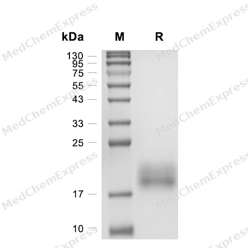
Purity - SDS-PAGE
-
Bioactivity - ELISA
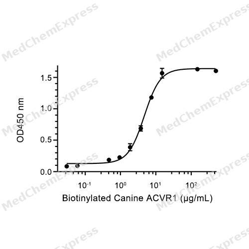
Bioactivity - ELISA

Assay Procedure
Materials
ACVR1 Protein, Canine (HEK293, His) (HY-P76715)
Biotin
Pre-chilled sterile water
Wash buffer: 1X PBST
Blocking buffer: 1% BSA in PBST
TMB substrate solution: Mix substrate solutions A and B in a 1:1 ratio immediately before use
Stop solution (2M H2SO4)
Procedure
Biotinylation
1. Dissolution of Biotin reagent: Remove Biotin from -10 to -30℃ and immediately dissolve 1 vial of Biotin in 180 μL of cold distilled water. Gently pipette up and down to mix, yielding a 10 mM Biotin solution.
2. Calculate the volume of EZ-LinkTM Sulfo-NHS-LC-LC-Biotin, No-WeighTM Format to be added.
3. Incubate at room temperature in the dark for 45 minutes.
4. Quantification of biotinylated protein: Use the BCA assay to quantify the protein concentration. The concentration of biotinylated Canine ACVR1 is 300 μg/mL.
ELISA Detection
1. Coating: Dilute BMP-6 protein to 0.25 μg/mL in 1X PBS, 100 μL per well, and incubate overnight at 4℃.
2. Washing: Wash the plate with wash buffer, 260 μL/well, wash once, pat dry.
3. Blocking: Block with 1% BSA in 1X PBST (blocking buffer) , 150 μL per well, at 25℃ with shaking at 450 rpm for 4 hours.
4. Dilute Canine ACVR1 to the following concentrations: 0, 0.03, 0.06, 0.12, 0.24, 0.47, 0.94, 1.875, 3.75, 7.5, 15, 150, 500 μg/mL.
5. Add Canine ACVR1 protein: 100 μL/well, incubate at 25℃ with shaking at 450 rpm for 2 hours.
6. Washing: Wash the plate with wash buffer, 260 μL/well, wash 5 times, with each wash lasting 1 minute, then pat dry.
7. Add secondary antibody: Dilute the secondary antibody at a 1:2000 ratio, add 100 μL/well; incubate at 25℃ with shaking at 450 rpm for 1 hour.
8. Washing: Wash the plate with wash buffer, 260 μL/well, wash 3 times, with each wash lasting 1 minute, then pat dry.
9. Add TMB substrate solution: 100 μL/well; incubate at room temperature in the dark for 15 minutes.
10. Terminate the reaction: Add 2 M H2SO4, 100 μL/well.
11. Plate reading: Read at 450 nm.
12. Use GraphPad Prism software to plot a curve with the OD value as the y-axis and the Biotinylated Canine ACVR1 concentration as the x-axis, then calculate the ED50 value.


Technical Parameters
-
Species Canine
-
Source HEK293
-
Accession
A0A811ZNW1/XP_549615.2 (D23-E123)
-
Molecular Construction
-
N-term
-
ACVR1 (D23-E123)
Accession # A0A811ZNW1/XP_549615.2 -
His
-
C-term
-
-
Protein Length
Partial
-
Synonyms
ACVR1; Activin A Receptor, Type II-Like Kinase 2; Prev. ACVRLK2; Receptor Protein Serine/Threonine Kinase; SKR1; Hydroxyalkyl-Protein Kinase; TGF-B Superfamily Receptor Type I; Alternative Protein ACVR1; Activin Receptor Type-1; ACTR-I; Activin Receptor T
-
AA Sequence
DEKPKVNPKLYMCVCEGLSCGNEDHCEGQQCFSSLSINDGFHVYQKGCFQVYEQGKMTCKTPPSPGQAVECCQGDWCNRNITAQLPTKGKSFPETQNFHLE
-
Molecular Weight
Approximately 19 kDa, based on SDS-PAGE under reducing conditions, due to the glycosylation.
-
Glycosylation
Yes
-
Purity
≥ 95%, as determined by reducing SDS-PAGE.


Product Properties
Lyophilized powder.
Lyophilized from a 0.22 μm filtered solution of PBS, pH 7.4.
<1 EU/μg, determined by LAL method.
It is not recommended to reconstitute to a concentration less than 100 μg/mL in ddH2O. For long term storage it is recommended to add a carrier protein (0.1% BSA, 5% HSA, 10% FBS or 5% Trehalose).
Stored at -20°C for 2 years from date of receipt. After reconstitution, it is stable at 4°C for 1 week or -20°C for longer (with carrier protein). It is recommended to freeze aliquots at -20°C or -80°C for extended storage.
Room temperature in continental US; may vary elsewhere.


Documentation
-
Data Sheet (263 KB)
-
SDS (251 KB)
- English - EN (251 KB)
- Français - FR (251 KB)
- Deutsch - DE (251 KB)
- Norwegian - NO (251 KB)
- Español - ES (251 KB)
- Swedish - SV (251 KB)
- Italian - IT (251 KB)
- Korean - KR (251 KB)
- Portuguese - PT (251 KB)
-
Handling Instructions (2659 KB)


References
[1]. José Antonio Valer, et al. ACVR1 Function in Health and Disease. Cells. 2019 Oct 31;8(11):1366. [Content Brief]
[2]. Jing Pang, et al. ACVR1-Fc suppresses BMP signaling and chondro-osseous differentiation in an in vitro model of Fibrodysplasia ossificans progressive. Bone. 2016 Nov;92:29-36. [Content Brief]

Calculators
Concentration (start) × Volume (start) = Concentration (final) × Volume (final)


 Powered by Bioz

