



IL-10 Protein, Mouse
Based on 7 publication(s) in Google Scholar
IL-10 protein is a key immunomodulator that, by binding to its heterotetrameric receptors (IL10RA and IL10RB), activates JAK1 and STAT2 (the latter phosphorylates STAT3), thereby triggering potent anti-inflammatory effects. Phosphorylated STAT3 translocates to the nucleus and promotes the expression of anti-inflammatory mediators. IL-10 Protein, Mouse is the recombinant mouse-derived IL-10 protein, expressed by E. coli , with tag free.
- Species: Mouse
- Source: E. coli
-
Storage:Stored at -20°C for 2 years from date of receipt. After reconstitution, it is stable at 4°C for 1 week or -20°C for longer (with carrier protein). It is recommended to freeze aliquots at -20°C or -80°C for extended storage.


To place orders, for customer services and technical support, please contact: MedChemExpress USA
Tel: 609-228-6898 E-mail: [email protected] [email protected]


-
 Biological Activity
-
Technical Parameters
-
Product Properties
- Documentation
- Help & FAQs


-

HY-P7080
IL-4 Protein, Mouse (CHO) -
HY-P7071
IFN-gamma Protein, Mouse -
HY-P70460
IL-13 Protein, Mouse -
HY-P7063
IL-6 Protein, Mouse -
HY-P70644
IL-4 Protein, Mouse -
HY-D1056
Lipopolysaccharides, from E. coli O55:B5 -
HY-P7090
TNF-alpha/TNFSF2 Protein, Mouse (156a.a) -
HY-P70653
IL-4 Protein, Mouse (HEK293, His) -
HY-P7117
TGF beta 1/TGFB1 Protein, Mouse/Rat (HEK293, Tag Free) -
HY-P70648
TGF beta 1/TGFB1 Protein, Mouse/Rat (HEK293) -
HY-P7085
M-CSF Protein, Mouse -
HY-P7361
GM-CSF Protein, Mouse


Biological Activity
Description
IL-10 protein is a key immunomodulator that, by binding to its heterotetrameric receptors (IL10RA and IL10RB), activates JAK1 and STAT2 (the latter phosphorylates STAT3), thereby triggering potent anti-inflammatory effects. Phosphorylated STAT3 translocates to the nucleus and promotes the expression of anti-inflammatory mediators. IL-10 Protein, Mouse is the recombinant mouse-derived IL-10 protein, expressed by E. coli , with tag free.
Background
IL-10, a major immune regulatory cytokine, exerts profound anti-inflammatory functions, mitigating excessive tissue disruption caused by inflammation. Upon binding to its heterotetrameric receptor, comprising IL10RA and IL10RB, IL-10 initiates a signaling cascade involving JAK1 and STAT2-mediated phosphorylation of STAT3. Subsequently, phosphorylated STAT3 translocates to the nucleus, where it drives the expression of anti-inflammatory mediators. IL-10 specifically targets antigen-presenting cells (APCs), such as macrophages and monocytes, inhibiting their release of pro-inflammatory cytokines, including GM-CSF, G-CSF, IL-1 alpha, IL-1 beta, IL-6, IL-8, and TNF-alpha. Additionally, IL-10 interferes with antigen presentation by reducing the expression of MHC-class II and co-stimulatory molecules, thereby dampening their ability to induce T cell activation. Furthermore, IL-10 controls the inflammatory response of macrophages by reprogramming essential metabolic pathways, including mTOR signaling. IL-10 exists as a homodimer and interacts with IL10RA and IL10RB.
Verified Bioactivity
Mouse IL-10 inhibits LPS-induced secretion of IL-6 by RAW264.7 cells. The ED50 for this effect is <5 ng/mL in the presence of 2 μg/mL LPS.
MCE Validation Data
-
Purity - SDS-PAGE
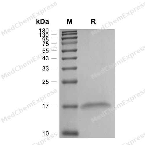
Purity - SDS-PAGE
-
Bioactivity - Cell-Based Assay
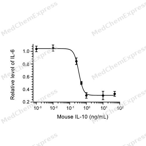
Bioactivity - Cell-Based Assay


Publications (7)
-
Journal Impact Factor
-
Most Recent
-
Cell Host Microbe
Enterococcus faecalis-derived lactic acid suppresses macrophage activation to facilitate persistent and polymicrobial wound infections. [Abstract]2026 Feb 11;34(2):245-262.e8. PMID: 41605216 -
Adv Sci (Weinh)
Functionalized Biomimetic Nanoparticles Targeting the IL-10/IL-10Rα/Glycolytic Axis in Synovial Macrophages Alleviate Cartilage Degeneration in Osteoarthritis. [Abstract]2025 Jul;12(27):e2504768. PMID: 40317692 -
J Transl Med
Staphylococcus aureus induces mitophagy via the HDAC11/IL10 pathway to sustain intracellular survival. [Abstract]2025 Feb 4;23(1):156. PMID: 39905391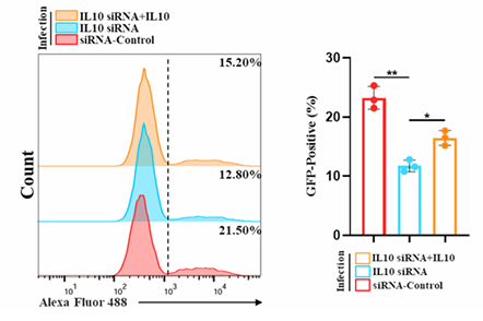
IL-10 Protein, Mouse purchased from MedChemExpress. Usage Cited in: J Transl Med. 2025 Feb 4;23(1):156. [Abstract]
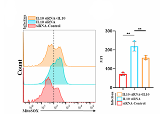
IL-10 (1 ng/mL, 24 h) reversed the increase in intracellular S. aureus growth within BMDMs induced by IL-10 siRNA treatment.
IL-10 Protein, Mouse purchased from MedChemExpress. Usage Cited in: J Transl Med. 2025 Feb 4;23(1):156. [Abstract]
Flow cytometry analysis of mtROS levels in BMDMs 24 h post-infection with GFP-S. aureus (MOI = 10), following transfection with either control siRNA or IL-10 siRNA, with or without the addition of IL-10 (1 ng/mL). Quantitative analysis of the Mean Fluorescence Intensity (MFI) reflects the intracellular mtROS levels.
IL-10 Protein, Mouse purchased from MedChemExpress. Usage Cited in: J Transl Med. 2025 Feb 4;23(1):156. [Abstract]
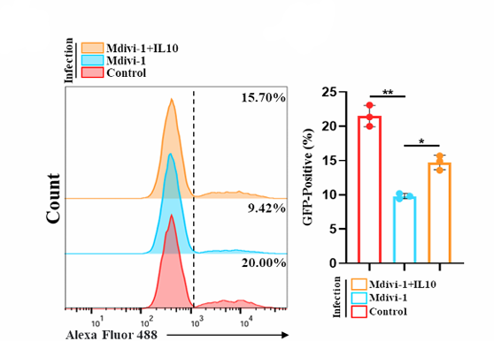
Flow cytometry analysis of the impact of Mdivi-1 (20µM) pretreatment, with or without added IL10 (1ng/ml), on the bacterial load of BMDMs infected with GFP-S. aureus (MOI = 10) for 24 h. Quantitative analysis of the proportion of BMDMs infected with GFP-S. aureus.
IL-10 Protein, Mouse purchased from MedChemExpress. Usage Cited in: J Transl Med. 2025 Feb 4;23(1):156. [Abstract]
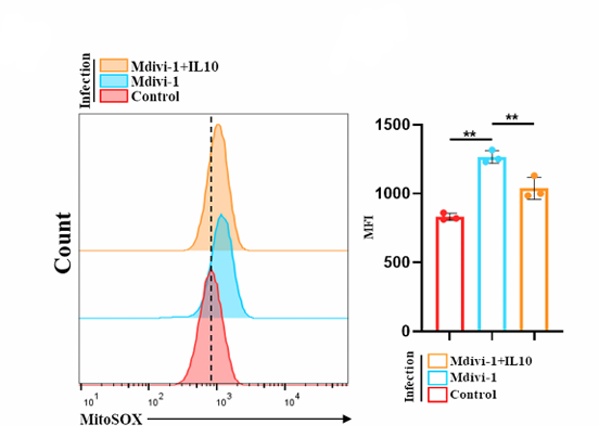
Flow cytometry analysis of mtROS levels in BMDMs 24 h after infection with S. aureus (MOI = 10) in various treatment groups. Quantitative analysis of intracellular mtROS levels (IL-10:1 ng/mL, 24 h,Mdivi-1:20 µM, 24 h).
-
Cell Rep
4-octyl itaconate inhibits cytokine-mediated inflammation via alkylation of TYK2 and JAK1. [Abstract]2026 Mar 30;45(4):117179. PMID: 41915469 -
Cell Signal
IL-10 alleviates ulcerative colitis by regulating mitochondrial function through reducing ISG15 expression. [Abstract]2025 Oct:134:111932. PMID: 40541818 -
BMC Cancer
Low-dose adropin stimulates inflammasome activation of macrophage via mitochondrial ROS involved in colorectal cancer progression. [Abstract]2023 Oct 30;23(1):1042. PMID: 37904094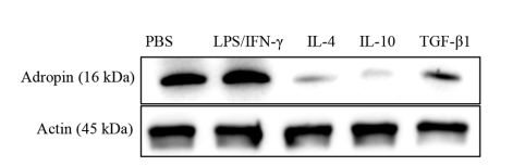
IL-10 Protein, Mouse purchased from MedChemExpress. Usage Cited in: BMC Cancer. 2023 Oct 30;23(1):1042. [Abstract]
M1 macrophages had increased adropin production as stimulated by LPS/IFN-γ, but M2 macrophages had decreased adropin production after being treated by IL-4 (50 ng/mL), IL-10 (50 ng/mL), or TGF-β1
-
Immunol Lett
Mechanism of M2 macrophages modulating astrocyte polarization through the TGF-β/PI3K/Akt pathway. [Abstract]2023 Jul:259:1-8. PMID: 37244460


Technical Parameters
-
Species Mouse
-
Source E. coli
-
Tag Tag Free
-
Accession
P18893 (S19-S178)
-
Molecular Construction
-
N-term
-
IL-10 (S19-S178)
Accession # P18893 -
C-term
-
-
Protein Length
Full Length of Mature Protein
-
Synonyms
IL10; TGIF; Interleukin 10; T-Cell Growth Inhibitory Factor; IL-10; Interleukin-10; CSIF; Interleukin Family Protein; Cytokine Synthesis Inhibitory Factor; GVHDS; IL10A; IF2A
-
AA Sequence
SRGQYSREDNNCTHFPVGQSHMLLELRTAFSQVKTFFQTKDQLDNILLTDSLMQDFKGYLGCQALSEMIQFYLVEVMPQAEKHGPEIKEHLNSLGEKLKTLRMRLRRCHRFLPCENKSKAVEQVKSDFNKLQDQGVYKAMNEFDIFINCIEAYMMIKMKS
-
Predicted Molecular Mass
18.9 kDa
-
Molecular Weight
Approximately 16 kDa, based on SDS-PAGE under reducing conditions.
-
Purity
≥ 95%, as determined by reducing SDS-PAGE.


Product Properties
Lyophilized powder
1.Lyophilized from a 0.22 μm filtered solution of 4 mM HCl.
2.Lyophilized from a 0.22 μm filtered solution of PBS, pH 7.4, 8% trehalose.
Please refer to the lot-specific COA for specific buffer information.
<0.01 EU/μg, determined by LAL method.
It is not recommended to reconstitute to a concentration less than 100 μg/mL in ddH2O. For long term storage it is recommended to add a carrier protein (0.1% BSA, 5% HSA, 10% FBS or 5% Trehalose).
Stored at -20°C for 2 years from date of receipt. After reconstitution, it is stable at 4°C for 1 week or -20°C for longer (with carrier protein). It is recommended to freeze aliquots at -20°C or -80°C for extended storage.
Room temperature in continental US; may vary elsewhere.


Documentation
-
Data Sheet (236 KB)
-
SDS (252 KB)
- English - EN (252 KB)
- Français - FR (252 KB)
- Deutsch - DE (252 KB)
- Norwegian - NO (252 KB)
- Español - ES (252 KB)
- Swedish - SV (252 KB)
- Italian - IT (252 KB)
- Korean - KR (252 KB)
- Portuguese - PT (252 KB)
-
Handling Instructions (2659 KB)

Calculators
Concentration (start) × Volume (start) = Concentration (final) × Volume (final)


 Powered by Bioz

