



TGF beta 1/TGFB1 Protein, Mouse/Rat (HEK293)
Based on 32 publication(s) in Google Scholar
TGF beta 1/TGFB1 Protein belongs to the TGF-β superfamily. TGF beta 1/TGFB1 Protein plays a key role in various physiological and pathological processes such as cell growth, differentiation, apoptosis, immune regulation, and extracellular matrix formation. TGF beta 1/TGFB1 Protein, Mouse/Rat (HEK293) is a recombinant TGF beta 1/TGFB1 Protein expressed by HEK293 without a tag.
- Species: Rat; Mouse
- Source: HEK293
-
Storage:Stored at -20°C for 2 years from date of receipt. After reconstitution, it is stable at 4°C for 1 week or -20°C for longer (with carrier protein). It is recommended to freeze aliquots at -20°C or -80°C for extended storage.


To place orders, for customer services and technical support, please contact: MedChemExpress USA
Tel: 609-228-6898 E-mail: [email protected] [email protected]


-
 Biological Activity
-
Technical Parameters
-
Product Properties
- Documentation
- References
- Help & FAQs


-

HY-10431
SB-431542 -

HY-P7118
TGF beta 1/TGFB1 Protein, Human (CHO) -
HY-P7063
IL-6 Protein, Mouse -
HY-P7080
IL-4 Protein, Mouse (CHO) -
HY-P70517
IL-10 Protein, Mouse -
HY-P7077
IL-2 Protein, Mouse -
HY-P7073
IL-1 beta Protein, Mouse -
HY-P7361
GM-CSF Protein, Mouse -
HY-P7085
M-CSF Protein, Mouse -

HY-D1056
Lipopolysaccharides, from E. coli O55:B5 -
HY-P7071
IFN-gamma Protein, Mouse -
HY-P70709
IL-23 Protein, Mouse (HEK293)


Biological Activity
Description
TGF beta 1/TGFB1 Protein belongs to the TGF-β superfamily. TGF beta 1/TGFB1 Protein plays a key role in various physiological and pathological processes such as cell growth, differentiation, apoptosis, immune regulation, and extracellular matrix formation. TGF beta 1/TGFB1 Protein, Mouse/Rat (HEK293) is a recombinant TGF beta 1/TGFB1 Protein expressed by HEK293 without a tag[1][2][3].
Background
TGF beta 1/TGFB1 is a multifunctional cytokine. In immune regulation, TGF beta 1/TGFB1 can inhibit the activation of T/B cells, induce the differentiation of regulatory T cells, and promote the polarization of macrophages toward an anti-inflammatory phenotype. TGF beta 1/TGFB1 can also affect the homeostasis of the extracellular matrix by regulating the synthesis and degradation of collagen. When it is overactivated, it will lead to fibrosis of organs such as the liver and lungs. In addition, TGF beta 1/TGFB1 is also involved in the occurrence and development of tumors. In the early stage, it inhibits the proliferation of epithelial cells, induces cell apoptosis, and plays an anti-cancer role. In the late stage, it promotes tumor progression by promoting epithelial-mesenchymal transformation, tumor angiogenesis, and immune escape[1][2][3].
In Vitro
TGF beta 1/TGFB1 Protein (Mouse/Rat; 10 ng/mL; 48 h) can be used to establish a fibrotic cell model. It activates SMAD2/3 phosphorylation, upregulates the levels of fibrotic proteins (α-SMA and Col1a1), and inhibits the levels of Acan and Col2a1 in rat nucleus pulposus cells[4].
Verified Bioactivity
The ability to inhibit IL-4-dependent proliferation of TF-1 human erythroleukemic cells has an ED50 value of 5-25 pg/mL.
MCE Validation Data
-
Purity - SDS-PAGE
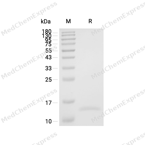
Purity - SDS-PAGE
-
Bioactivity - Cell-Based Assay
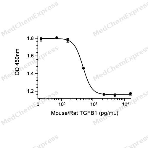
Bioactivity - Cell-Based Assay


Publications (32)
-
Journal Impact Factor
-
Most Recent
-
Adv Mater
Piezoelectric Injectable Anti-Adhesive Hydrogel to Promote Endogenous Healing of Tendon Injuries. [Abstract]2025 Jul 14:e2501306. PMID: 40658817 -
-
J Nanobiotechnology
Targeted inhibition of RGS19 alleviates renal fibrosis by restoring autophagy and modulating immune cell infiltration. [Abstract]2026 Feb 28;24(1):315. PMID: 41761314 -
J Clin Invest
2025 Jun 2;135(11):e184158. PMID: 40454481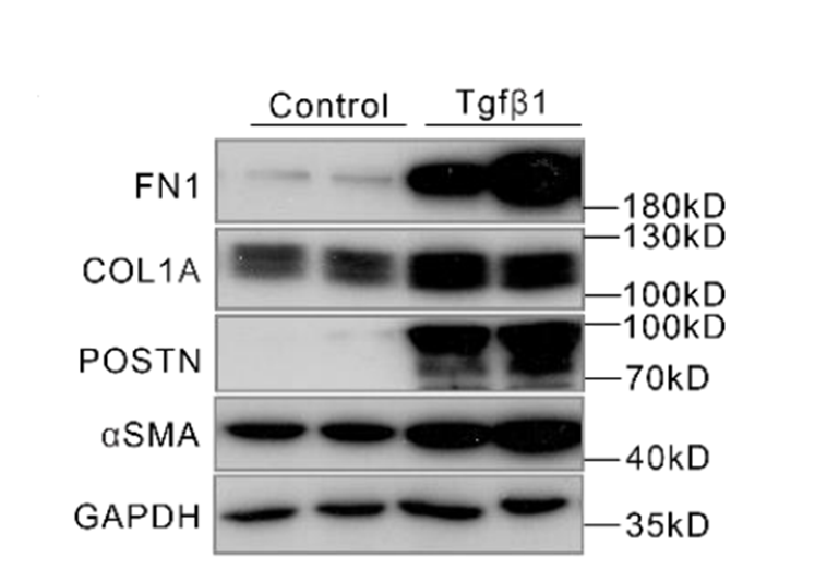
TGF beta 1/TGFB1 Protein, Mouse/Rat (HEK293) purchased from MedChemExpress. Usage Cited in: J Clin Invest. 2025 Jun 2;135(11):e184158. [Abstract]
Western blot analysis of the indicated protein levels in NIH3T3 cell with TGFβ (5 ng/mL) stimulation for 72 h.
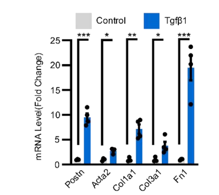
TGF beta 1/TGFB1 Protein, Mouse/Rat (HEK293) purchased from MedChemExpress. Usage Cited in: J Clin Invest. 2025 Jun 2;135(11):e184158. [Abstract]
Real-time qPCR analysis of Postn, Acta2, Col1a1, Col3a1 and Fn1 mRNA expression levels in NIH3T3 cell with TGFβ (5 ng/mL) stimulation for 72 h.
-
Mol Biomed
Epithelial membrane protein 1 drives hepatic stellate cell activation via the TLN1/FAK cascade in MASLD donor liver transplantation. [Abstract]2025 Nov 24;6(1):116. PMID: 41284206 -
Phytomedicine
Integrated UHPLC-Q-exactive orbitrap HRMS and serum pharmacochemistry for the investigation of anti-hepatic fibrosis effect of Baoganning Decoction. [Abstract]2025 Feb:137:156363. PMID: 39799893 -
Mater Today Bio
Uniform and controllable surface nano-structure on polyetheretherketone implants can regulate mechanical property to enhance soft tissue integration through Piezo1/TGF-β1 signaling axis. [Abstract]2025 Mar 8:31:101645. PMID: 40151615 -
Adv Healthc Mater
Antifibrotic Effects of Tetrahedral Framework Nucleic Acids by Inhibiting Macrophage Polarization and Macrophage-Myofibroblast Transition in Bladder Remodeling. [Abstract]2023 Apr;12(11):e2203076. PMID: 36603196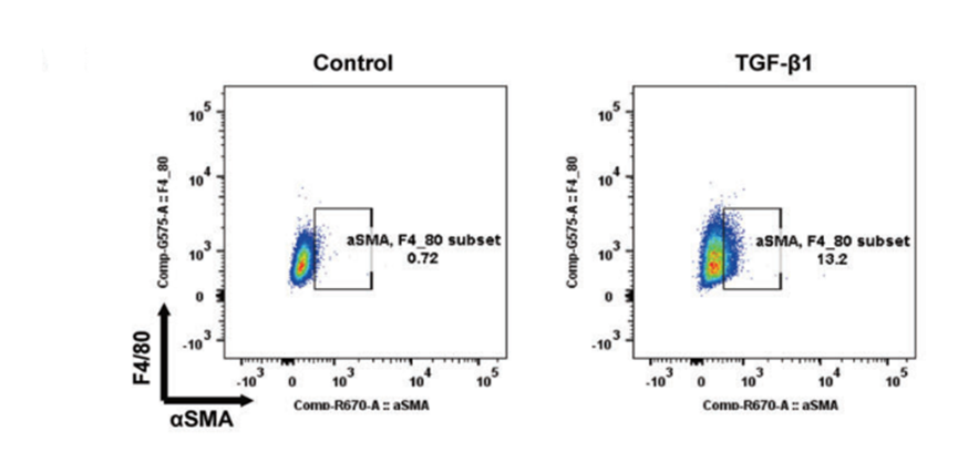
TGF beta 1/TGFB1 Protein, Mouse/Rat (HEK293) purchased from MedChemExpress. Usage Cited in: Adv Healthc Mater. 2023 Apr;12(11):e2203076. [Abstract]
The proportions of MMT cells were significantly elevated by TGF-β1 (10 ng/mL, 48 h).
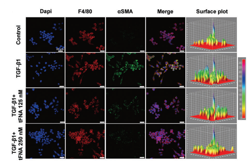
TGF beta 1/TGFB1 Protein, Mouse/Rat (HEK293) purchased from MedChemExpress. Usage Cited in: Adv Healthc Mater. 2023 Apr;12(11):e2203076. [Abstract]
Representative immunofluorescence images of α-SMA, the protein expression levels of α-SMA were upregulated by TGF-β1 (10 ng/mL, 48 h).
-
J Orthop Translat
Physalin A alleviates intervertebral disc degeneration via anti-inflammatory and anti-fibrotic effects. [Abstract]2023 Jan 25:39:74-87. PMID: 36788965 -
Int J Nanomedicine
Inhalable Albumin Nanoparticles Co-Delivering Dihydroartemisinin and Nintedanib Attenuate Pulmonary Fibrosis by Suppressing TGF-β1/Smad2/3 Signaling. [Abstract]2026 Feb 26:21:576680. PMID: 41777379 -
Neural Regen Res
Small extracellular vesicles derived from hair follicle neural crest stem cells enhance perineurial cell proliferation and migration via the TGF-β/SMAD/HAS2 pathway. [Abstract]2025 Sep 3. PMID: 40903975 -
Clin Transl Med
Multi-omics reveals a novel Cxcr4+ subpopulation of alveolar macrophages and therapeutic effect of AMD3100 in mice with advanced silicosis. [Abstract]2026 Jun;16(6):e70705. PMID: 42204838 -
Clin Sci
The HDAC2/SP1/miR-205 feedback loop contributes to tubular epithelial cell extracellular matrix production in diabetic kidney disease. [Abstract]2022 Feb 11;136(3):223-238. PMID: 35084460 -
Biochem Pharmacol
TTC3-mediated ubiquitination of APPL1 is suppression involved in the anti-asthmatic effects of the substance P receptor antagonist WIN62577. [Abstract]2026 Apr:246:117701. PMID: 41520735 -
Biochem Pharmacol
Theaflavin reduces Achilles tendon heterotopic ossification in mice through the TGF-β/Smad signaling pathway. [Abstract]2025 Dec;242(Pt 4):117387. PMID: 41047029 -
Life Sci
STC-1 improves scar remodeling and is associated with PI3K/AKT signaling and immune modulation. [Abstract]2026 Mar 15:389:124253. PMID: 41644046 -
Life Sci
MMP3 as a new target of Danshensu/tetramethylpyrazine derivative for attenuating cardiac fibrosis post-myocardial infarction. [Abstract]2025 Jun 1:370:123570. PMID: 40113078 -
Food Funct
Ameliorative effect of docosahexaenoic acid-acylated astaxanthin ester on diabetic nephropathy: association with the trehalose/HSP90AA1 and colon-kidney axis. [Abstract]2026 May 18. PMID: 42149022 -
Cell Biol Toxicol
SENP3 mediated DeSUMOylation of macrophage derived CCL17 accelerates atherosclerosis via regulation of Treg. [Abstract]2025 Nov 21;41(1):151. PMID: 41266856 -
Biomolecules
Glycogen Hydrogel Loaded with Schistosoma japonicas Peptide SJMHE1 Improves Skin Wound Healing. [Abstract]2026 Mar 5;16(3):392. PMID: 41897329 -
Stem Cell Rev Rep
Investigating the Therapeutic Efficacy of Quality-Controlled, miR-146a-5p-Enriched Small Extracellular Vesicles Derived From MSCs Against Idiopathic Pulmonary Fibrosis. [Abstract]2025 Sep 24. PMID: 40987919 -
Cell Signal
AKR1C3 promotes aerobic glycolysis in hepatic stellate cells via the AKT/mTOR pathway to induce liver fibrosis. [Abstract]2026 May:141:112369. PMID: 41554490 -
Naunyn Schmiedebergs Arch Pharmacol
Integrative bioinformatics and machine learning to explore the mechanism of Scutellaria baicalensis Georgi in TGF-β1-induced ASMCs proliferation during asthma airway remodeling. [Abstract]2025 Dec 4. PMID: 41345301 -
Toxicol Appl Pharmacol
Artesunate attenuates pulmonary fibrosis by suppressing fibroblast senescence through inhibition of the STAT3/p53 signaling pathway. [Abstract]2026 Jan:506:117646. PMID: 41271031 -
Exp Cell Res
MG53-mediated membrane repair attenuates pulmonary fibrosis by antagonizing TGF-β1-driven epithelial mesenchymal transition. [Abstract]2026 Jun 19;461(1):115108. PMID: 42320722 -
J Integr Neurosci
Atractylodin Suppresses Fibrotic Scar Formation and Enhances Functional Recovery Following Spinal Cord Injury. [Abstract]2026 Apr 22;25(4):47565. PMID: 42052760 -
Tissue Cell
Comparative phenotypic analysis of primary aortic VSMCs versus TGF - β/PDGF - BB differentiated MSCs in rat. [Abstract]2026 Jun 20:103:103714. PMID: 42335648 -
Cardiovasc Ther
Fibroblast Activation Protein-Targeted CAR-T Cells Induce Apoptosis in Murine Cardiac Myofibroblasts. [Abstract]2025 Sep 13:2025:7230505. PMID: 40977952 -
Burns
Aloe vera barbadensis extract regenerator with metabolic regulatory functions promotes skin regeneration and reduces fibrosis deposition in diabetic burn model pigs. [Abstract]2026 Jun 9;52(8):108107. PMID: 42385476 -
Connect Tissue Res
E3 ubiquitin ligase Peli3 regulates M2 macrophage-to-myofibroblast transition and fibrosis in wound healing via IRF4. [Abstract]2026 May;67(3):308-320. PMID: 42018282 -
Folia Histochem Cytobiol
HIPK2 attenuates bleomycin-induced pulmonary fibrosis by suppressing the Wnt/β-catenin signaling pathway. [Abstract]2022;60(3):247-259. PMID: 36004621 -
Tohoku J Exp Med
From Carbonic Anhydrase 12 to TGFβ1/Smad Pathway: A Key Mechanism for Isoorientin in Pulmonary Fibrosis Therapy. [Abstract]2026 Jan 22. PMID: 41565266


Technical Parameters
-
Species Rat; Mouse
-
Source HEK293
-
Tag Tag Free
-
Accession
P04202/P17246 (A279-S390)
-
Molecular Construction
-
N-term
-
TGFB1 (A279-S390)
Accession # P04202 -
C-term
-
-
Protein Length
Full Length of TGFB1 Chain
-
Synonyms
TGFB1; Transforming Growth Factor Beta1; Prev. TGFB; Camurati-Engelmann Disease; Prev. DPD1; Latency-Associated Peptide; TGFbeta; TGF-Beta-1; CED; TGF-Beta1; Transforming Growth Factor Beta-1 Proprotein; IBDIMDE; Prepro-Transforming Growth Factor Beta-1;
-
AA Sequence
ALDTNYCFSSTEKNCCVRQLYIDFRKDLGWKWIHEPKGYHANFCLGPCPYIWSLDTQYSKVLALYNQHNPGASASPCCVPQALEPLPIVYYVGRKPKVEQLSNMIVRSCKCS
-
Predicted Molecular Mass
12.8 kDa
-
Molecular Weight
Approximately 13 kDa, based on SDS-PAGE under reducing conditions.
-
Structure/Form
Disulfide-linked homodimer
-
Purity
≥ 95%, as determined by reducing SDS-PAGE.


Product Properties
Lyophilized powder
1.Lyophilized from a 0.22 μm filtered solution of 4 mM HCl.
2.Lyophilized from a 0.22 μm filtered solution of 4 mM HCl, 4%Mannitol.
Please refer to the lot-specific COA for specific buffer information.
<1 EU/μg, determined by LAL method.
It is not recommended to reconstitute to a concentration less than 100 μg/mL in 4mM HCl. For long term storage it is recommended to add a carrier protein (0.1% BSA, 5% HSA, 10% FBS or 5% Trehalose).
Stored at -20°C for 2 years from date of receipt. After reconstitution, it is stable at 4°C for 1 week or -20°C for longer (with carrier protein). It is recommended to freeze aliquots at -20°C or -80°C for extended storage.
Room temperature in continental US; may vary elsewhere.


Documentation
-
Data Sheet (263 KB)
-
SDS (252 KB)
- English - EN (252 KB)
- Français - FR (252 KB)
- Deutsch - DE (252 KB)
- Norwegian - NO (252 KB)
- Español - ES (252 KB)
- Swedish - SV (252 KB)
- Italian - IT (252 KB)
- Korean - KR (252 KB)
- Portuguese - PT (252 KB)
-
Handling Instructions (2659 KB)


References
[1]. Kim KK, et al. TGF-β1 Signaling and Tissue Fibrosis. Cold Spring Harb Perspect Biol. 2018 Apr 2;10(4):a022293. [Content Brief]
[2]. Laudisi F, et al. TGF-β1 signaling and Smad7 control T-cell responses in health and immune-mediated disorders. Eur J Immunol. 2023 Nov;53(11):e2350460. [Content Brief]
[3]. Lodyga M, et al. TGF-β1 - A truly transforming growth factor in fibrosis and immunity. Semin Cell Dev Biol. 2020 May;101:123-139. [Content Brief]
[4]. Lu R, et al. Physalin A alleviates intervertebral disc degeneration via anti-inflammatory and anti-fibrotic effects. J Orthop Translat. 2023 Jan 25;39:74-87. [Content Brief]

Calculators
Concentration (start) × Volume (start) = Concentration (final) × Volume (final)


 Powered by Bioz

