



Rilmenidine

Based on 1 Customer Validation
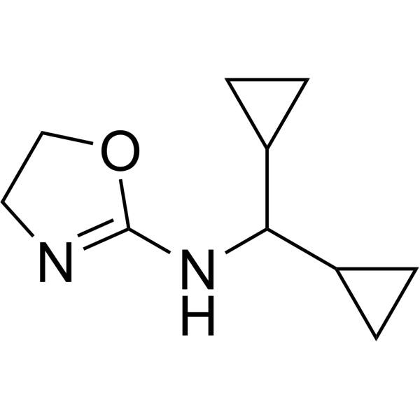
Rilmenidine, an innovative antihypertensive agent, is an orally active, selective I1 imidazoline receptor agonist. Rilmenidine is an alpha 2-adrenoceptor agonist. Rilmenidine induces autophagy. Rilmenidine acts both centrally by reducing sympathetic overactivity and in the kidney by inhibiting the Na+/H+ antiport. Rilmenidine modulates proliferation and stimulates the proapoptotic protein Bax thus inducing the perturbation of the mitochondrial pathway and apoptosis in human leukemic K562 cells.
For research use only. We do not sell to patients.
- Purity : 99.9%
- CAS No.: 54187-04-1
- Formula: C10H16N2O
- Molecular Weight:180.25
-
Storage:
-20°C, sealed storage, away from moisture and light
* In solvent : -80°C, 6 months; -20°C, 1 month (sealed storage, away from moisture and light)

To place orders, for customer services and technical support, please contact: MedChemExpress USA
Tel: 609-228-6898 E-mail: [email protected] [email protected]


-
 Biological Activity
-
Chemical Information
-
Solvent & Solubility
- Protocols
- Purity & Documentation
- References
-
 Help & FAQs


-
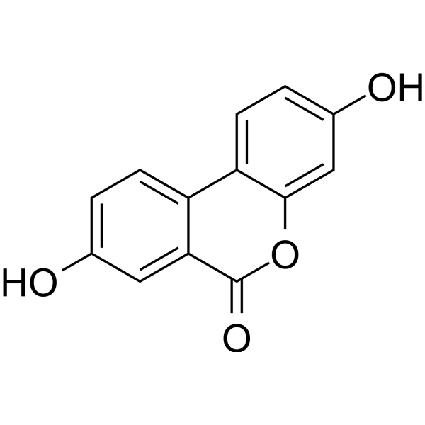
HY-100599
Urolithin AMitophagy Activator -

HY-100490R
Rilmenidine (Standard)I1 Imidazoline Receptor/α2-Adrenoceptor Agonist -
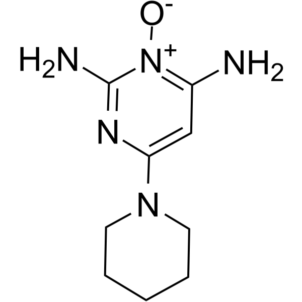
HY-B0112
MinoxidilEndogenous Metabolite -
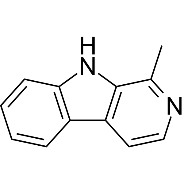
HY-101392
HarmaneMAO Inhibitor -
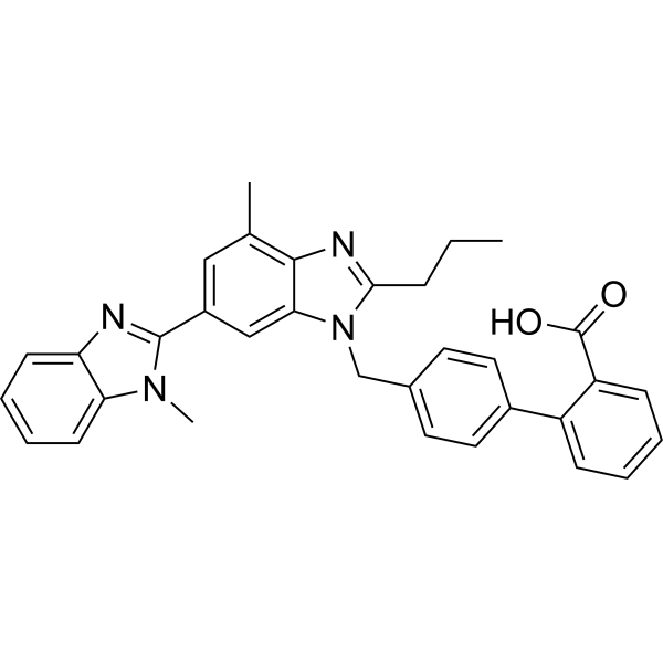
HY-13955
TelmisartanAngiotensin II Type 1 Receptor -
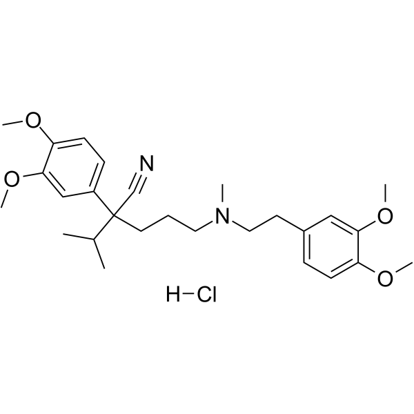
HY-A0064
Verapamil hydrochlorideCalcium Channel Blocker -
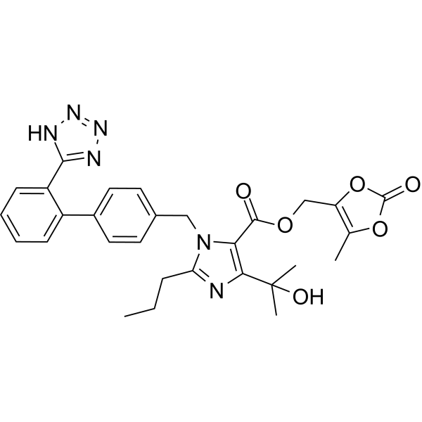
HY-17005
Olmesartan medoxomil -
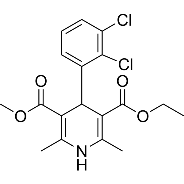
HY-B0309
FelodipineCalcium Channel Antagonist -
HY-B0193A
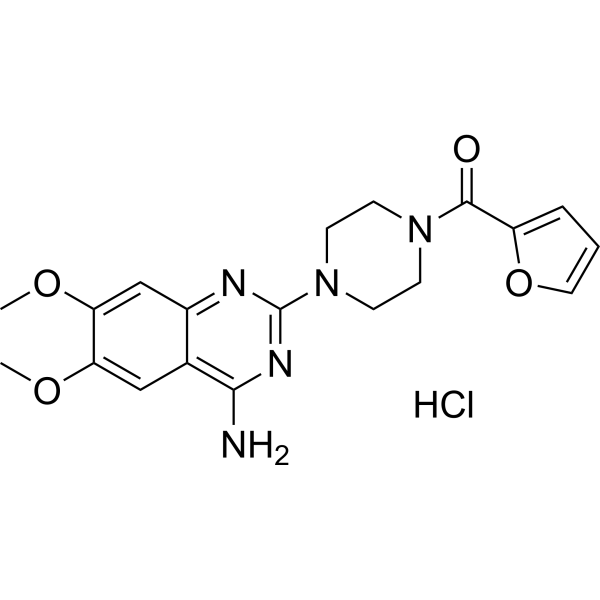
Prazosin hydrochlorideAdrenergic Receptor Antagonist -
HY-14284
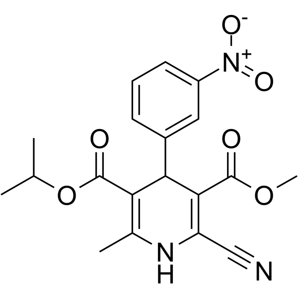
NilvadipineCalcium Channel Antagonist -
HY-17005R
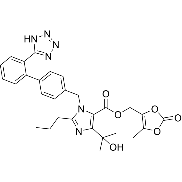
Olmesartan medoxomil (Standard) -
HY-B0209
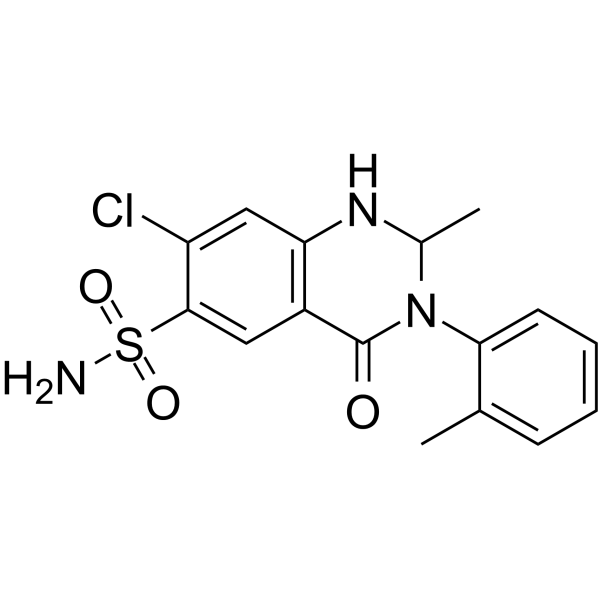
MetolazoneDiuretics
All Adrenergic Receptor Isoforms
More

Biological Activity
Description
Cellular Effect
|
Cell Line

|
Type | Value | Description | References |
|---|---|---|---|---|
| K562 | IC50 |
65 μM
Compound: ChEMBLID-289480
|
Antiproliferative activity against human K562 cells assessed as inhibition of cell survival after 48 hrs by MTT assay
Antiproliferative activity against human K562 cells assessed as inhibition of cell survival after 48 hrs by MTT assay
|
[PMID: 27265687] |
In Vitro
Rilmenidine provides antihypertensive efficacy comparable with that of diuretics, beta-blockers, calcium channel blockers, and angiotensin-converting enzyme (ACE) inhibitors[1].
Rilmenidine (25-100 μM; 24 hours) inhibits K562 cell proliferation[2].
MedChemExpress (MCE) has not independently confirmed the accuracy of these methods. They are for reference only. Further protocols information, click here.
-
Cell Line:K562 cells
-
Concentration:25, 50, 100 μM
-
Incubation Time:24 hours
-
Result:Dose-dependently inhibited K562 colony formation.
In Vivo
Rilmenidine decreases levels of mutant huntingtin[3].
MedChemExpress (MCE) has not independently confirmed the accuracy of these methods. They are for reference only.
Clinical Trial
| NCT Number | Sponsor | Condition | Start Date |
Phase
|
|---|---|---|---|---|
| NCT01329991 | Plexxikon| | 2011-05 | PHASE1 |


Chemical Information
-
CAS No. 54187-04-1
-
Appearance Solid
-
Molecular Weight 180.25
-
Formula C10H16N2O
-
Color White to off-white
-
SMILES
C1(NC(C2CC2)C3CC3)=NCCO1
-
Shipping
Room temperature in continental US; may vary elsewhere.
-
Storage
-20°C, sealed storage, away from moisture and light
* In solvent : -80°C, 6 months; -20°C, 1 month (sealed storage, away from moisture and light)


Solvent & Solubility
In Vitro:
DMSO : 100 mg/mL (554.79 mM; Need ultrasonic; Hygroscopic DMSO has a significant impact on the solubility of product, please use newly opened DMSO)
Please refer to the solubility information to select the appropriate solvent. Once prepared, please aliquot and store the solution to prevent product inactivation from repeated freeze-thaw cycles.
Storage method and period of stock solution: -80°C, 6 months; -20°C, 1 month (sealed storage, away from moisture and light). When stored at -80°C, please use it within 6 months. When stored at -20°C, please use it within 1 month.
Please refer to the solubility information to select the appropriate solvent. Once prepared, please aliquot and store the solution to prevent product inactivation from repeated freeze-thaw cycles.
Storage method and period of stock solution: -80°C, 6 months; -20°C, 1 month (sealed storage, away from moisture and light). When stored at -80°C, please use it within 6 months. When stored at -20°C, please use it within 1 month.
Concentration (start) × Volume (start) = Concentration (final) × Volume (final)

Protocols
-
Apoptosis
Apoptosis, also called programmed cell death, is generally characterized by distinct morphological characteristics.
-
TUNEL staining for apoptotic DNA fragmentation
TUNEL staining detects DNA strand breaks by using terminal deoxynucleotidyl transferase to add labeled nucleotides to exposed 3′-OH DNA termini, generating either microscopic staining in fixed cells or tissue sections, or fluorescence/cytometric signal in cell suspensions. TUNEL positivity reflects DNA fragmentation but should not be interpreted alone as definitive apoptosis, because TUNEL can also label necrotic, autolytic, mechanically damaged, or DNA-repair-associated DNA breaks.
-
Annexin V plus membrane-impermeant dye apoptosis staining
Annexin V-based apoptosis assays rely on the detection of phosphatidylserine (PS) externalization from the inner leaflet of the plasma membrane to the outer leaflet, an early biochemical hallmark of apoptosis. Fluorescently labeled Annexin V binds PS in a calcium-dependent manner, enabling identification of early apoptotic cells by flow cytometry or fluorescence microscopy. When combined with a membrane-impermeant DNA-binding dye (e. g. , propidium iodide), this approach allows discrimination between viable (Annexin V−/dye−), early apoptotic (Annexin V+/dye−), and late apoptotic or necrotic (Annexin V+/dye+) cell populations by assessing membrane integrity and PS exposure.
-
Autophagy
Autophagy is a process in which eukaryotic cells use lysosomes to degrade their own cytoplasmic proteins and damaged organelles under the regulation of autophagy related gene (Atg). Microtubule-associated proteins light chain 3 (LC3) is recognized as autophagy marker, which transfers from cytoplasmic LC3 (LC3-I) to membrane type (LC3-II). LC3-II/I ratio could be detected by Western Blot and fluorescence microscopy.
-
Apoptosis Solutions
Apoptosis is a regulated, generally non-lytic cell-death pathway that removes unwanted, damaged, infected, or abnormal cells through coordinated morphological changes, caspase activation, DNA fragmentation, and membrane remodeling. The intrinsic apoptosis pathway is controlled mainly by mitochondrial outer membrane permeabilization, BCL-2 family proteins, cytochrome c release, apoptosome formation, caspase-9 activation, and downstream executioner caspase-3/7 activation. The extrinsic apoptosis pathway is initiated by death receptors such as Fas, TNFR, and TRAIL receptors, which recruit adaptor proteins and activate caspase-8 before engaging executioner caspases or mitochondrial amplification through BID cleavage. Apoptosis is linked to many phenotypes, including cancer cell killing, tissue homeostasis, immune regulation, neurodegeneration, infection response, and treatment-induced cytotoxicity; unresolved questions include how apoptosis interacts with necroptosis, pyroptosis, ferroptos
-
Lysosome and acidic-vesicle live-cell staining
Lysosome and acidic-vesicle live-cell staining detects acidic intracellular compartments by using membrane-permeant acidotropic probes that accumulate in low-pH vesicles, including lysosomes, late endosomes, autolysosomes, and acidic phagosomes. LysoTracker staining is commonly used as an intensity-based readout of acidic lysosomal compartment abundance or enlargement, while acridine orange produces green fluorescence in less concentrated compartments and red fluorescence after concentration-dependent accumulation in acidic vesicular organelles. Loss or reduction of acridine-orange red signal can be used as a readout of lysosomal membrane permeabilization or reduced acidic-vesicle integrity. This protocol is designed for live cultured cells and can be adapted for fluorescence microscopy, high-content imaging, plate-reader readout, or flow cytometry when the selected literature supports the readout. Because these dyes report acidotropic accumulation rather than lysosome identity alone,
-
Macroautophagy Solutions
Macroautophagy is a conserved lysosome-dependent degradation pathway in which cytoplasmic material is sequestered into double-membrane autophagosomes and delivered to lysosomes for degradation and recycling. The pathway supports cellular homeostasis during nutrient limitation, organelle stress, protein-aggregate accumulation, infection, differentiation, and tissue remodeling by coupling cargo sequestration, autophagosome maturation, lysosomal fusion, and degradation of cargo-derived macromolecules. The core molecular sequence includes initiation by nutrient- and stress-regulated autophagy machinery, autophagosome nucleation, LC3/ATG8-family conjugation to autophagosomal membranes, cargo selection through receptors such as SQSTM1/p62, autophagosome-lysosome fusion, and lysosomal degradation. LC3 was identified as a mammalian homolog of yeast Atg8 that localizes to autophagosomal membranes after processing, and p62/SQSTM1 was shown to connect ubiquitinated cargo with autophagic degradati
-
Cell Viability Determination by MTT Colorimetric Assay
The following protocol uses the MTT colorimetric assay as a classic literature-established method for assessing cell viability/metabolic activity in cultured mammalian cells. MTT[3-(4,5-dimethylthiazol-2-yl)-2,5-diphenyltetrazolium bromide] is reduced by metabolically active cells to a colored formazan product; the amount of formazan is quantified spectrophotometrically and provides an indirect measure of metabolically active viable cells. Importantly, MTT reduction reflects cellular oxidoreductase/metabolic activity rather than an absolute direct count of living cells, so changes in cellular metabolism can alter the signal independently of cell number.


Purity & Documentation
-
Data Sheet (275 KB)
-
SDS (393 KB)
- English - EN (393 KB)
- Français - FR (393 KB)
- Deutsch - DE (393 KB)
- Norwegian - NO (393 KB)
- Español - ES (393 KB)
- Swedish - SV (393 KB)
- Italian - IT (393 KB)
- Korean - KR (393 KB)
- Portuguese - PT (393 KB)
-
Handling Instructions (2659 KB)
References
[1]. Reid JL. Rilmenidine: a clinical overview. Am J Hypertens. 2000;13(6 Pt 2):106S-111S. [Content Brief]
[2]. Srdic-Rajic T, et al. Rilmenidine suppresses proliferation and promotes apoptosis via the mitochondrial pathway in human leukemic K562 cells. Eur J Pharm Sci. 2016;81:172-180. [Content Brief]
[3]. Rose C, et al. Rilmenidine attenuates toxicity of polyglutamine expansions in a mouse model of Huntington's disease. Hum Mol Genet. 2010;19(11):2144-2153. [Content Brief]


Complete Stock Solution Preparation Table
Please refer to the solubility information to select the appropriate solvent. Once prepared, please aliquot and store the solution to prevent product inactivation from repeated freeze-thaw cycles.
Storage method and period of stock solution: -80°C, 6 months; -20°C, 1 month (sealed storage, away from moisture and light). When stored at -80°C, please use it within 6 months. When stored at -20°C, please use it within 1 month.
| Optional Solvent | Concentration Solvent Mass | 1 mg | 5 mg | 10 mg | 25 mg |
|---|---|---|---|---|---|
| DMSO | 1 mM | 5.5479 mL | 27.7393 mL | 55.4785 mL | 138.6963 mL |
| 5 mM | 1.1096 mL | 5.5479 mL | 11.0957 mL | 27.7393 mL | |
| 10 mM | 0.5548 mL | 2.7739 mL | 5.5479 mL | 13.8696 mL | |
| 15 mM | 0.3699 mL | 1.8493 mL | 3.6986 mL | 9.2464 mL | |
| 20 mM | 0.2774 mL | 1.3870 mL | 2.7739 mL | 6.9348 mL | |
| 25 mM | 0.2219 mL | 1.1096 mL | 2.2191 mL | 5.5479 mL | |
| 30 mM | 0.1849 mL | 0.9246 mL | 1.8493 mL | 4.6232 mL | |
| 40 mM | 0.1387 mL | 0.6935 mL | 1.3870 mL | 3.4674 mL | |
| 50 mM | 0.1110 mL | 0.5548 mL | 1.1096 mL | 2.7739 mL | |
| 60 mM | 0.0925 mL | 0.4623 mL | 0.9246 mL | 2.3116 mL | |
| 80 mM | 0.0693 mL | 0.3467 mL | 0.6935 mL | 1.7337 mL | |
| 100 mM | 0.0555 mL | 0.2774 mL | 0.5548 mL | 1.3870 mL |


Rilmenidine Related Classifications
HY-100490 Related Classifications
 Powered by Bioz