



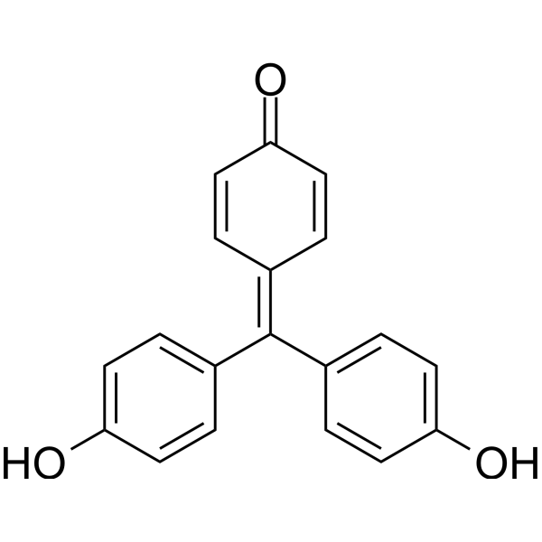
Rosolic acid
Rosolic acid is a Michael acceptor molecule isolated from the rhizomes of Plantago asiatica L. Rosolic acid inhibits Keap1, promotes Nrf2 nuclear translocation, and upregulates HO-1 and NQO-1. Rosolic acid reduces ER stress markers PERK, ATF-6, GRP78, and CHOP, as well as oxidative stress, ROS production, inflammation, and apoptosis. Rosolic acid upregulates SOD, CAT, and GPx activities, induces angiogenesis and insulin secretion, and restores endothelial function and pancreatic beta cell protection under ER stress. Rosolic acid can be used in research on diabetes and endothelial dysfunction.
For research use only. We do not sell to patients.
- CAS No.: 603-45-2
- Formula: C19H14O3
- Molecular Weight:290.31
-
Storage:
Please store the product under the recommended conditions in the Certificate of Analysis.

To place orders, for customer services and technical support, please contact: MedChemExpress USA
Tel: 609-228-6898 E-mail: [email protected] [email protected]


-
 Biological Activity
-
Chemical Information
- Protocols
- Purity & Documentation
- References
-
 Help & FAQs


-
HY-B0324A
Crystal VioletFluorochrome -
HY-W267364
Coumarin 343Fluorescent Probe -
HY-123630
Allura Red ACFood Colourant -

HY-DY2001
Crystal Violet (solution)Fluorochrome -
HY-W110794
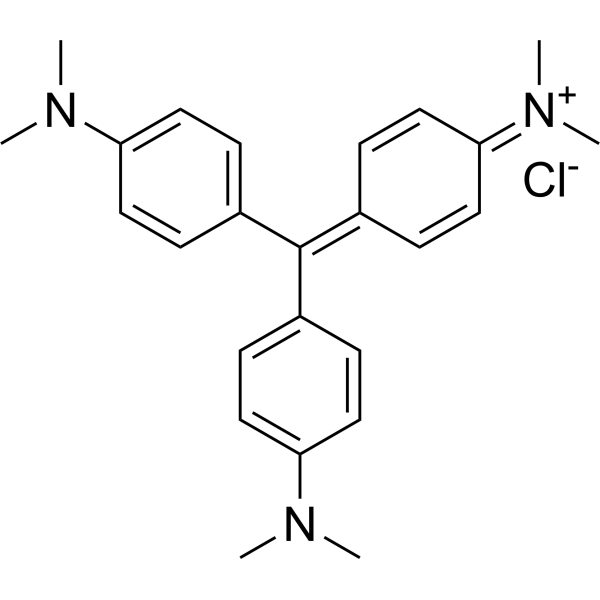
Crystal violet, IND (0.0-2.0)Biochemical Assay Reagent -
HY-112675
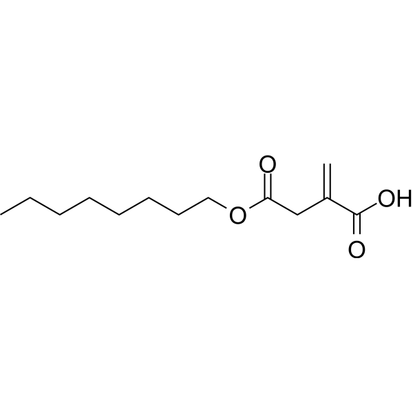
4-Octyl itaconateNrf2 Activator -
HY-D0958
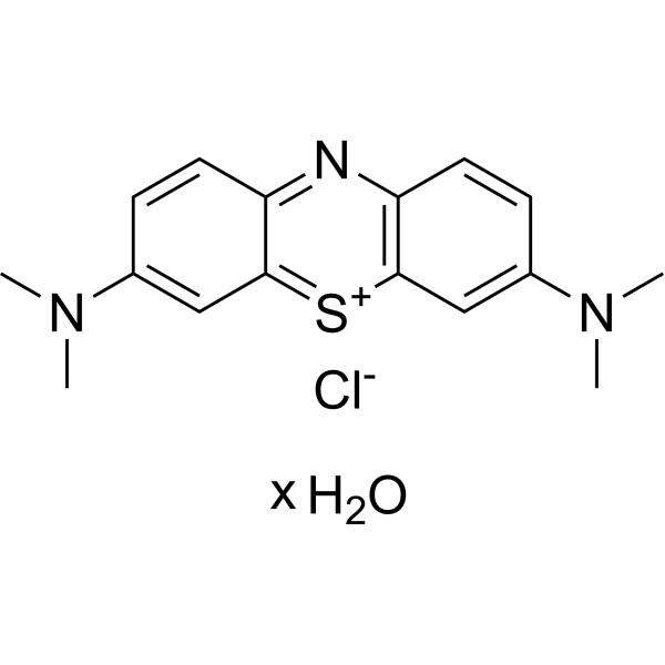
Methylene blue hydratesGC Inhibitor -

HY-W110909
Thiazole Yellow G, for microscopyBiochemical Reagent -

HY-123630R
Allura Red AC (Standard)Food Colourant -
HY-D0252
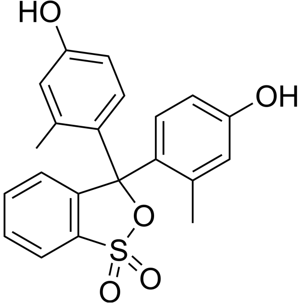
m-Cresol purplepH Indicator Dye -
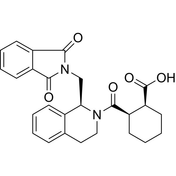
HY-110258
ML334NRF2 Activator -
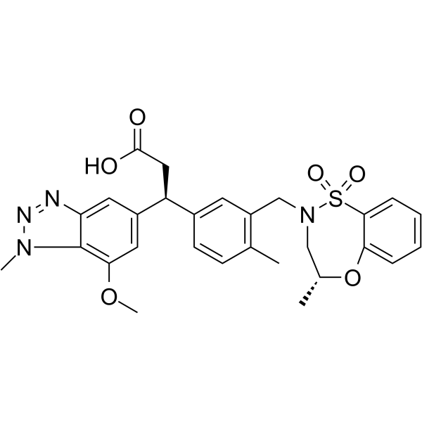
HY-101140
KI696KEAP1/NRF2 Protein-protein Interaction Inhibitor


Biological Activity
Description
In Vitro
Rosolic acid pretreatment (10 μM; 24 h) reduces Thapsigargin-induced ER stress markers GRP78 and CHOP in EA.hy926 cells in the co-culture setup[1].
Rosolic acid pretreatment (10 μM; 24 h) decreases GRP-78 and CHOP protein levels in EA.hy926 cells under Thapsigargin-induced ER stress in the co-culture setup[1].
Rosolic acid pretreatment (10 μM; 24 h) of EA.hy926 cells restores BCL2 expression in MIN6 pancreatic beta cells under Thapsigargin-induced ER stress in the co-culture setup[1].
Rosolic acid pretreatment (10 μM; 24 h) of EA.hy926 cells rescues PDX1 expression in MIN6 pancreatic beta cells under Thapsigargin-induced ER stress in the co-culture setup[1].
Rosolic acid pretreatment (10 μM; 24 h) of EA.hy926 cells normalizes GRP78 and CHOP expression in MIN6 pancreatic beta cells under Thapsigargin-induced ER stress in the co-culture setup[1].
Rosolic acid pretreatment (10 μM; 24 h) of EA.hy926 cells restores intracellular insulin levels in MIN6 pancreatic beta cells under Thapsigargin-induced ER stress in the co-culture setup[1].
Rosolic acid pretreatment (10 μM; 24 h) of EA.hy926 cells upregulates Nrf2 and NQO-1 expression in MIN6 pancreatic beta cells under Thapsigargin-induced ER stress in the co-culture setup[1].
Rosolic acid pretreatment (10 μM; 24 h) of EA.hy926 cells suppresses increased MDA levels in MIN6 pancreatic beta cells under Thapsigargin-induced ER stress in the co-culture setup[1].
MedChemExpress (MCE) has not independently confirmed the accuracy of these methods. They are for reference only.
-
Cell Line:EA.hy926 endothelial cells
-
Concentration:2.5 μM; 5 μM; 10 μM; 25 μM
-
Incubation Time:24 h
-
Result:Observed cytotoxicity at doses higher than 25 μM.\n
Protected cells against aThapsigargin-induced cytotoxicity at 5 and 10 μM in a dose-dependent manner.
-
Cell Line:EA.hy926 endothelial cells
-
Concentration:10 μM
-
Incubation Time:24 h
-
Result:Significantly repressed Thapsigargin-induced GRP78 and CHOP expression to levels comparable to control.\nUpregulated Nrf2, HO-1, and NQO-1 expression.
-
Cell Line:EA.hy926 endothelial cells
-
Concentration:10 μM
-
Incubation Time:24 h
-
Result:Decreased GRP-78 and CHOP protein levels compared to Thapsigargin-exposed cells.
-
Cell Line:MIN6 pancreatic beta cells
-
Concentration:10 μM
-
Incubation Time:24 h
-
Result:Restored BCL2 expression to control level.\nRescued PDX1 expression.\nUpregulated Nrf2 and NQO-1 gene expression.\nNormalized GRP78 and CHOP expression.
-
Cell Line:MIN6 pancreatic beta cells
-
Concentration:10 μM
-
Incubation Time:24 h
-
Result:Showed an intracellular insulin level comparable to control.
-
Cell Line:MIN6 pancreatic beta cells
-
Concentration:10 μM
-
Incubation Time:24 h
-
Result:Protected MIN6 pancreatic beta cells from Thapsigargin-induced loss of viability in a co-culture system.
-
Cell Line:Bovine aortic endothelial cells
-
Concentration:15 μM
-
Incubation Time:6 h
-
Result:Increased heme oxygenase activity to 2676 pmol bilirubin/mg protein/h compared to 453 pmol bilirubin/mg protein/h in control cells.
Achieved cell viability of 94% of control.
-
Cell Line:Bovine aortic endothelial cells
-
Concentration:15 μM
-
Incubation Time:6 h; 18 h
-
Result:Was the most potent inducer of HO-1 at 6 h among the compounds tested.
Further enhanced HO-1 expression at 18 h, whereas it was gradually decreasing with curcumin, CAPE, or 2'-hydroxychalcone.
-
Cell Line:Bovine aortic endothelial cells
-
Concentration:15 μM
-
Incubation Time:24 h; 48 h
-
Result:Did not produce any evident cytotoxicity at 24 or 48 h, unlike curcumin or CAPE, which caused a time-dependent decrease in cell viability.
-
Cell Line:Bovine aortic endothelial cells
-
Concentration:15 μM
-
Incubation Time:6 h pretreatment; 2 h
-
Result:Significantly attenuated hydrogen peroxide-mediated cytotoxicity in endothelial cells.
In Vivo
MedChemExpress (MCE) has not independently confirmed the accuracy of these methods. They are for reference only.
-
Animal Model:Wistar Albino rats (male, 100-150 g, 5-7 weeks old, high-fat diet-fed and streptozotocin-induced type-2 diabetic model)[2]
-
Dosage:5 mg/kg b.w.; 10 mg/kg b.w.
-
Administration:i.p.; once daily; 21 days
-
Result:Reduced cholesterol to 255.36 ± 6.83 mg/dL, triglycerides to 56.82 ± 5.63 mg/dL, VLDL to 11.38 ± 2.27 mg/dL, and LDL to 181.35 ± 11.25 mg/dL, and increased HDL to 61.28 ± 5.19 mg/dL at 10 mg/kg.
Decreased blood glucose levels starting from Day 7 at 10 mg/kg.
Improved glucose tolerance in OGTT starting from 60 minutes with peak reduction at 180 minutes.
Restored aortic tissue architecture at 5 and 10 mg/kg.
Increased NRF2 and downstream targets NQO1 and HO-1 gene expression, with a 1.8-fold increase in NRF2 protein levels at 10 mg/kg.
Dose-dependently reduced ER stress markers GRP78 and CHOP, inflammatory markers ICAM-1 and VCAM-1, and endothelin-1 expression with 5- and 2-fold reductions at 5 and 10 mg/kg, respectively.
-
Animal Model:Wistar rats (female)[2]
-
Dosage:1 mg/kg b.w.; 3 mg/kg b.w.; 10 mg/kg b.w.; 30 mg/kg b.w.
-
Administration:i.p.; daily; 21 days
-
Result:Induced hepatic toxicity characterized by cellular damage and structural alterations in liver tissues and significant impairment of serum liver function parameters at 30 mg/kg b.w.
Induced pronounced renal cell damage including structural alterations in renal tubules and glomeruli and a significant increase in blood urea nitrogen (BUN) levels at 30 mg/kg b.w.
No toxicity or functional impairments were observed at 1, 3, and 10 mg/kg.
- Human pluripotent stem cell endothelial-cell differentiation
- Vascular/Branching Fractal Analysis
- Annexin V plus membrane-impermeant dye apoptosis staining
- ROS/oxidative-stress fluorescent staining
- Human Islet Cell Culture
- Research Protocol for Inflammation-related Diseases
- Research Protocol for Metabolic Diseases
- Cytoplasmic-Nuclear Fractionated Protein Extraction
- Detection of Apoptosis
- TUNEL staining for apoptotic DNA fragmentation
- Research Protocol for Endocrine Diseases
- Endothelial Tube Formation Assay
- Apoptosis Solutions
- Endothelial Cell Migration/Angiogenic Sprouting Assay


Chemical Information
-
CAS No. 603-45-2
-
Appearance Solid
-
Molecular Weight 290.31
-
Formula C19H14O3
-
Color White to off-white
-
SMILES
C1=C(C=CC(=C1)O)C(=C2C=CC(=O)C=C2)C3=CC=C(C=C3)O
-
Shipping
Room temperature in continental US; may vary elsewhere.
-
Storage
Please store the product under the recommended conditions in the Certificate of Analysis.


Purity & Documentation
-
Data Sheet (292 KB)
-
SDS (252 KB)
- Français - FR (252 KB)
- Deutsch - DE (252 KB)
- Norwegian - NO (252 KB)
- Español - ES (252 KB)
- Swedish - SV (252 KB)
- Italian - IT (252 KB)
- Korean - KR (252 KB)
- Portuguese - PT (252 KB)
-
Handling Instructions (2659 KB)
References

Calculators
Concentration (start) × Volume (start) = Concentration (final) × Volume (final)


Rosolic acid Related Classifications
HY-W104821 Related Classifications
 Powered by Bioz