



RPI-1

Based on 1 Customer Validation
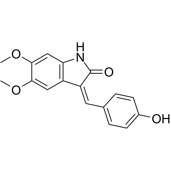
RPI-1 is a specific, orally available 2-indolinone Ret tyrosine kinase inhibitor. RPI-1 inhibits proliferation, Ret tyrosine phosphorylation, Ret protein expression, and the activation of PLCgamma, ERKs and AKT in human medullary thyroid carcinoma TT cells. Antitumor activity.
For research use only. We do not sell to patients.
- Purity : 98.47%
- CAS No.: 269730-03-2
- Formula: C17H15NO4
- Molecular Weight:297.31
-
Storage:Powder -20°C, 3 years , 4°C, 2 years ; In solvent -80°C, 6 months , -20°C, 1 month

To place orders, for customer services and technical support, please contact: MedChemExpress USA
Tel: 609-228-6898 E-mail: [email protected] [email protected]


-
 Biological Activity
-
Chemical Information
-
Solvent & Solubility
- Protocols
- Purity & Documentation
- References
-
 Help & FAQs


-
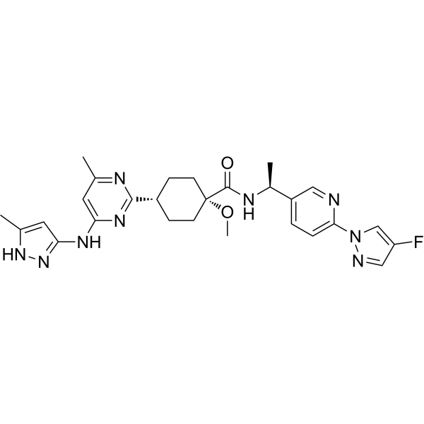
HY-112301
PralsetinibRET Inhibitor -
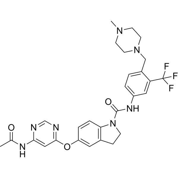
HY-18840
BBT594RET Inhibitor -

HY-112301R
Pralsetinib (Standard)RET Inhibitor -
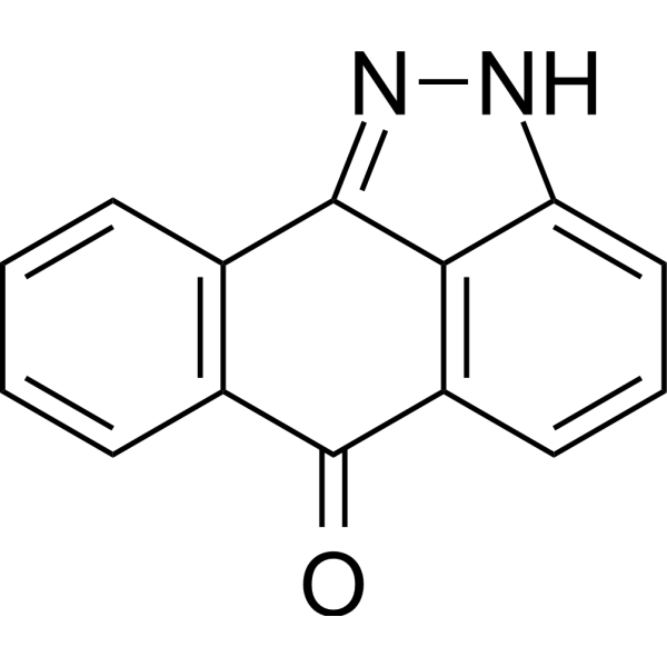
HY-12041
SP600125JNK Inhibitor -
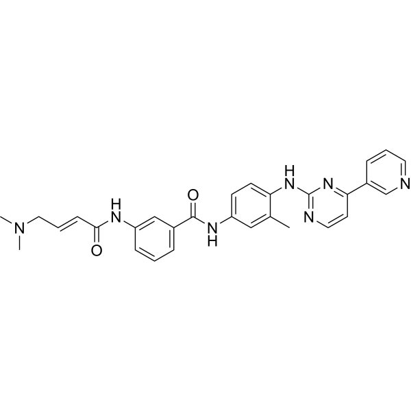
HY-13319
JNK-IN-8JNK Inhibitor -
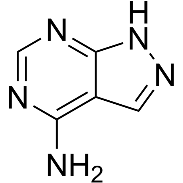
HY-34595
PyrazoloadenineRET Inhibitor -
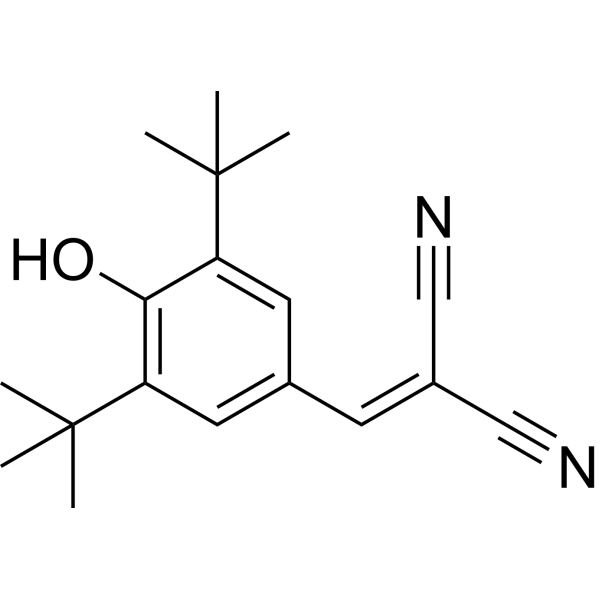
HY-15511
Tyrphostin A9PDGFR Inhibitor -
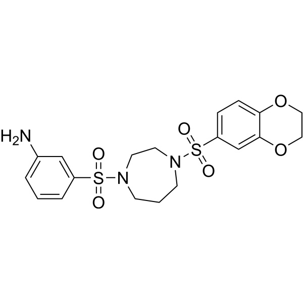
HY-19330
DASA-58Pyruvate Kinase Isozyme (PKM2) Allosteric Activator -
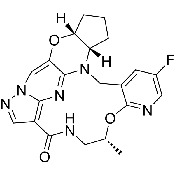
HY-148015
Enbezotinib (enantiomer)Enbezotinib Enantiomer -
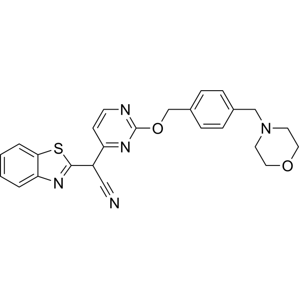
HY-14761
BentamapimodJNK Inhibitor -
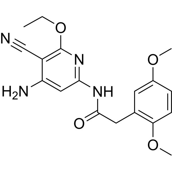
HY-107598
JNK Inhibitor VIIIJNK Inhibitor


Biological Activity
Description
In Vitro
TPC-1 cells are sensitive to the growth inhibitory effect of RPI-1 with an IC50 of 5.1 μM, following 72 hours of treatment. RPI-1 (7.5-60 µM) inhibits Ret/Ptc1 autophosphorylation in TPC-1 cells. RPI-1 inhibitory effects in the TPC-1 cell culture conditions lead to inhibition of pathways involving JNK2 and AKT[1].
The RPI-1 IC50 value for cell proliferation is 3.6 µM in NIH3T3 cells expressing the Ret mutant compared with 16 µM in non-transfected NIH3T3 cells, and that for colony formation in soft agar was 2.4 µM and 26 µM in RET mutant-transfected and H-RAS-transfected NIH3T3 cells, respectively. In NIH3T3 cells expressing the Ret mutant, Ret protein and tyrosine phosphorylation were undetectable after 24 hours of RPI-1 treatment. In TT cells, RPI-1 inhibits proliferation, Ret tyrosine phosphorylation, Ret protein expression, and the activation of PLCgamma, ERKs and AKT[2].
MedChemExpress (MCE) has not independently confirmed the accuracy of these methods. They are for reference only. Further protocols information, click here.
In Vivo
MedChemExpress (MCE) has not independently confirmed the accuracy of these methods. They are for reference only.
-
Animal Model:8- to 11-week-old female athymic nude CD-1 mice (bearing TT cells)[2]
-
Dosage:50, 100 mg/kg
-
Administration:P.o.; twice a day for 10 days
-
Result:A dose-dependent effect on tumor growth was observed, with a dose of 2*50 mg/kg/day resulting in less tumor weight inhibition than the 2*100mg/kg/day dose after 10 days of treatment.


Chemical Information
-
CAS No. 269730-03-2
-
Appearance Solid
-
Molecular Weight 297.31
-
Formula C17H15NO4
-
Color Yellow to brown
-
SMILES
O=C1NC2=C(C=C(OC)C(OC)=C2)/C1=C/C3=CC=C(O)C=C3
-
Shipping
Room temperature in continental US; may vary elsewhere.
-
Storage
Powder -20°C 3 years 4°C 2 years In solvent -80°C 6 months -20°C 1 month


Solvent & Solubility
In Vitro:
DMSO : 125 mg/mL (420.44 mM; Need ultrasonic; Hygroscopic DMSO has a significant impact on the solubility of product, please use newly opened DMSO)
Please refer to the solubility information to select the appropriate solvent. Once prepared, please aliquot and store the solution to prevent product inactivation from repeated freeze-thaw cycles.
Storage method and period of stock solution: -80°C, 6 months; -20°C, 1 month. When stored at -80°C, please use it within 6 months. When stored at -20°C, please use it within 1 month.
Please refer to the solubility information to select the appropriate solvent. Once prepared, please aliquot and store the solution to prevent product inactivation from repeated freeze-thaw cycles.
Storage method and period of stock solution: -80°C, 6 months; -20°C, 1 month. When stored at -80°C, please use it within 6 months. When stored at -20°C, please use it within 1 month.
Concentration (start) × Volume (start) = Concentration (final) × Volume (final)
In Vivo:
Select the appropriate dissolution method based on your experimental animal and administration route.
- For the following dissolution methods, please ensure to first prepare a clear stock solution using an In Vitro approach and then sequentially add co-solvents:
- To ensure reliable experimental results, the clarified stock solution can be appropriately stored based on storage conditions. As for the working solution for In Vivo experiments, it is recommended to prepare freshly and use it on the same day.
- The percentages shown for the solvents indicate their volumetric ratio in the final prepared solution. If precipitation or phase separation occurs during preparation, heat and/or sonication can be used to aid dissolution.
Add each solvent one by one: 10% DMSO 90% (20% SBE-β-CD in Saline)
Solubility: 2.08 mg/mL (7.00 mM); Suspended solution; Need ultrasonic
This protocol yields a suspended solution of 2.08 mg/mL. Suspended solution can be used for oral and intraperitoneal injection.
Taking 1 mL working solution as an example, add 100 μL DMSO stock solution (20.8 mg/mL) to 900 μL 20% SBE-β-CD in Saline, and mix evenly.
Preparation of 20% SBE-β-CD in Saline (4°C, storage for one week): 2 g SBE-β-CD powder is dissolved in 10 mL Saline, completely dissolve until clear.
In Vivo Dissolution Calculator
Please enter the basic information of animal experiments:
-
-
-
-
Recommended: Prepare an additional quantity of animals to account for potential losses during experiments.
Please enter your animal formula composition:
-
%DMSO +
Recommended: Keep the proportion of DMSO in working solution below 2% if your animal is weak.
-
%+
-
+%Tween-80 + +
-
%Saline +
The co-solvents required include: DMSO, . All of co-solvents are available by MedChemExpress (MCE). , Tween 80. All of co-solvents are available by MedChemExpress (MCE).
Working solution concentration: 0.22 mg/mL
Method for preparing stock solution: mg drug dissolved in μL DMSO. Stock solution concentration: mg/mL.
1. Take μL DMSO stock solution;
2. Add μL .
μL , mix evenly;
3. Then add μL Tween 80, mix evenly;
4. Then add μL
Please ensure that the stock solution in the first step is dissolved to a clear state, and add co-solvents in sequence. You can use ultrasonic heating (ultrasonic cleaner, recommended frequency 20-40 kHz), vortexing, etc. to assist dissolution.

Protocols
-
Kinase activity and phosphorylation assays
Kinase activity assays measure the ability of kinases to transfer phosphate groups from ATP to specific substrates, while phosphorylation assays detect the presence and levels of phosphorylated proteins. Common methods include radiolabeled ATP incorporation (e. g. ,), ADP release detection via bioluminescence (e. g. ,[3]), enzyme-linked immunosorbent assays (ELISA) for phospho-specific epitopes (e. g. ,[6]), and microtiter-based formats for high-throughput screening (e. g. ,[8]). The ADP-Glo assay quantifies kinase activity by measuring ADP produced during phosphorylation using a luciferase-based system. Radiometric assays involve autoradiography or scintillation counting after incorporation of 32P-labeled ATP into substrate proteins. ELISA-based approaches rely on phospho-specific antibodies to detect activated kinases in cell lysates or purified samples.
-
Western Blot
Western blotting (WB) is a commonly used experimental method in molecular biology, biochemistry, and immunogenetics for identifying and quantifying target proteins. It combines gel electrophoresis with immunoassay, enabling researchers to analyze protein expression, post-translational modifications, and molecular weight.
-
RNA extraction experimental
By lysing cells, releasing RNA, and removing impurities such as proteins and DNA, high-purity RNA products are finally obtained. The commonly used traditional method is the guanidine isothiocyanate/phenol/chloroform method (Trizol), which is suitable for a variety of animal materials including animal tissues, microorganisms, cultured cells, etc., and most plant materials.
-
Research Protocol for Endocrine Diseases
Endocrine diseases often arise from disrupted hormone production, hormone signaling, or target-tissue responsiveness; for diabetes-focused endocrine disease models, insulin signaling regulates glucose uptake, hepatic glucose output, lipid metabolism, and β-cell compensation. Type 2 diabetes develops through interacting defects in insulin resistance, β-cell dysfunction, adipose inflammation, hepatic glucose overproduction, altered incretin signaling, and ectopic lipid metabolism. A major unresolved question is whether endocrine dysfunction is driven primarily by target-tissue insulin resistance, intrinsic β-cell failure, immune/inflammatory stress, or combined multi-organ failure that differs by disease stage.
-
Protocol for Kinase activity and phosphorylation assays
Kinase activity assays measure transfer of phosphate from ATP to a protein or peptide substrate, generating phosphorylated substrate, ADP, or incorporated radiolabeled phosphate as the readout; phosphorylation assays measure site-specific phosphorylation in cells or tissues as a proxy for kinase-pathway activation, inhibition, or substrate regulation. Phosphorylation can be detected by phospho-specific Western blot, immunoprecipitation kinase assay, phospho-immunofluorescence, phospho-flow cytometry, luminescent ADP detection, radiolabeled ATP incorporation, or reporter-based pathway assays, and these readouts can be applied to cancer cells, primary neurons, mouse tumors, organoids, inflammatory macrophages, ferroptosis studies, and mitophagy studies when the kinase target is biologically relevant.
-
Subcutaneous Cell-Line-Derived Xenograft
Subcutaneous cell-line-derived xenograft (CDX) models are established by implanting cultured human cancer cell lines into immunodeficient mice, where the injected cells form localized tumors that can be monitored in vivo as a measure of tumorigenic potential, growth kinetics, and treatment response. These models are widely used in oncology research because they allow reproducible tumor formation and enable comparative assessment of tumor growth between different cell lines or genetic manipulations in a controlled in vivo microenvironment. Subcutaneous implantation of cancer cells in immunodeficient mice is a standard approach for evaluating tumor growth behavior and therapeutic response across multiple cancer types, including prostate, esophageal, pancreatic, and colon cancer models.
-
Cell Viability Determination by MTT Colorimetric Assay
The following protocol uses the MTT colorimetric assay as a classic literature-established method for assessing cell viability/metabolic activity in cultured mammalian cells. MTT[3-(4,5-dimethylthiazol-2-yl)-2,5-diphenyltetrazolium bromide] is reduced by metabolically active cells to a colored formazan product; the amount of formazan is quantified spectrophotometrically and provides an indirect measure of metabolically active viable cells. Importantly, MTT reduction reflects cellular oxidoreductase/metabolic activity rather than an absolute direct count of living cells, so changes in cellular metabolism can alter the signal independently of cell number.
-
Protocol For Protein Expression And Purification
Recombinant protein expression in Escherichia coli followed by purification of a His-tagged soluble protein by immobilized metal affinity chromatography (IMAC), with optional MBP fusion and TEV tag removal when the construct includes these elements. The biological readout is production of the encoded target protein, detected as an inducible band at the expected molecular mass by SDS-PAGE and quantified by total protein assay or chromatographic absorbance; the purification readout is enrichment of the target protein in elution fractions after selective binding of polyhistidine residues to immobilized Ni2+/metal-chelate resin and elution by imidazole-containing buffer. Expression is driven by an inducible bacterial expression system, commonly T7/lac-based, in which IPTG or lactose/auto-induction activates transcription and translation of the cloned gene; lower induction temperature, lower inducer concentration, induction timing, and solubility-enhancing fusion tags can influence the frac


Purity & Documentation
-
Data Sheet (274 KB)
-
SDS (398 KB)
- English - EN (398 KB)
- Français - FR (398 KB)
- Deutsch - DE (398 KB)
- Norwegian - NO (398 KB)
- Español - ES (398 KB)
- Swedish - SV (398 KB)
- Italian - IT (398 KB)
- Korean - KR (398 KB)
- Portuguese - PT (398 KB)
-
Handling Instructions (2659 KB)
References
[1]. Lanzi C, et al. Inactivation of Ret/Ptc1 oncoprotein and inhibition of papillary thyroid carcinoma cell proliferation by indolinone RPI-1. Cell Mol Life Sci. 2003;60(7):1449-1459. [Content Brief]
[2]. Cuccuru G, et al. Cellular effects and antitumor activity of RET inhibitor RPI-1 on MEN2A-associated medullary thyroid carcinoma. J Natl Cancer Inst. 2004;96(13):1006-1014. [Content Brief]


Complete Stock Solution Preparation Table
Please refer to the solubility information to select the appropriate solvent. Once prepared, please aliquot and store the solution to prevent product inactivation from repeated freeze-thaw cycles.
Storage method and period of stock solution: -80°C, 6 months; -20°C, 1 month. When stored at -80°C, please use it within 6 months. When stored at -20°C, please use it within 1 month.
| Optional Solvent | Concentration Solvent Mass | 1 mg | 5 mg | 10 mg | 25 mg |
|---|---|---|---|---|---|
| DMSO | 1 mM | 3.3635 mL | 16.8175 mL | 33.6349 mL | 84.0873 mL |
| 5 mM | 0.6727 mL | 3.3635 mL | 6.7270 mL | 16.8175 mL | |
| 10 mM | 0.3363 mL | 1.6817 mL | 3.3635 mL | 8.4087 mL | |
| 15 mM | 0.2242 mL | 1.1212 mL | 2.2423 mL | 5.6058 mL | |
| 20 mM | 0.1682 mL | 0.8409 mL | 1.6817 mL | 4.2044 mL | |
| 25 mM | 0.1345 mL | 0.6727 mL | 1.3454 mL | 3.3635 mL | |
| 30 mM | 0.1121 mL | 0.5606 mL | 1.1212 mL | 2.8029 mL | |
| 40 mM | 0.0841 mL | 0.4204 mL | 0.8409 mL | 2.1022 mL | |
| 50 mM | 0.0673 mL | 0.3363 mL | 0.6727 mL | 1.6817 mL | |
| 60 mM | 0.0561 mL | 0.2803 mL | 0.5606 mL | 1.4015 mL | |
| 80 mM | 0.0420 mL | 0.2102 mL | 0.4204 mL | 1.0511 mL | |
| 100 mM | 0.0336 mL | 0.1682 mL | 0.3363 mL | 0.8409 mL |


RPI-1 Related Classifications
HY-101246 Related Classifications
 Powered by Bioz