



SAR-442257

Based on 1 Customer Validation
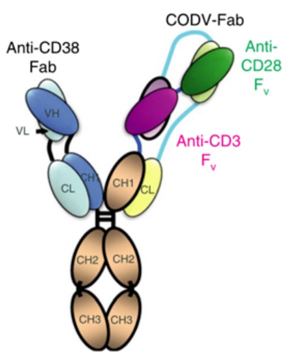
SAR-442257 is a trispecific T-cell engager and monoclonal antibody that binds to CD38, CD3 and CD28. SAR-442257 can target and bind to CD3 and CD28 on T cells, as well as CD38 expressed on tumor cells. SAR-442257 mediates T-cell activation, enhances T-cell activity, induces T-cell degranulation, and drives cytotoxicity against multiple myeloma cells. SAR-442257 functions via CD38 expression, and its in vitro activity correlates with CD38 expression on the surface of multiple myeloma cells. SAR-442257 can be used in studies related to multiple myeloma and relapsed/refractory multiple myeloma.
For research use only. We do not sell to patients.
- Purity : ≥99.0%
- Molecular Weight:145.44 kDa
-
Storage:
Please store the product under the recommended conditions in the Certificate of Analysis.

To place orders, for customer services and technical support, please contact: MedChemExpress USA
Tel: 609-228-6898 E-mail: [email protected] [email protected]


-
 Biological Activity
-
Chemical Information
- Protocols
- Purity & Documentation
- References
-
 Help & FAQs




Biological Activity
Description
Isotype
Human IgG4 kappa
Recommend Isotype Controls
Species Reactivity
Human
IC50 & Target
CD28 & CD38
In Vitro
SAR-442257 (0.001-10 nM; 48 h) potently reduces primary multiple myeloma cell viability ex vivo with picomolar EC50 valuesl[1].
SAR-442257 (1 nM; 48 h) induces significant degranulation of endogenous CD3+ T cells in bone marrow mononuclear cells (MNCs) derived from multiple myeloma samples[1].
SAR-442257 (1 nM; 48 h)-mediated cytotoxicity against primary multiple myeloma cells is dependent on the presence of endogenous CD3+ T cells[1].
SAR-442257 (1 nM; 48 h) response in primary multiple myeloma samples has surface CD38 expression (6-fold higher MFI on multiple myeloma cells vs. non-myeloma cells) as the strongest biomarker, while a very low E:T ratio may contribute to resistance[1].
SAR-442257 (1 nM, 10 nM, 100 nM, 1000 nM; 7.5 min) enhances T cell-to-multiple myeloma cell binding avidity, inducing a 1.5-fold higher frequency of bound T cells in U266 and MM1S cell lines compared to the CD38-CD3 bispecific antibody[2].
SAR-442257 (0.01-16 nM; 24 h) potently induces dose-dependent cytotoxicity against 10 human multiple myeloma cell lines, with EC50 values of 0.02 nM (MM1S) and 0.2 nM (U266), and reduces cell viability by 58-97% at 1 nM after 24 h[2].
MedChemExpress (MCE) has not independently confirmed the accuracy of these methods. They are for reference only. Further protocols information, click here.
-
Cell Line:MM1S, U266, AMO, AMO-TP53, RPMI 8226, INA-6, OPM-2, LP-1, KMS-11BM, KMS-11 (human multiple myeloma cell lines); healthy donor CD3-positive T cells
-
Concentration:0.01 nM, 0.02 nM, 0.1 nM, 0.2 nM, 0.5 nM, 1 nM, 10 nM, 16 nM; 1 nM (10 cell line panel)
-
Incubation Time:24 h
-
Result:Induced dose-dependent myeloma cell death, with EC50 values of 0.02 nM for MM1S and 0.2 nM for U266, and EC75 values of 16 nM for MM1S and 0.5 nM for U266.
Significantly reduced viability in all 10 tested myeloma cell lines at 1 nM, with viability reduction ranging from 58% to 97% after 24 h.
In Vivo
SAR442257 stimulates memory/effector T cell proliferation, reduces regulatory T cells, and is well-tolerated in non-human primates[2].
MedChemExpress (MCE) has not independently confirmed the accuracy of these methods. They are for reference only.
Clinical Trial
| NCT Number | Sponsor | Condition | Start Date |
Phase

|
|---|---|---|---|---|
| NCT01329991 | Plexxikon| | 2011-05 | PHASE1 |
Gene ID
Accession
Conjugated
Unconjugated
Reconsititution
The product can be reconstituted/diluted with sterile PBS or saline.
Format
-
trispecific antibody
Application
ELISA, FACS, Functional assay
Verified Bioactivity
-
 Flow cytometric analysis of 1X10⁶ Raji cells with SAR-442257 (HY-P991183, red). Cells were fixed with 4% paraformaldehyde and permeabilised with 90% methanol. Then stained with the primary antibody at 1/50 dilution for an hour at 4℃. Alexa Fluor 488-conjugated AffiniPure Goat Anti-Human IgG H&L (HY-P83776) was used as the secondary antibody at 1/1,000 dilution for 30 minutes at 4℃. Human IgG4 kappa (HY-P99003, blue) was used as the isotype control, cells without incubation with primary antibody were used as the unlabeled control (black).
 -
 Flow cytometric analysis of 1X10⁶ Jurkat cells with SAR-442257 (HY-P991183, red). Cells were fixed with 4% paraformaldehyde. Then stained with the primary antibody at 1/200 dilution for an hour at 4℃. Alexa Fluor 488-conjugated AffiniPure Goat Anti-Human IgG H&L (HY-P83776) was used as the secondary antibody at 1/1,000 dilution for 30 minutes at 4℃. Human IgG4 kappa (HY-P99003, blue) was used as the isotype control, cells without incubation with primary antibody were used as the unlabeled control (black).
 -
 Immobilized CD28-mFC can bind SAR-442257. The EC50 for this effect is 300.8 ng/mL.


Chemical Information
-
Appearance Liquid
-
Molecular Weight 145.44 kDa
-
Color Colorless to light yellow
-
SMILES
[*]S[R442257]
-
Shipping
Shipping with dry ice.
-
Formulation
Please refer to the lot-specific COA for specific buffer information.
-
Storage
Please store the product under the recommended conditions in the Certificate of Analysis.

Protocols
-
RNA extraction experimental
By lysing cells, releasing RNA, and removing impurities such as proteins and DNA, high-purity RNA products are finally obtained. The commonly used traditional method is the guanidine isothiocyanate/phenol/chloroform method (Trizol), which is suitable for a variety of animal materials including animal tissues, microorganisms, cultured cells, etc., and most plant materials.
-
Cell Cytotoxicity Assay
Cytotoxicity assays are usually based on the assessment of cell membrane damage, which can also be indirectly detected by measuring cell viability. Detection methods include MTT assay, CKK-8 assay, LDH assay and ATP assay, etc.
-
Mammalian live/dead viability and cytotoxicity staining
Live/dead viability and cytotoxicity staining assays are based on the simultaneous detection of intracellular esterase activity in metabolically active (viable) cells and membrane integrity loss in non-viable cells. In commonly used dual-staining approaches, membrane-permeant fluorogenic substrates are converted by intracellular esterases into fluorescent products in live cells, while impermeant DNA-binding dyes selectively enter cells with compromised plasma membranes and label nucleic acids in dead or dying cells, enabling discrimination between viable and non-viable populations by fluorescence microscopy or flow cytometry.
-
Cell Viability Determination by MTT Colorimetric Assay
The following protocol uses the MTT colorimetric assay as a classic literature-established method for assessing cell viability/metabolic activity in cultured mammalian cells. MTT[3-(4,5-dimethylthiazol-2-yl)-2,5-diphenyltetrazolium bromide] is reduced by metabolically active cells to a colored formazan product; the amount of formazan is quantified spectrophotometrically and provides an indirect measure of metabolically active viable cells. Importantly, MTT reduction reflects cellular oxidoreductase/metabolic activity rather than an absolute direct count of living cells, so changes in cellular metabolism can alter the signal independently of cell number.


Purity & Documentation
-
Data Sheet (262 KB)
-
SDS (251 KB)
- English - EN (251 KB)
- Français - FR (251 KB)
- Deutsch - DE (251 KB)
- Norwegian - NO (251 KB)
- Español - ES (251 KB)
- Swedish - SV (251 KB)
- Italian - IT (251 KB)
- Korean - KR (251 KB)
- Portuguese - PT (251 KB)
-
Inhibitory Antibodies User Guide (603 KB)
References
[1]. Keller AL, et al. Ex Vivo Efficacy of SAR442257 Anti-CD38 Trispecific T-cell Engager in Multiple Myeloma Relapsed After Daratumumab and BCMA-targeted Therapies. Cancer research communications. 2024 Mar 12;4(3):757-764. [Content Brief]
[2]. Grab AL, et al. Pre-Clinical Assessment of SAR442257, a CD38/CD3xCD28 Trispecific T Cell Engager in Treatment of Relapsed/Refractory Multiple Myeloma. Cells. 2024 May 20;13(10):879. [Content Brief]

Calculators
Concentration (start) × Volume (start) = Concentration (final) × Volume (final)


SAR-442257 Related Classifications
HY-P991183 Related Classifications
 Powered by Bioz