



ODN 2216
Based on 1 publication(s) in Google Scholar
ODN 2216 is a type A CpG oligodeoxynucleotide vaccine adjuvant and a TLR9 agonist. ODN 2216 interacts with TLR9 in the lysosomes of CD4+ T cells and activates feedback-dependent signaling pathways. ODN 2216 induces the production of type I interferons, IL-6 and TGF-β via the IRAK4/IRF7 axis, while increasing intracellular ATP levels. ODN 2216 not only induces the differentiation of CD4+ T cells into anti-inflammatory Th3-like regulatory phenotypes to inhibit autologous proliferation, but also enhances the specific CD8+ T cell-mediated cytotoxicity against Mammaglobin-A in breast cancer cells. ODN 2216 is widely used in studies related to breast cancer and systemic lupus erythematosus.
For research use only. We do not sell to patients.
- Purity : 99.34%
- CAS No.: 332437-00-0
- Molecular Weight:6432.00
-
Storage:
-20°C, sealed storage, away from moisture
* In solvent : -80°C, 6 months; -20°C, 1 month (sealed storage, away from moisture)


To place orders, for customer services and technical support, please contact: MedChemExpress USA
Tel: 609-228-6898 E-mail: [email protected] [email protected]


-
 Biological Activity
-
Chemical Information
- Protocols
- Purity & Documentation
- References
-
 Help & FAQs


-
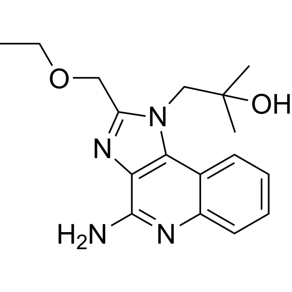
HY-13740
ResiquimodTLR7/8 Agonist -

HY-146244
AgatolimodTLR9 Agonist -

HY-150725
ODN 1585IFN/TNFα Inducer -
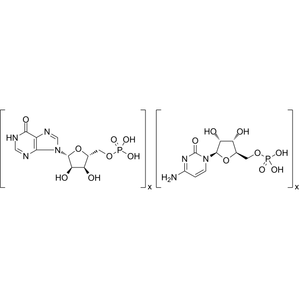
HY-107202
Polyinosinic-polycytidylic acidTLR-3 Agonist -
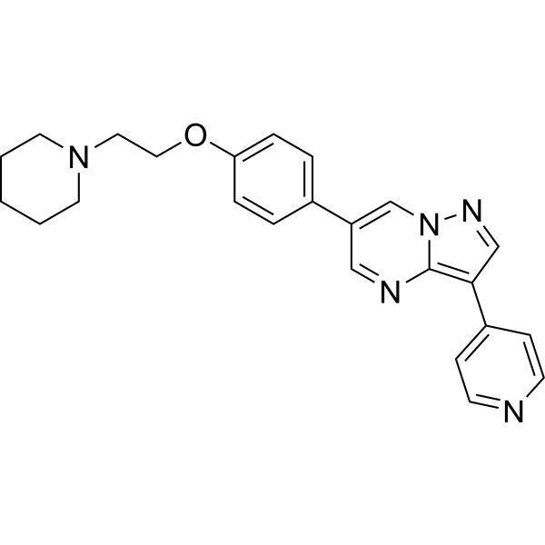
HY-13418A
DorsomorphinAMPK Inhibitor -
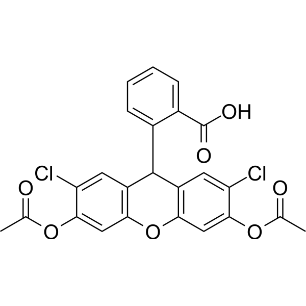
HY-D0940
H2DCFDAROS-Sensitive Probe -
HY-18749
SC79Akt Activator -
HY-150738
ODN 2088TLR3/7/9 Inhibitor -
HY-12512
cGAMPSTING Activator -
HY-B0180
ImiquimodTLR7 Agonist -
HY-Y0873
PEG300Neutral Polymer -
HY-112754A
DOTAP chlorideCationic Lipid
Publications Citing Use of MedChemExpress (MCE) ODN 2216
More

Biological Activity
Description
IC50 & Target
|
TLR9 |
IL-12 |
In Vitro
ODN 2216 (10 μM; 16 h pre-treatment, 9 days co-culture, 48 h cytotoxicity assay) does not significantly enhance MamA2.1-specific activation of primary HLA-A2+ CD8+ T cells or subsequent MamA-specific cytotoxicity against AU565 breast cancer cells compared to MamA2.1 peptide alone[1].
ODN 2216 (3 μM; 24 h) induces TLR9-mediated IFN-α secretion by human PBMCs after 24 hours of incubation at a concentration of 3 μM[2].
ODN 2216 (0.5-10 μM; 4 days) dose-dependently increases intracellular ATP and IL-6 levels in human PBMCs[2].
ODN 2216 (500 ng/mL; 14 hours) physically interacts with intracellular TLR9 in lysosomes of human CD4+ CD25- T cells following 14-hour incubation with 500 ng/mL ODN 2216 FITC[3].
ODN 2216 (500 ng/mL; 72 hours) induced increased proliferation of human CD4+ CD25- T cells, which is dependent on TLR9 signaling via TLR9, MyD88, IRAK1, and TRAF3, but not IRF7[3].
ODN 2216 (500 ng/mL; 48 hours) treated human CD4+ CD25- T cells acquire dose-dependent suppressive activity against untreated autologous CD4+ T cells, with maximum suppression (48.57%) at a 4:1 suppressor:responder ratio[3].
MedChemExpress (MCE) has not independently confirmed the accuracy of these methods. They are for reference only. Further protocols information, click here.
-
Cell Line:Thymic DC, pDC
-
Concentration:2 μM
-
Incubation Time:15 h
-
Result:Significantly decreased apoptosis of thymic DC (23.1 ± 0.9%), and naturally induced apoptosis of thymic pDC was not decreased (72.8 ± 2.9%).
In Vivo
MedChemExpress (MCE) has not independently confirmed the accuracy of these methods. They are for reference only.


Chemical Information
-
CAS No. 332437-00-0
-
Appearance Solid
-
Molecular Weight 6432.00
-
Color White to off-white
-
SMILES
[DNA, d(G-sp-G-sp-G-G-G-A-C-G-A-T-C-G-T-C-G-sp-G-sp-G-sp-G-sp-G-sp-G)]
-
Sequence
DNA, d(G-sp-G-sp-G-G-G-A-C-G-A-T-C-G-T-C-G-sp-G-sp-G-sp-G-sp-G-sp-G)
-
Shipping
Room temperature in continental US; may vary elsewhere.
-
Storage
-20°C, sealed storage, away from moisture
* In solvent : -80°C, 6 months; -20°C, 1 month (sealed storage, away from moisture)


Publications (1)
-
Journal Impact Factor
-
Most Recent
-
Immunity
Polyamine metabolism controls B-to-Z DNA transition to orchestrate DNA sensor cGAS activity. [Abstract]2023 Nov 14;56(11):2508-2522.e6. PMID: 37848037

Protocols
-
Cell Cytotoxicity Assay
Cytotoxicity assays are usually based on the assessment of cell membrane damage, which can also be indirectly detected by measuring cell viability. Detection methods include MTT assay, CKK-8 assay, LDH assay and ATP assay, etc.
-
Mammalian live/dead viability and cytotoxicity staining
Live/dead viability and cytotoxicity staining assays are based on the simultaneous detection of intracellular esterase activity in metabolically active (viable) cells and membrane integrity loss in non-viable cells. In commonly used dual-staining approaches, membrane-permeant fluorogenic substrates are converted by intracellular esterases into fluorescent products in live cells, while impermeant DNA-binding dyes selectively enter cells with compromised plasma membranes and label nucleic acids in dead or dying cells, enabling discrimination between viable and non-viable populations by fluorescence microscopy or flow cytometry.
-
Research Protocol for Inflammation-related Diseases
The NLRP3 inflammasome is a cytosolic innate immune signaling platform that integrates priming signals and danger-signal activation to promote caspase-1 activation, maturation of IL-1β and IL-18, and gasdermin D-mediated pyroptotic cell death. The core experimental logic is to determine whether inflammatory disease phenotypes are driven by increased NLRP3 expression, ASC-containing inflammasome assembly, caspase-1 cleavage, GSDMD cleavage, and extracellular release of IL-1β/IL-18 rather than by nonspecific cell injury alone. The pathway is strongly linked to inflammation-related disease phenotypes because monosodium urate crystals activate NALP3/NLRP3 inflammasome signaling in gout-like crystal inflammation, cholesterol crystals activate NLRP3 inflammasomes in atherogenesis models, and DSS-induced intestinal inflammation has been reported to involve NLRP3 inflammasome activity. However, experimental colitis studies also show context-dependent protective effects of NLRP3 inflammasome co
-
Breast Cancer Modeling
Breast cancer is a heterogeneous cancer, and it has been distinguished into four subtypes: luminal A, luminal B, HER2-positive and basal-like. Molecular mutations, epigenetic alterations, hormone exposure and immune microenvironment are related to the progression of breast cancer.
-
Research Protocol for Cancer Immunology
Cancer immunology studies how the immune system recognizes, suppresses, edits, or fails to eliminate malignant cells through tumor antigen release, antigen presentation, T-cell priming, immune trafficking, tumor-cell killing, and feedback inhibition in the tumor microenvironment. The cancer-immunity cycle links tumor antigenicity, dendritic-cell priming, CD8+ T-cell infiltration, cytotoxic function, and immune-checkpoint regulation to tumor rejection or immune escape. Immune-checkpoint pathways such as PD-1/PD-L1 and CTLA-4 suppress antitumor T-cell activity and can be therapeutically blocked, but many tumors remain resistant because of poor antigen presentation, weak T-cell infiltration, suppressive myeloid cells, regulatory T cells, and tumor-intrinsic immune-exclusion programs. Unresolved questions include which immune-cell states predict response, how tumor-intrinsic pathways exclude immune cells, how myeloid suppression limits checkpoint blockade, and which combination strategies
-
Cell Viability Determination by MTT Colorimetric Assay
The following protocol uses the MTT colorimetric assay as a classic literature-established method for assessing cell viability/metabolic activity in cultured mammalian cells. MTT[3-(4,5-dimethylthiazol-2-yl)-2,5-diphenyltetrazolium bromide] is reduced by metabolically active cells to a colored formazan product; the amount of formazan is quantified spectrophotometrically and provides an indirect measure of metabolically active viable cells. Importantly, MTT reduction reflects cellular oxidoreductase/metabolic activity rather than an absolute direct count of living cells, so changes in cellular metabolism can alter the signal independently of cell number.


Purity & Documentation
-
Data Sheet (271 KB)
-
SDS (251 KB)
- English - EN (251 KB)
- Français - FR (251 KB)
- Deutsch - DE (251 KB)
- Norwegian - NO (251 KB)
- Español - ES (251 KB)
- Swedish - SV (251 KB)
- Italian - IT (251 KB)
- Korean - KR (251 KB)
- Portuguese - PT (251 KB)
-
Handling Instructions (2242 KB)
References
[1]. Krug A, et al. Identification of CpG oligonucleotide sequences with high induction of IFN-alpha/beta in plasmacytoid dendritic cells. Eur J Immunol. 2001 Jul;31(7):2154-63. [Content Brief]
[2]. Okada T, et al. Murine thymic plasmacytoid dendritic cells. Eur J Immunol. 2003 Apr;33(4):1012-9. [Content Brief]
[3]. Huang L, et al. CpG-based immunotherapy impairs antitumor activity of BRAF inhibitors in a B-cell-dependent manner. Oncogene. 2017 Jul 13;36(28):4081-4086. [Content Brief]
[4]. He Y, et al. Hepatic mitochondrial DNA/Toll-like receptor 9/MicroRNA-223 forms a negative feedback loop to limit neutrophil overactivation and acetaminophen hepatotoxicity in mice. Hepatology. 2017 Jul;66(1):220-234. [Content Brief]
[5]. Li Y, et al. A novel antagonist of TLR9 blocking all classes of immunostimulatory CpG-ODNs. Vaccine. 2011 Mar 3;29(11):2193-8. [Content Brief]

Calculators
Concentration (start) × Volume (start) = Concentration (final) × Volume (final)


ODN 2216 Related Classifications
HY-150741 Related Classifications
 Powered by Bioz