
56 Results for "

Protein staining

" in MedChemExpress (MCE) Product Catalog:
Products (56)

56 Results for "Protein staining" in MCE Product Catalog:

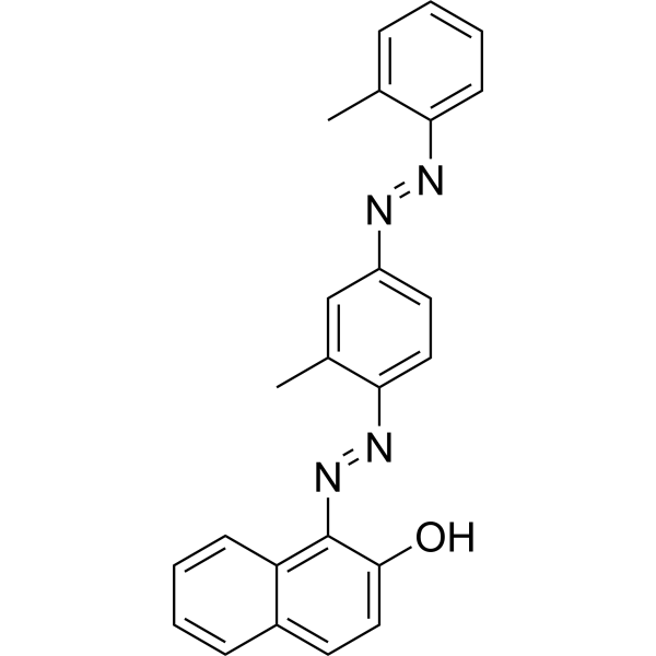
Cat. No.: HY-D0932
CAS No.: 85-83-6
Synonyms: Solvent Red 24; C.I. 26105
Sudan IV is an agonist of the aryl hydrocarbon receptor (AhR) that activates downstream signaling pathways and induces CYP1A1 expression. Sudan IV promotes CYP1A1 gene transcription by activating AhR-ARNT heterodimers and binding to exogenous response elements (XREs) on DNA, thereby enhancing drug metabolizing enzyme activity. Sudan IV can be used to study the toxicity mechanisms of industrial dyes and the effects of interactions with serum proteins (such as bovine serum albumin (BSA)) on their distribution in vivo. Sudan IV is a fat-soluble diazo dye that can be used to stain lipids, triglycerides, and lipoproteins on frozen sections .
loading...
    loading...
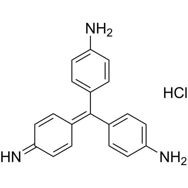
Cat. No.: HY-W127770
CAS No.: 569-61-9
Synonyms: Basic red 9
Target:  

Fluorescent Dye

Research Areas:  

Infection

Pararosaniline hydrochloride (Basic red 9) is a pH-responsive basic dye, as a biological stain to track certain proteins. The pH of the acidified Pararosaniline hydrochloride reagent has a significant effect on the color and the maximum absorption wavelength (λmax) of the reaction system, with its optimum pH 0.48 and a λmax at 549 nm. Pararosaniline hydrochloride is also a strong modifier of RNA splicing. Pararosaniline hydrochloride has been used in the analysis of SO2 and formaldehyde and staining of bacteria or other organisms. Pararosaniline hydrochloride is extensively used in industries like textile, printing, paper, cosmetic, and leather .
loading...
    loading...
Cat. No.: HY-W088068
CAS No.: 68988-92-1
Wright's stain is a composite cell stain that mainly binds to intracellular nucleic acids, proteins and other components through thiazine dyes (such as methylene blue) and eosin. Wright's stain is pH-dependent (optimal pH 6.4-6.7) and achieves cell morphology resolution by differentially staining the cytoplasm and nucleus. Under alkaline conditions, thiazine dyes bind to nucleic acids to form purple, and acidic eosin binds to cytoplasmic proteins to form red, which can form contrasting cell morphological features. Wright's stain can clearly display the fine structures of blood cells and bone marrow cells (such as nuclear chromatin and granules) and quickly evaluate cell morphological abnormalities .
loading...
    loading...
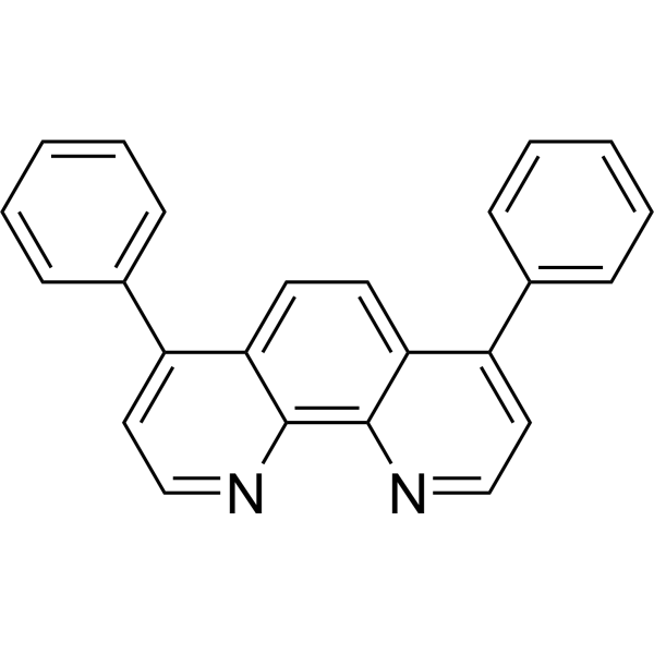
Cat. No.: HY-D0006
CAS No.: 1662-01-7
Bathophenanthroline is a nitrogen-containing organic ligand that can form stable complexes with transition metal ions such as Ru 2+. In proteomics, Bathophenanthroline is often used for in-gel protein staining. The negatively charged sulfonic acid groups of Bathophenanthroline interact electrostatically with the positively charged amino acid residues of proteins, thereby achieving specific binding to proteins and enhancing the fluorescent signal of detection. Bathophenanthroline can be used for protein separation and detection in SDS-PAGE gels in the field of proteomics, as well as other fluorescence studies[1]. Ex/Em=532 nm/610 nm[1].
loading...
    loading...
Cat. No.: HY-DY1032
CAS No.: 201860-17-5
Mito-Tracker Green (solution) is a green fluorescent dye that selectively accumulates in the mitochondrial matrix. MitoTracker Green FM covalently binds mitochondrial proteins by reacting with free mercaptan of cysteine residues, allowing staining of mitochondrial membrane potential independent of membrane potential. Excitation/emission wavelength 490/523 nm.
Solvent and concentration: DMSO: 1 mM
loading...
    loading...
Cat. No.: HY-D1805
Vari Fluor 647-Streptavidin is a dye marker of Vari Fluor-streptavidin consisting of labeling streptavidin with a Vari Fluor series of fluorescent probes. Streptavidin is a high-affinity tetramer protein, each tetramer consisting of four identical streptavidin subunits. Streptavidin binds to biotin specifically via a reversible non-covalent effect. Streptavidin can achieve rapid and efficient detection of biotin markers, and is often used in immunofluorescence (IF), enzyme-linked immunosorbent assay (ELISA), immunohistochemical staining (IFH), in situ hybridization (ISH) and other experiments. Ex/Em=650 nm/665 nm.
Product Concentration: 0.8 mg/mL
loading...
    loading...
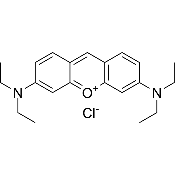
Cat. No.: HY-D1543
CAS No.: 2150-48-3
Pyronin B is an organic cationic dye used for the staining of bacteria, mycobacteria and ribonucleic acids. Pyronin B is also used as a small hydrophobic (SH) protein channel inhibitor .
loading...
    loading...
Cat. No.: HY-W133919
CAS No.: 28631-66-5
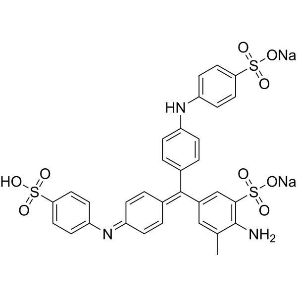
Aniline Blue sodium is a water-soluble dye commonly used as a biological stain for the detection of nucleic acids and proteins in various laboratory procedures such as electrophoresis and microscopy. Aniline Blue sodium has unique chemical properties that allow it to bind to specific cellular components, producing a color change that facilitates their visualization and analysis.
loading...
    loading...
Cat. No.: HY-W250147
CAS No.: 2580-56-5
Synonyms: Victoria blue B
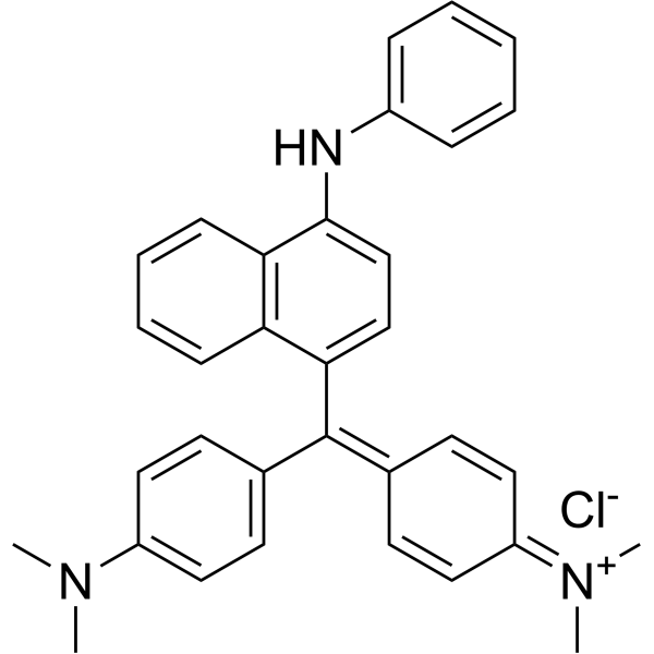
Basic blue 26 (Victoria blue B) is a synthetic cationic dye belonging to the class of triarylmethane dyes. It has a bright blue color and is commonly used as a colorant for a variety of applications, including textiles, paper and leather. Basic Blue 26 is also used as a biological stain for DNA and protein detection in laboratories. Due to its ability to bind negatively charged materials, it can be used as an indicator of the presence of specific molecules in biological samples. However, Basic blue 26 has been reported to have potentially harmful effects on human health and the environment and its use is regulated in some countries. Proper handling and disposal procedures are necessary to minimize its impact on the environment.
loading...
    loading...
Cat. No.: HY-DY1009
CAS No.: 150347-59-4
CFDA-SE (solution) is a fluorescent dye that can penetrate the cell membrane. It can react with the free amine group in the cytoskeleton protein inside the cell, and finally form a protein complex with fluorescence. After entering the cell, CFDA-SE locates in the cell membrane, cytoplasm and nucleus, and the fluorescence staining is strongest in the nucleus . CFDA-SE dye can be uniformly inherited by the cells with cell division and proliferation, and its attenuation is proportional to the number of cell divisions. This phenomenon can be detected and analyzed by flow cytometry under the excitation light of 488 nm, and can be used to detect the proliferation of cells .
Solvent and concentration: DMSO: 5 mM
loading...
    loading...
Cat. No.: HY-D1807
Vari Fluor 555-Streptavidin is a dye marker of Vari Fluor-streptavidin consisting of labeling streptavidin with a Vari Fluor series of fluorescent probes. Streptavidin is a high-affinity tetramer protein, each tetramer consisting of four identical streptavidin subunits. Streptavidin binds to biotin specifically via a reversible non-covalent effect. Streptavidin can achieve rapid and efficient detection of biotin markers, and is often used in immunofluorescence (IF), enzyme-linked immunosorbent assay (ELISA), immunohistochemical staining (IFH), in situ hybridization (ISH) and other experiments. Ex/Em=555 nm/565 nm.
Product Concentration: 0.8 mg/mL
loading...
    loading...
Cat. No.: HY-D2170
AF488 streptavidin is a fluorescently labeled streptavidin. AF488 streptavidin is a streptavidin conjugated to Alexa Fluor 488, with 4 fluorophores per protein molecule, enabling stoichiometric fluorescent labeling of cell surface targets. AF488 streptavidin can form a complex with biotinylated E07 aptamer to stain cells expressing EGFR, and the staining is reversible after treatment with mA9 detoxifying oligonucleotides (Ex/Em = 470/520 nm) .
loading...
    loading...
Cat. No.: HY-DY1015
CAS No.: 104821-25-2
Dihydroethidium (solution) , also known as DHE, is a peroxide indicator. Dihydroethidium penetrates cell membranes to form a fluorescent protein complex with blue fluoresces. After entering the cells, Dihydroethidium is mainly localized in the cell membrane, cytoplasm and nucleus, and the staining effect is the strongest in the nucleus. Dihydroethidium produces inherent blue fluorescence with a maximum excitation wavelength of 370 nm and a maximum emission wavelength of 420 nm; after dehydrogenation, Dihydroethidium combines with RNA or DNA to produce red fluorescence with a maximum excitation wavelength of 300 nm and a maximum emission wavelength of 610 nm. 535 nm can also be used as the excitation wavelength for actual observation .
Solvent and concentration: DMSO: 5 mM
loading...
    loading...
Cat. No.: HY-D1544
CAS No.: 14541-90-3
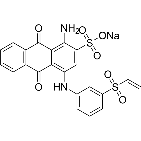
Uniblue A sodium is a reactive protein stain that can be used in the covalent pre-gel staining of the protein (Ex=594 nM) .
loading...
    loading...
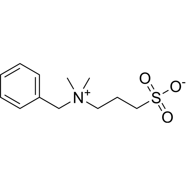
Cat. No.: HY-W011831
CAS No.: 81239-45-4
Synonyms: 3-(Benzyldimethylammonio)propane-1-sulfonate; PDA
NDSB-256 is a non-stain remover sulfabetaine. NDSB-256 prevents protein aggregation and promotes denaturation of chemically and thermally denatured proteins .
loading...
    loading...
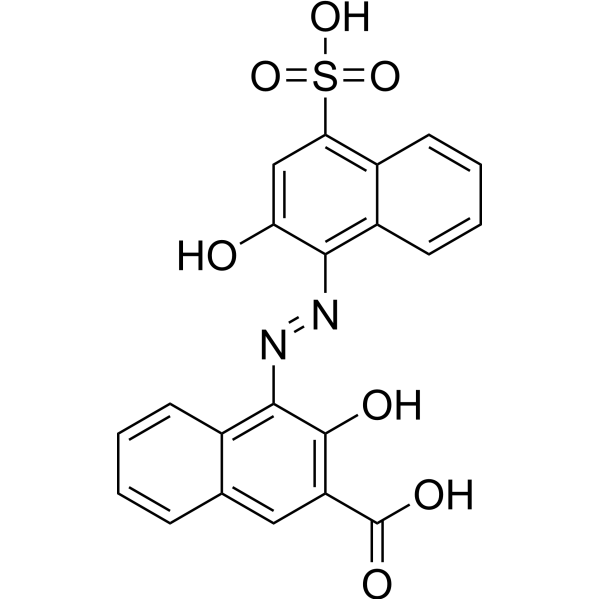
Cat. No.: HY-Y0700
CAS No.: 3737-95-9
Calconcarboxylic acid, an azo dye, acts as a silver-ion sensitizer to stain protein in SDS-PAGE gels. Calconcarboxylic acid increases silver binding on protein bands or spots by the formation of a silver-dye complex and also increases the reducing power o
loading...
    loading...
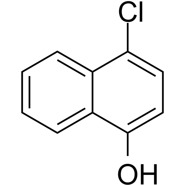
Cat. No.: HY-W007967
CAS No.: 604-44-4
Research Areas:  

Others

4-Chloronaphthalen-1-ol is a substrate of horseradish peroxidase. 4-Chloronaphthalen-1-ol can be used to visualize protein bands in western blotting and immunohistochemical staining .
loading...
    loading...
Cat. No.: HY-D1804
Vari Fluor 680-Streptavidin is a dye marker of Vari Fluor-streptavidin consisting of labeling streptavidin with a Vari Fluor series of fluorescent probes. Streptavidin is a high-affinity tetramer protein, each tetramer consisting of four identical streptavidin subunits. Streptavidin binds to biotin specifically via a reversible non-covalent effect. Streptavidin can achieve rapid and efficient detection of biotin markers, and is often used in immunofluorescence (IF), enzyme-linked immunosorbent assay (ELISA), immunohistochemical staining (IFH), in situ hybridization (ISH) and other experiments. Ex/Em=680 nm/701 nm.
Product Concentration: 0.8 mg/mL
loading...
    loading...
Cat. No.: HY-D1809
Vari Fluor 405-Streptavidin is a dye marker of Vari Fluor-streptavidin consisting of labeling streptavidin with a Vari Fluor series of fluorescent probes. Streptavidin is a high-affinity tetramer protein, each tetramer consisting of four identical streptavidin subunits. Streptavidin binds to biotin specifically via a reversible non-covalent effect. Streptavidin can achieve rapid and efficient detection of biotin markers, and is often used in immunofluorescence (IF), enzyme-linked immunosorbent assay (ELISA), immunohistochemical staining (IFH), in situ hybridization (ISH) and other experiments. Ex/Em=405 nm/431 nm.
Product Concentration: 0.8 mg/mL
loading...
    loading...
Cat. No.: HY-D0932R
CAS No.: 85-83-6
Synonyms: Solvent Red 24 (Standard); C.I. 26105 (Standard)
Sudan IV (Standard) (Solvent Red 24 (Standard)) is the analytical standard of Sudan IV (HY-D0932). This product is intended for research and analytical applications. Sudan IV is an agonist of the aryl hydrocarbon receptor (AhR) that activates downstream signaling pathways and induces CYP1A1 expression. Sudan IV promotes CYP1A1 gene transcription by activating AhR-ARNT heterodimers and binding to exogenous response elements (XREs) on DNA, thereby enhancing drug metabolizing enzyme activity. Sudan IV can be used to study the toxicity mechanisms of industrial dyes and the effects of interactions with serum proteins (such as bovine serum albumin (BSA)) on their distribution in vivo. Sudan IV is a fat-soluble diazo dye that can be used to stain lipids, triglycerides, and lipoproteins on frozen sections.
loading...
    loading...

