
1301 Results for "

two-photon confocal fluorescence microscope

" in MedChemExpress (MCE) Product Catalog:
Products (1301)

1301 Results for "two-photon confocal fluorescence microscope" in MCE Product Catalog:

Cat. No.: HY-D3075
CAS No.: 2490504-23-7
Target:  

Fluorescent Dye

Research Areas:  

Others

TPE-2E N-oxide is a fluorescent probe used for hypoxia detection and tumor hypoxia imaging. Due to its active intramolecular motion, TPE-2E N-oxide shows no fluorescence in aqueous solution. In hypoxic environments, its N-oxide group undergoes two-electron reduction by CYP450 reductase and other heme protein reductases expressed under hypoxia, forming hydrophobic aggregates that trigger aggregation-induced emission through the restriction of intramolecular motion. TPE-2E N-oxide exhibits selective lipid droplet localization in cells. The absorption wavelength of TPE-2E N-oxide in dichloromethane is 313 nm; in a 99% hexane/dichloromethane mixed system, its excitation wavelength is 330 nm and emission wavelength is 460 nm. For in vitro cell imaging, excitation is performed at 405 nm, with an emission wavelength range of 430-560 nm .
loading...
    loading...
Cat. No.: HY-D3210
CAS No.: 2098632-04-1
Target:  

Fluorescent Dye

Research Areas:  

Inflammation/Immunology

DCM-KPV is a fluorescent probe targeting the human intestinal oligopeptide transporter PEPT1/SLC15A1 receptor (λex=480 nm, λem=620-670 nm). DCM-KPV specifically binds to PepT1 via its KPV domain and mediates receptor-targeted internalization, thus effectively accumulating in the cytoplasm and nucleus of cells overexpressing this receptor. DCM-KPV has the advantages of long emission wavelength, high emission efficiency, low photobleaching, and negligible cytotoxicity. DCM-KPV maintains stable fluorescence intensity under continuous illumination, exhibiting extremely high live cell compatibility. DCM-KPV can specifically accumulate at colonic inflammatory sites through the intestinal mucosa, enabling direct non-invasive visual differentiation between chronic and acute ulcerative colitis groups and the normal group .
loading...
    loading...
Cat. No.: HY-D3276
Target:  

Fluorescent Dye

Research Areas:  

Others

PE-Cy7 is a tandem dye conjugate based on fluorescence resonance energy transfer (FRET). Composed of phycoerythrin (PE) as the donor and Cy7 as the acceptor fluorescent dye, PE-Cy7 is suitable for multicolor flow cytometry, laser scanning cytometry and immunofluorescence applications. PE-Cy7 can be conjugated to antibodies or used in the biotin-streptavidin system to label cell surface antigens in immunophenotyping and multiparametric cell analysis. PE-Cy7 undergoes photon-induced oxidative degradation, whose rate depends on light intensity and storage conditions, leading to reduced FRET efficiency and increased PE leakage. PE-Cy7 can be excited at wavelengths of 488 nm, 532 nm or ~760 nm, and its emission signal can be detected using 780/60 nm, 810/90 nm or 785/50 nm bandpass filters, with a Stokes shift of 300 nm .
loading...
    loading...
Cat. No.: HY-D3525
Target:  

Fluorescent Dye

Research Areas:  

Others

BDL-E5 is a fluorescent probe for detecting live induced pluripotent stem cells at the early reprogramming stage. Its detection mechanism involves specific binding to authentic reprogramming cells, which exhibit increased expression of pluripotency and epithelial genes and decreased expression of mesenchymal genes compared to non-reprogramming cells; it appears to co-localize more significantly with the Golgi complex than other organelle markers in reprogramming cells, and it does not require washing after staining to reduce background signals; fluorescence is generated upon binding to these early reprogramming cells, with positive cells appearing 7 days before iPS colonies are visible and stain positive for the conventional pluripotent marker TRA-1-60, and sorting BDL-E5-positive cells enriches populations that generate higher numbers and quality of iPS colonies. Its detection wavelength is Ex/Em = 578/599 nm .
loading...
    loading...
Cat. No.: HY-DY1007
CAS No.: 3326-32-7
Target:  

Fluorescent Dye

Research Areas:  

Inflammation/Immunology

FITC (Fluorescein Isothiocyanate) (solution) , is one of the green fluorescein derivatives widely used in biology. FITC has the characteristics of high absorptivity and excellent fluorescence quantum yield. The isothiocyanate group of FITC can be combined with amino, sulfhydryl, imidazole, tyrosyl, carbonyl and other groups on the protein, so as to achieve protein labeling including antibodies and lectins. In addition to its use as a protein marker, FITC can also be used as a fluorescent protein tracer to rapidly identify pathogens by labeling antibodies, or for microsequencing of proteins and peptides (HPLC). The maximum excitation wavelength of FITC is 494 nm. Once excited, it fluoresces yellow-green at a maximum emission wavelength of 520 nm. In addition, FITC is also a hapten that can induce contact hypersensitivity (CHS) and induce an atopic dermatitis model .
Solvent and concentration: DMSO: 20 mM
The 1 mL volume is defined as the base specification. All larger sizes correspond to incremental volumes of this base.
loading...
    loading...
Cat. No.: HY-108667
CAS No.: 61368-63-6
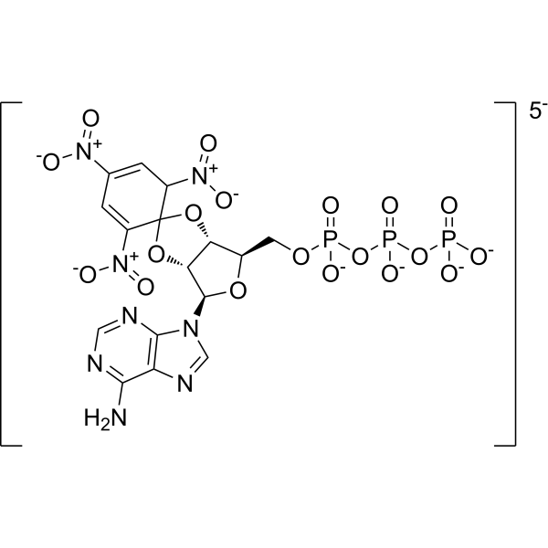
TNP-ATP triethylammonium is a P2X receptor antagonist with an IC50 of 0.010 μM for P2X3 and an IC50 of 0.062 μM for P2X2/3. TNP-ATP triethylammonium acts as an inhibitor of CheA autophosphorylation, with a Ki of 0.7 µM. TNP-ATP triethylammonium blocks the functional activation of P2X1-7 receptors. TNP-ATP triethylammonium attenuates hypoxia-induced IL-1β expression and release. TNP-ATP triethylammonium alleviates visceral pain, and improves hypoxia-induced cognitive impairment, insufficient myelination and neuroinflammation. Binding of TNP-ATP triethylammonium to CheA enhances the fluorescence of the TNP group. TNP-ATP triethylammonium can be used in studies related to visceral pain. NP-ATP triethylammonium can be used in studies related to hypoxia-induced insufficient myelination and cognitive decline .
loading...
    loading...
Cat. No.: HY-111137
CAS No.: 916890-10-3
Synonyms: XC-302 free base
Target:  

Akt

Research Areas:  

Cancer

Puquitinib (XC-302 free base) is a multi-target inhibitor with the activity of inducing autophagy in nasopharyngeal carcinoma cells by inhibiting the PI3K/AKT/mTOR signaling pathway. Puquitinib was able to inhibit the proliferation of CNE-2 cells, showing a dose-dependent antiproliferative effect. Puquitinib induced the formation of autophagosomes and autolysosomes in CNE-2 cells, which were observed by fluorescence microscopy and electron microscopy. Puquitinib promoted the formation of LC3-II and increased the expression of beclin 1, while reducing the level of p62. Puquitinib inhibited the PI3K/AKT/mTOR pathway by reducing the expression of p-AKT and p-mTOR. Puquitinib also induced apoptosis in CNE-2 cells, and when autophagy was inhibited, the apoptosis rate was reduced, which means that autophagy may interact with apoptosis to induce cell death .
loading...
    loading...
Cat. No.: HY-184603
Bovine serum albumin-modified gold nanoclusters (BSA-Au NCs) are nanomaterials combining gold nanoclusters and bovine serum albumin (BSA). Composed of several to dozens of gold atoms forming the core of the gold nanoclusters, BSA acts as a protective ligand, coating the surface of the gold nanoclusters. BSA-Au NCs are widely used due to their high luminescence properties and stability over a wide pH range. When Au(III) ions are introduced into a BSA solution, BSA acts as a scaffold protein, isolating and capturing Au ions, similar to the biomineralization behavior of inorganic ions in organisms in nature. The captured Au ions are then reduced in situ by BSA to form Au NCs. The synthesized Au NCs consist of 25-Au atoms and are stable in BSA as BSA-Au NCs, exhibiting strong red fluorescence. BSA-Au NCs possess good biocompatibility and their surface is easily modified or functionalized, making them attractive for many biomedical applications.
loading...
    loading...
Cat. No.: HY-D3003
CAS No.: 2522921-17-9
M-H2S is an endoplasmic reticulum-targeted ratio-type near-infrared fluorescent probe (Ex/Em : 480 nm/560 nm) specifically designed for the detection of hydrogen sulfide (H2S) in living cells and zebrafish. M-H2S exhibits detection limit for H2S of 39.1 nM, and the fluorescence intensity ratio (F₆₅₀/F₅₆₀) within the range of 0-40 μM shows a linear relationship with the H2S concentration. M-H2S responds best under pH = 7.2 (the normal physiological pH of the endoplasmic reticulum) conditions, making it suitable for the detection of the microenvironment of the endoplasmic reticulum. M-H2S can be used to study the role of H2S in endoplasmic reticulum function, stress response, and related diseases .
loading...
    loading...
Cat. No.: HY-D3072
Research Areas:  

Neurological Disease

P6-Aggrate is a fluorescent probe for aggregated proteome detection. P6-Aggrate specifically recognizes amorphous aggregated proteomes through non-covalent reversible binding, and its fluorescence enhances after heat-induced protein aggregation. P6-Aggrate reflects the polarity and compactness heterogeneity within aggregated proteomes via emission wavelength shift: short-wavelength emission (blue shift) corresponds to large aggregates with high compactness, while long-wavelength emission (red shift) corresponds to small spots with low compactness (Ex/Em = 488/520-580 nm). P6-Aggrate enables reversible monitoring of the dynamic processes of formation and clearance of stress-induced proteome aggregation such as that induced by MG132 (HY-13259) in living cells. P6-Aggrate can be used in studies related to protein homeostasis imbalance, neurodegenerative diseases and protein aggregation .
loading...
    loading...
Cat. No.: HY-D3074
Research Areas:  

Others

TCM-4COOH is an AIE-active fluorescent probe for calcium ions. TCM-4COOH relies on aggregation-induced emission activity, and its carboxyl chelating group can bind to Ca 2+ and Mg 2+ to form insoluble aggregates, triggering fluorescence enhancement via restricting intramolecular motion (Ex/Em = 477/603 nm). TCM-4COOH shows pH independence in physiological environments (pH 4-9), can be strongly activated at Ca 2+ concentrations ranging from low nM to mM, and has a stronger binding ability to Ca 2+ than to Mg 2+. TCM-4COOH, as its acetoxymethyl ester derivative TCM-AM, can penetrate cell membranes for intracellular detection. TCM-4COOH can be used for imaging of cellular calcium overload, detection of bone microcracks, and studies related to calcium-rich biological environments .
loading...
    loading...
Cat. No.: HY-D3401
CAS No.: 3121511-17-6
Research Areas:  

Others

LHA585 is a fluorescent probe targeting the lipid hydrogen abstraction process for the early detection of ferroptosis (Ferroptosis). Its detection mechanism relies on the reactivity of its own 4-phenyl-3-methylbut-2-enyl moiety with biologically relevant oxidative radicals, including peroxyl radicals (ROO·), alkoxyl radicals (RO·), hydroxyl radicals (HO·), and peroxynitrite anions (ONOO−); upon hydrogen abstraction from this structure, radical rearrangement and radical cleavage occur, releasing a xanthene radical which converts into a highly fluorescent rhodamine dye in biological environments, thereby achieving a fluorescence turn-on ratio of up to 360-fold. LHA585 is non-fluorescent in its native state, possesses excellent membrane permeability due to its non-polar property, and the rhodamine dye generated after oxidation diffuses into mitochondria. The excitation wavelength of LHA585 is 561 nm, and its emission wavelength ranges from 570 to 620 nm .
loading...
    loading...
Cat. No.: HY-DY1114
CAS No.: 53213-81-3
DiOC5(3) (solution) is a mitochondrial Fluorescent dye that alters membrane potential, and it also acts as an inhibitor of NADH-ubiquinone reductase (respiratory complex I). DiOC5(3) (solution) increases the NADH/NAD ratio and induces excessive production of ROS. DiOC5(3) (solution) induces Apoptosis. DiOC5(3) accumulates in the mitochondria of leukemia stem-like cells via organic anion-transporting polypeptides, specifically labels these cells, and inhibits their proliferation. DiOC5(3) (solution) enhances the cellular uptake of Hoechst 33342 (HY-15559), reduces Hoechst 33342-associated cytotoxicity, and improves the resolution of DNA content analysis by flow cytometry. DiOC5(3) (solution) shows no detectable fluorescence signal when excited in the 400-500 nm ultraviolet light range. DiOC5(3) (solution) is applicable to leukemia-related research .
Solvent and concentration: DMSO: 10 mM
loading...
    loading...
Cat. No.: HY-160062
Target:  

Mucin

Research Areas:  

Cancer

S2.2 aptamer sodium is a nucleic acid-based MUC1-binding aptamer with high affinity and low toxicity. Upon binding to its target, S2.2 aptamer sodium undergoes a conformational switch and restores fluorescence signal, serving as a targeted imaging agent for MUC1-positive cancer cells. S2.2 aptamer sodium enables targeted delivery to breast cancer cells with overexpressed MUC1. When formulated as the S2.2-PEG-MZF molecular probe, S2.2 aptamer sodium possesses the functions of T2 signal inhibition, magnetic field-induced hyperthermia and targeted magnetic resonance molecular imaging. In the S2.2-PEG-MZF/DOX nanoliposome, S2.2 aptamer sodium supports targeted thermochemotherapy, effectively inhibiting cancer cell proliferation and invasion as well as inducing apoptosis, and is widely used in studies related to breast cancer .
loading...
    loading...
Cat. No.: HY-D3187
CAS No.: 2484831-02-7
HMRef-αMan is a substrate-based green fluorescent probe (Ex/Em=465 nm/515 nm) targeting MAN2C1 (α-mannosidase). HMRef-αMan can be specifically cleaved by MAN2C1 to generate a highly fluorescent product, which thus gets activated to produce green fluorescence in malignant breast tissues, benign lesions and living cancer cells. The signal intensity of HMRef-αMan is directly correlated with MAN2C1 activity, and it can effectively detect tiny breast cancer lesions with a diameter of less than 1 mm. When used in combination with the red-emitting γ-glutamyl transpeptidase (GGT) probe gGlu-2OMe SiR600 (HY-D3188), HMRef-αMan enables precise optical differentiation of breast tissue types via a dual-color imaging strategy. HMRef-αMan has been widely used in the research of breast diseases such as breast cancer, fibroadenoma, phyllodes tumor and various types of papilloma .
loading...
    loading...
Cat. No.: HY-D3416
Synonyms: TAMRA-2,4-dinitroaniline
Target:  

Fluorescent Dye

Research Areas:  

Others

TMR-DN (TAMRA-2,4-dinitroaniline) is a fluorescent probe for RNA imaging that consists of a covalently coupled fluorophore (5-carboxy tetramethylrhodamine (TMR)) and quencher (dinitroaniline (DN)). The Kd value of TMR-DN with SRB-2 is 35 nM, and its Kd value with RhoBAST is approximately 30 nM. TMR-DN has a contact quenching function and forms a non-fluorescent intramolecular ground-state dimer in the free state; the excitation/emission wavelengths of the free probe are Ex/Em = 557/579 nm, those of the complex formed by binding to SRB-2 are Ex/Em = 561/587 nm, and those of the probe bound to the DNB aptamer are Ex/Em = 555/582 nm. TMR-DN reduces non-specific binding to genomic nucleic acids via negatively charged carboxyl-functionalized aromatic rings. Fluorescence dequenching occurs when TMR-DN specifically binds to SRB-2, RhoBAST or the DNB aptamer, among which the binding of RhoBAST requires Mg 2+ for proper folding .
loading...
    loading...
Cat. No.: HY-N16300
CAS No.: 3091879-75-0
Target:  

Fluorescent Dye

Research Areas:  

Others

Mito-laurdan (bromide) is a Fluorescent dye for mitochondrial membrane fluidity and packing measurement. It depends on membrane potential for targeting: it contains a cationic triphenylphosphonium moiety that accumulates at the inner mitochondrial membrane due to the membrane's negative potential, connected via a 3-carbon linker. As a solvatochromic dye derived from Laurdan (HY-D0080), it retains the characteristic mechanism where fluorescence emission shifts based on membrane packing: in more fluid, loosely packed membranes, greater water penetration leads to increased dipolar relaxation and a bathochromic red shift in emission, while more ordered membranes show a blue-shifted emission; these shifts are quantified using generalized polarization, calculated from emission intensities in ordered and disordered spectral windows. Its excitation wavelength is 352 nm, with emission detected in ordered (409-463 nm) and disordered (473-516 nm) spectral windows. It localizes specifically to mitochondria in HeLa cells, showing high signal overlap with MitoTracker Deep Red, and can detect changes in inner mitochondrial membrane fluidity .
loading...
    loading...
Cat. No.: HY-W327027
CAS No.: 314742-00-2
Target:  

Fluorescent Dye

Research Areas:  

Others

7-(2,4-Dinitrophenoxy)-4-methyl-2H-chromen-2-one (Compound 1) is a fluorescent probe for the detection of hydrogen sulfide (H2S). 7-(2,4-Dinitrophenoxy)-4-methyl-2H-chromen-2-one has a low detection limit (4×10 -6 mol/L), good selectivity and high sensitivity. 7-(2,4-Dinitrophenoxy)-4-methyl-2H-chromen-2-one shows almost no cytotoxicity at concentrations of 150 µg/mL. 7-(2,4-Dinitrophenoxy)-4-methyl-2H-chromen-2-one has the excitation peak of 331 nm, and the emission peak about 385 nm in DMSO solvent. Upon the addition of increasing amounts of HS -, the fluorescence intensity increases obviously at about 392 nm .
loading...
    loading...
Cat. No.: HY-170316
Target:  

Ras

Research Areas:  

Cancer

Ibetazol is a Importin β1 (KPNB1) inhibitor and nucleocytoplasmic transport disruptor. Ibetazol binds covalently to Cys585 of Importin β1, blocks both Importin β1-mediated direct transport and Importin α-dependent nuclear import processes, without affecting transport mediated by other nucleocytoplasmic transport proteins. Ibetazol induces cytoplasmic accumulation of Importin α1, and inhibits nuclear import of substrates carrying nuclear localization signals (NLS), including the NLS-cMyc reporter gene, RelA/p65 and SREBP1. Ibetazol triggers spindle malformation and chromosome misalignment by disrupting the mitotic function of Importin β1. Ibetazol inhibits the proliferation of cells expressing wild-type Importin β1. Ibetazol has a high activity-cytotoxicity window, lacks intrinsic fluorescence, and acts rapidly on nucleocytoplasmic transport processes. Ibetazol serves as a tool compound for investigating nuclear import processes specifically mediated by Importin β1 .
loading...
    loading...
Cat. No.: HY-W585442
CAS No.: 105528-25-4
5,9,14,18,23,27,32,36-Octabutoxy-2,3-naphthalocyanine is a photosensitive material with excellent light absorption and electron conduction activity. 5,9,14,18,23,27,32,36-Octabutoxy-2,3-naphthalocyanine is widely used in optoelectronic devices and is considered to be an effective photocatalyst. 5,9,14,18,23,27,32,36-Octabutoxy-2,3-naphthalocyanine can be used to improve the performance of solar cells and increase the photoelectric conversion efficiency. 5,9,14,18,23,27,32,36-Octabutoxy-2,3-naphthalocyanine also has potential anti-tumor activity and can inhibit the proliferation of certain cancer cells. 5,9,14,18,23,27,32,36-Octabutoxy-2,3-naphthalocyanine exhibits excellent fluorescence properties in medical imaging, which helps to improve the clarity and accuracy of imaging. 5,9,14,18,23,27,32,36-Octabutoxy-2,3-naphthalocyanine is studied as a component of a novel compound delivery system to improve the targeting and release effect of the compound.
loading...
    loading...

