



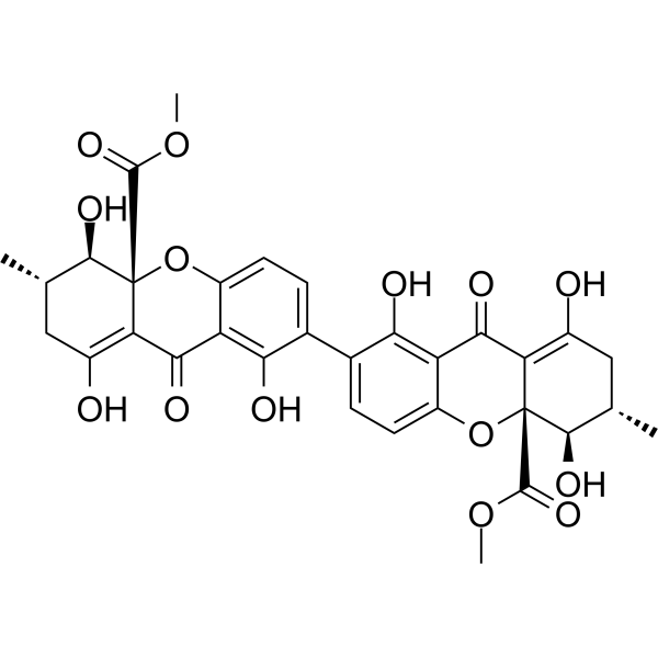
Secalonic acid D

Based on 1 Customer Validation
Secalonic acid D is a toxic compound against tumor cells. Secalonic acid D can be isolated from the metabolites of Aspergillus aculeatus. Secalonic acid D activates GSK3-β, and degrades β-catenin. Thus, Secalonic acid D down-regulates c-Myc expression, arrests cell cycle at G1 phase, induces cell apoptosis.
商品は「研究用試薬」です。人や動物の医療用・臨床診断用・食品用の製品ではありません。
研究用途以外に使用した場合、当社は一切の責任を負いかねます。
- 純度 : 99.63%
- CAS 番号: 35287-69-5
- 分子式: C32H30O14
- 分子量:638.57
-
保管条件:
-20°C, protect from light
* In solvent : -80°C, 6 months; -20°C, 1 month (protect from light)

To place orders, for customer services and technical support, please contact: MedChemExpress USA
Tel: 609-228-6898 E-mail: [email protected] [email protected]


-
 生物活性
生物活性
-
化学情報
-
溶剤 & 溶解度
- プロトコル
- 純度とドキュメンテーション
- 参考文献
-
 Help & FAQs




生物活性
製品説明
Cellular Effect
|
Cell Line

|
Type | Value | Description | References |
|---|---|---|---|---|
| A549 | IC50 |
2.1 μM
Compound: 7
|
Cytotoxicity against human A549 cells after 72 hrs by MTT assay
Cytotoxicity against human A549 cells after 72 hrs by MTT assay
|
[PMID: 26506221] |
| HCT-116 | IC50 |
0.9 μM
Compound: 7
|
Cytotoxicity against human HCT116 cells after 72 hrs by MTT assay
Cytotoxicity against human HCT116 cells after 72 hrs by MTT assay
|
[PMID: 26506221] |
| HL-60 | IC50 |
1 μM
Compound: 7
|
Cytotoxicity against human HL60 cells after 72 hrs by MTT assay
Cytotoxicity against human HL60 cells after 72 hrs by MTT assay
|
[PMID: 26506221] |
| K562 | IC50 |
2 μM
Compound: 7
|
Cytotoxicity against human K562 cells after 72 hrs by MTT assay
Cytotoxicity against human K562 cells after 72 hrs by MTT assay
|
[PMID: 26506221] |
| MGC-803 | IC50 |
1.5 μM
Compound: 7
|
Cytotoxicity against human MGC803 cells after 72 hrs by MTT assay
Cytotoxicity against human MGC803 cells after 72 hrs by MTT assay
|
[PMID: 26506221] |
体外実験
Secalonic acid D (0.1-1 μM; 72 h) inhibits cell proliferation in a concentration-dependent manner in both HL60 and K562, with IC50s of 0.38 μM and 0.43 μM, respectively[1].
Secalonic acid D (0.3 μM; 12 h, 24 h) causes cell cycle arrest of G1 phase and (1.2 μM; 0-72 h) induces apoptosis in HL60 and K562 cells[1].
MedChemExpress (MCE) has not independently confirmed the accuracy of these methods. They are for reference only.
-
Cell Line:HL60 and K562 cells
-
Concentration:1.2 μM
-
Incubation Time:0, 24, 48 and 72 h
-
Result:Induced cell apoptosis via arresting cell cycle at G1 phase. As well as, down-regulated the expression level of c-Myc.
-
Cell Line:HL60 and K562 cells
-
Concentration:1.2 μM
-
Incubation Time:0, 24, 48 and 72 h
-
Result:Dose-dependently decreases the level of Caspase-1.


化学情報
-
CAS 番号 35287-69-5
-
性状 Solid
-
分子量 638.57
-
分子式 C32H30O14
-
Color Light yellow to yellow
-
SMILES
COC([C@]12C(C(C3=C(O)C(C4=CC=C(O[C@@]5(C(C6=O)=C(C[C@@H]([C@H]5O)C)O)C(OC)=O)C6=C4O)=CC=C3O1)=O)=C(C[C@@H]([C@H]2O)C)O)=O
-
Structure Classification
-
Initial Source
aspergillus
-
輸送条件
Room temperature in continental US; may vary elsewhere.
-
保管条件
-20°C, protect from light
* In solvent : -80°C, 6 months; -20°C, 1 month (protect from light)


溶剤 & 溶解度
体外:
DMSO : ≥ 100 mg/mL (156.60 mM; Hygroscopic DMSO has a significant impact on the solubility of product, please use newly opened DMSO)
* "≥" means soluble, but saturation unknown.
Please refer to the solubility information to select the appropriate solvent. Once prepared, please aliquot and store the solution to prevent product inactivation from repeated freeze-thaw cycles.
Storage method and period of stock solution: -80°C, 6 months; -20°C, 1 month (protect from light). When stored at -80°C, please use it within 6 months. When stored at -20°C, please use it within 1 month.
Please refer to the solubility information to select the appropriate solvent. Once prepared, please aliquot and store the solution to prevent product inactivation from repeated freeze-thaw cycles.
Storage method and period of stock solution: -80°C, 6 months; -20°C, 1 month (protect from light). When stored at -80°C, please use it within 6 months. When stored at -20°C, please use it within 1 month.
濃度 (開始) × 体積 (開始) = 濃度 (終了) × 体積 (終了)


純度とドキュメンテーション
-
データシート (278 KB)
-
SDS (252 KB)
- English - EN (252 KB)
- Français - FR (252 KB)
- Deutsch - DE (252 KB)
- Norwegian - NO (252 KB)
- Español - ES (252 KB)
- Swedish - SV (252 KB)
- Italian - IT (252 KB)
- Korean - KR (252 KB)
- Portuguese - PT (252 KB)
-
取扱説明書 (2659 KB)
参考文献
[1]. Andersen R, et al. Secalonic acids D and F are toxic metabolites of Aspergillus aculeatus. J Org Chem. 1977 Jan 21;42(2):352-3. [Content Brief]
[2]. Zhang JY, et al. Secalonic acid D induced leukemia cell apoptosis and cell cycle arrest of G(1) with involvement of GSK-3beta/beta-catenin/c-Myc pathway. Cell Cycle. 2009 Aug;8(15):2444-50. [Content Brief]


Complete Stock Solution Preparation Table
Please refer to the solubility information to select the appropriate solvent. Once prepared, please aliquot and store the solution to prevent product inactivation from repeated freeze-thaw cycles.
Storage method and period of stock solution: -80°C, 6 months; -20°C, 1 month (protect from light). When stored at -80°C, please use it within 6 months. When stored at -20°C, please use it within 1 month.
| Optional Solvent | Concentration Solvent Mass | 1 mg | 5 mg | 10 mg | 25 mg |
|---|---|---|---|---|---|
| DMSO | 1 mM | 1.5660 mL | 7.8300 mL | 15.6600 mL | 39.1500 mL |
| 5 mM | 0.3132 mL | 1.5660 mL | 3.1320 mL | 7.8300 mL | |
| 10 mM | 0.1566 mL | 0.7830 mL | 1.5660 mL | 3.9150 mL | |
| 15 mM | 0.1044 mL | 0.5220 mL | 1.0440 mL | 2.6100 mL | |
| 20 mM | 0.0783 mL | 0.3915 mL | 0.7830 mL | 1.9575 mL | |
| 25 mM | 0.0626 mL | 0.3132 mL | 0.6264 mL | 1.5660 mL | |
| 30 mM | 0.0522 mL | 0.2610 mL | 0.5220 mL | 1.3050 mL | |
| 40 mM | 0.0391 mL | 0.1957 mL | 0.3915 mL | 0.9787 mL | |
| 50 mM | 0.0313 mL | 0.1566 mL | 0.3132 mL | 0.7830 mL | |
| 60 mM | 0.0261 mL | 0.1305 mL | 0.2610 mL | 0.6525 mL | |
| 80 mM | 0.0196 mL | 0.0979 mL | 0.1957 mL | 0.4894 mL | |
| 100 mM | 0.0157 mL | 0.0783 mL | 0.1566 mL | 0.3915 mL |


Secalonic acid D 関連分類
HY-N11576 関連分類
 Powered by Bioz