



Setomimycin
Setomimycin is a potent antibiotic. Setomimycin inhibits the SARS-CoV-2 Mpro enzyme with an IC50 value of 12.02 µM. Setomimycin shows anti-inflammatory and antioxidant properties. Setomimycin shows antiproliferative and antitumor activity.
For research use only. We do not sell to patients.
- CAS No.: 69431-87-4
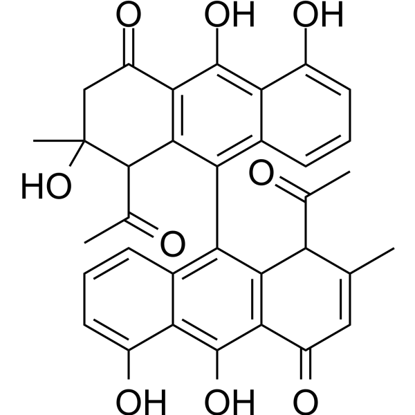
- Formula: C34H28O9
- Molecular Weight:580.58
-
Storage:
Please store the product under the recommended conditions in the Certificate of Analysis.

To place orders, for customer services and technical support, please contact: MedChemExpress USA
Tel: 609-228-6898 E-mail: [email protected] [email protected]


-
 Biological Activity
-
Chemical Information
- Protocols
- Purity & Documentation
- References
-
 Help & FAQs


All Antibiotic Isoforms
More

Biological Activity
Description
IC50 & Target
IC50: 12.02 µM (SARS-CoV-2 Mpro)[1]
In Vitro
Setomimycin (0.01-1 µM) inhibits the release of cytokines IL-1β, IL-6 and TNF-α and nitric oxide release from LPS stimulated RAW 264.7 cells in a dose-dependent manner[1].
Setomimycin (compound 1) shows antimicrobial activity with MICs of 8, 4, 16, 4 μg/mL for Staphylococcus aureus, Bacillus cereus, Bacillus subtilis, Micrococcus luteus, respectively[2].
Setomimycin (0-100 µM; 5 days) shows antiproliferative activity and inhibits colony formation[2].
Setomimycin (4, 5.5, 7 µM) decreases the protein expression of p-MEK, p-ERK, Bcl-2, increases the expression of Par-4[2].
MedChemExpress (MCE) has not independently confirmed the accuracy of these methods. They are for reference only. Further protocols information, click here.
-
Cell Line:A549; HOP-92; Panc-1; MiaPaca-2 cells
-
Concentration:0-100 µM
-
Incubation Time:44 h
-
Result:Showed antiproliferative activity with IC50s of 11.45, >100, 48, 4.57 µM for A549; HOP-92; Panc-1; MiaPaca-2 cells, respectively.
-
Cell Line:MCF-7, HCT-116 cells
-
Concentration:4, 5.5, 7 µM
-
Incubation Time:
-
Result:Decreased the protein expression of p-MEK, p-ERK, Bcl-2, increased the expression of Par-4 in a dose-dependent manner.
In Vivo
Pharmacokinetic Parameters of Setomimycin in female BALB/c mice[2].
| Pharmacokinetic parameters | Value |
| Cmax (ng/ml) | 694 ± 62 |
| Tmax (h) | 0.3 ± 0.1 |
| T1/2 (h) | 2.3 ± 0.5 |
| AUC0-t (ng.h/mL) | 2613 ± 111 |
| AUC0-α (ng.h/mL) | 2734 ± 108 |
| Vd (L/Kg) | 24 ± 4 |
| Cl (L/h/Kg) | 7.4 ± 0.3 |
MedChemExpress (MCE) has not independently confirmed the accuracy of these methods. They are for reference only.
-
Animal Model:6 weeks, 25-30 g, female BALB/c mice (4T1 cells)[2]
-
Dosage:20 mg/kg
-
Administration:I.p.; every other day for two weeks
-
Result:Decreased primary tumor weight (76%) and volume (90.5%).


Chemical Information
-
CAS No. 69431-87-4
-
Molecular Weight 580.58
-
Formula C34H28O9
-
SMILES
O=C1CC(C)(O)C(C(C)=O)C2=C1C(O)=C3C(O)=CC=CC3=C2C4=C5C=CC=C(O)C5=C(O)C6=C4C(C(C)=O)C(C)=CC6=O
-
Shipping
Room temperature in continental US; may vary elsewhere.
-
Storage
Please store the product under the recommended conditions in the Certificate of Analysis.

Protocols
-
Cell Cytotoxicity Assay
Cytotoxicity assays are usually based on the assessment of cell membrane damage, which can also be indirectly detected by measuring cell viability. Detection methods include MTT assay, CKK-8 assay, LDH assay and ATP assay, etc.
-
Research Protocol for Inflammation-related Diseases
The NLRP3 inflammasome is a cytosolic innate immune signaling platform that integrates priming signals and danger-signal activation to promote caspase-1 activation, maturation of IL-1β and IL-18, and gasdermin D-mediated pyroptotic cell death. The core experimental logic is to determine whether inflammatory disease phenotypes are driven by increased NLRP3 expression, ASC-containing inflammasome assembly, caspase-1 cleavage, GSDMD cleavage, and extracellular release of IL-1β/IL-18 rather than by nonspecific cell injury alone. The pathway is strongly linked to inflammation-related disease phenotypes because monosodium urate crystals activate NALP3/NLRP3 inflammasome signaling in gout-like crystal inflammation, cholesterol crystals activate NLRP3 inflammasomes in atherogenesis models, and DSS-induced intestinal inflammation has been reported to involve NLRP3 inflammasome activity. However, experimental colitis studies also show context-dependent protective effects of NLRP3 inflammasome co
-
Subcutaneous Cell-Line-Derived Xenograft
Subcutaneous cell-line-derived xenograft (CDX) models are established by implanting cultured human cancer cell lines into immunodeficient mice, where the injected cells form localized tumors that can be monitored in vivo as a measure of tumorigenic potential, growth kinetics, and treatment response. These models are widely used in oncology research because they allow reproducible tumor formation and enable comparative assessment of tumor growth between different cell lines or genetic manipulations in a controlled in vivo microenvironment. Subcutaneous implantation of cancer cells in immunodeficient mice is a standard approach for evaluating tumor growth behavior and therapeutic response across multiple cancer types, including prostate, esophageal, pancreatic, and colon cancer models.
-
Cell Viability Determination by MTT Colorimetric Assay
The following protocol uses the MTT colorimetric assay as a classic literature-established method for assessing cell viability/metabolic activity in cultured mammalian cells. MTT[3-(4,5-dimethylthiazol-2-yl)-2,5-diphenyltetrazolium bromide] is reduced by metabolically active cells to a colored formazan product; the amount of formazan is quantified spectrophotometrically and provides an indirect measure of metabolically active viable cells. Importantly, MTT reduction reflects cellular oxidoreductase/metabolic activity rather than an absolute direct count of living cells, so changes in cellular metabolism can alter the signal independently of cell number.


Purity & Documentation
References
[1]. Manhas RS, et al. Setomimycin as a potential molecule for COVID‑19 target: in silico approach and in vitro validation. Mol Divers. 2023 Apr;27(2):619-633. [Content Brief]
[2]. Manhas RS, et al. Isolation and anticancer activity evaluation of rare Bisaryl anthraquinone antibiotics from novel Streptomyces sp. strain of NW Himalayan region. Chem Biol Interact. 2022 Sep 25;365:110093. [Content Brief]

Calculators
Concentration (start) × Volume (start) = Concentration (final) × Volume (final)


Setomimycin Related Classifications
HY-124439 Related Classifications
 Powered by Bioz