



Thiocoraline
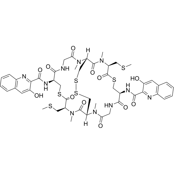
Thiocoraline is a depsipeptide that can be isolated by marine Micromonospora. Thiocoraline has antitumor activity with IC50s of 0.002, 0.002, 0.01 and 0.002 μg/mL in P388, A549, HT-29 and MEL-28 cells, respectively. Thiocoraline also a strong antimicrobial activity against Gram-positive microorganisms.
For research use only. We do not sell to patients.
- CAS No.: 173046-02-1
- Formula: C48H56N10O12S6
- Molecular Weight:1157.41
-
Storage:
Please store the product under the recommended conditions in the Certificate of Analysis.

To place orders, for customer services and technical support, please contact: MedChemExpress USA
Tel: 609-228-6898 E-mail: [email protected] [email protected]


-
 Biological Activity
-
Chemical Information
- Protocols
- Purity & Documentation
- References
-
 Help & FAQs





Biological Activity
Description
Cellular Effect
|
Cell Line

|
Type | Value | Description | References |
|---|---|---|---|---|
| A549 | EC50 |
0.01 μM
Compound: 1
|
Cytotoxicity against human A549 cells
Cytotoxicity against human A549 cells
|
[PMID: 26444379] |
| A549 | GI50 |
4.8 nM
Compound: thiocoraline A
|
Cytotoxicity against human A549 cells after 72 hrs by SRB assay
Cytotoxicity against human A549 cells after 72 hrs by SRB assay
|
[PMID: 17571868] |
| DU-145 | GI50 |
1.9 nM
Compound: thiocoraline A
|
Cytotoxicity against human DU145 cells after 72 hrs by SRB assay
Cytotoxicity against human DU145 cells after 72 hrs by SRB assay
|
[PMID: 17571868] |
| HeLa | GI50 |
1.3 nM
Compound: thiocoraline A
|
Cytotoxicity against human HeLa cells after 72 hrs by SRB assay
Cytotoxicity against human HeLa cells after 72 hrs by SRB assay
|
[PMID: 17571868] |
| HT-29 | GI50 |
3.9 nM
Compound: thiocoraline A
|
Cytotoxicity against human HT29 cells after 72 hrs by SRB assay
Cytotoxicity against human HT29 cells after 72 hrs by SRB assay
|
[PMID: 17571868] |
| K562 | GI50 |
1.3 nM
Compound: thiocoraline A
|
Cytotoxicity against human K562 cells after 72 hrs by SRB assay
Cytotoxicity against human K562 cells after 72 hrs by SRB assay
|
[PMID: 17571868] |
| LNCaP | GI50 |
3 nM
Compound: thiocoraline A
|
Cytotoxicity against human LNCaP cells after 72 hrs by SRB assay
Cytotoxicity against human LNCaP cells after 72 hrs by SRB assay
|
[PMID: 17571868] |
| LoVo | GI50 |
212.3 nM
Compound: thiocoraline A
|
Cytotoxicity against doxorubicin resistant human LoVo cells after 72 hrs by SRB assay
Cytotoxicity against doxorubicin resistant human LoVo cells after 72 hrs by SRB assay
|
[PMID: 17571868] |
| LoVo | GI50 |
7.2 nM
Compound: thiocoraline A
|
Cytotoxicity against human LoVo cells after 72 hrs by SRB assay
Cytotoxicity against human LoVo cells after 72 hrs by SRB assay
|
[PMID: 17571868] |
| PANC-1 | GI50 |
2.5 nM
Compound: thiocoraline A
|
Cytotoxicity against human PANC1 cells after 72 hrs by SRB assay
Cytotoxicity against human PANC1 cells after 72 hrs by SRB assay
|
[PMID: 17571868] |
| SK-BR-3 | GI50 |
2.2 nM
Compound: thiocoraline A
|
Cytotoxicity against human SK-BR3 cells after 72 hrs by SRB assay
Cytotoxicity against human SK-BR3 cells after 72 hrs by SRB assay
|
[PMID: 17571868] |
| SK-MEL-28 | GI50 |
3 nM
Compound: thiocoraline A
|
Cytotoxicity against human SK-MEL-28 cells after 72 hrs by SRB assay
Cytotoxicity against human SK-MEL-28 cells after 72 hrs by SRB assay
|
[PMID: 17571868] |


Chemical Information
-
CAS No. 173046-02-1
-
Molecular Weight 1157.41
-
Formula C48H56N10O12S6
-
Shipping
Room temperature in continental US; may vary elsewhere.
-
Storage
Please store the product under the recommended conditions in the Certificate of Analysis.

Protocols
-
Research Protocol for Infectious Diseases
Infectious-disease experiments test how pathogens interact with host barriers, innate immune receptors, inflammatory signaling, pathogen replication, and tissue injury; pattern-recognition receptors such as TLRs, RIG-I-like receptors, NOD-like receptors, and inflammasomes detect microbial molecules and activate NF-κB, interferon, and cytokine responses. The central hypothesis is that infection severity reflects the balance between pathogen burden and host response: protective inflammation restricts pathogen growth, whereas excessive or mislocalized inflammation contributes to tissue damage and disease phenotype. Unresolved questions include which host pathways are protective versus pathogenic, why some infection models fail to translate to human disease, and which combined readouts best predict clinically relevant infection outcomes.
-
Gram Staining of Tissue Sections
Gram staining of tissue sections is a histochemical technique used to differentiate Gram-positive and Gram-negative bacteria within histological specimens based on differences in bacterial cell wall structure and dye retention, adapted from classical bacteriological Gram staining into tissue-compatible “histological Gram stain” variants. In tissue applications, modifications of the Brown-Hopps and Brown-Brenn methods are commonly used to improve differentiation of microorganisms embedded within host connective tissue and to reduce overstaining or loss of Gram-negative signal, which are known limitations of earlier approaches. The principle relies on crystal violet-iodine complex retention in Gram-positive organisms and subsequent decolorization and counterstaining steps that allow contrast visualization of Gram-negative organisms against tissue background.
-
Subcutaneous Cell-Line-Derived Xenograft
Subcutaneous cell-line-derived xenograft (CDX) models are established by implanting cultured human cancer cell lines into immunodeficient mice, where the injected cells form localized tumors that can be monitored in vivo as a measure of tumorigenic potential, growth kinetics, and treatment response. These models are widely used in oncology research because they allow reproducible tumor formation and enable comparative assessment of tumor growth between different cell lines or genetic manipulations in a controlled in vivo microenvironment. Subcutaneous implantation of cancer cells in immunodeficient mice is a standard approach for evaluating tumor growth behavior and therapeutic response across multiple cancer types, including prostate, esophageal, pancreatic, and colon cancer models.


Purity & Documentation
References

Calculators
Concentration (start) × Volume (start) = Concentration (final) × Volume (final)


Thiocoraline Related Classifications
HY-P3116 Related Classifications
 Powered by Bioz