



M47
Based on 1 publication(s) in Google Scholar
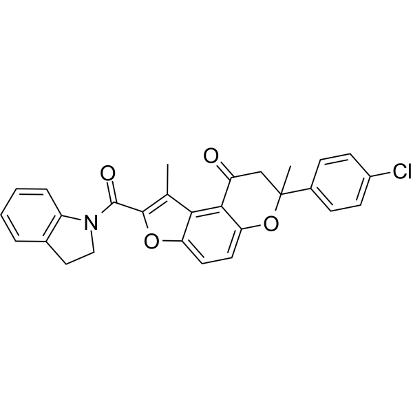
M47 is a molecular glue that selectively destabilizes Cryptochrome 1 (CRY1) and increases degradation of the CRY1 in the nucleus. M47 enhances apoptosis in Ras-transformed P53-deficient mouse skin fibroblast lines and enhances life span in p53 knockout mice. M47 can be used in research of cancer.
For research use only. We do not sell to patients.
- Purity : 99.96%
- CAS No.: 890808-56-7
- Formula: C28H22ClNO4
- Molecular Weight:471.93
-
Storage:Powder -20°C, 3 years , 4°C, 2 years ; In solvent -80°C, 6 months , -20°C, 1 month

To place orders, for customer services and technical support, please contact: MedChemExpress USA
Tel: 609-228-6898 E-mail: [email protected] [email protected]


-
 Biological Activity
-
Chemical Information
-
Solvent & Solubility
- Protocols
- Purity & Documentation
- References
-
 Help & FAQs


Publications Citing Use of MedChemExpress (MCE) M47
More

Biological Activity
Description
IC50 & Target
[1]|
CRY1 |
FXBL3 |
In Vitro
M47 (0-10 μM; 0-120 h; U2OS Bmal1-dLuc cells) reduces the half-life of CRY1 in a dose-dependent manner and causes circadian changes. M47 selectively enhances the degradation rate of CRY1 by increasing its ubiquitination and resulted in increasing the circadian period length of U2OS Bmal1-dLuc cells[1].
M47 (10 μM; 24-44 h; U2OS cells) decreases the level of CRY1 in U2OS cells[1].
MedChemExpress (MCE) has not independently confirmed the accuracy of these methods. They are for reference only.
-
Cell Line:U2OS cells
-
Concentration:10 μM
-
Incubation Time:24-44 hours
-
Result:Decreased CRY1 protein levels between 24 and 48 h.
In Vivo
MedChemExpress (MCE) has not independently confirmed the accuracy of these methods. They are for reference only.
-
Animal Model:C57BL/6 J mice and p53 gene knockout C57BL/6 J mice[1]
-
Dosage:5, 50, 300 and 1000 mg/kg
-
Administration:intraperitoneal injection, daily, for 5 days
-
Result:Prolonged the life span of p53 gene knockout mice.


Chemical Information
-
CAS No. 890808-56-7
-
Appearance Solid
-
Molecular Weight 471.93
-
Formula C28H22ClNO4
-
Color Off-white to light yellow
-
SMILES
O=C(N1C2=C(CC1)C=CC=C2)C(OC3=CC=C4OC(C)(C5)C6=CC=C(C=C6)Cl)=C(C)C3=C4C5=O
-
Shipping
Room temperature in continental US; may vary elsewhere.
-
Storage
Powder -20°C 3 years 4°C 2 years In solvent -80°C 6 months -20°C 1 month


Publications (1)
-
Journal Impact Factor
-
Most Recent
-
bioRxiv
Stage-Specific Regulation of DNA Damage Repair by the Circadian Regulator, CRY1, in Prostate Cancer. [Abstract]2026 May 21:2026.05.19.726303. PMID: 42239372


Solvent & Solubility
In Vitro:
DMSO : 5 mg/mL (10.59 mM; Need ultrasonic; Hygroscopic DMSO has a significant impact on the solubility of product, please use newly opened DMSO)
Please refer to the solubility information to select the appropriate solvent. Once prepared, please aliquot and store the solution to prevent product inactivation from repeated freeze-thaw cycles.
Storage method and period of stock solution: -80°C, 6 months; -20°C, 1 month. When stored at -80°C, please use it within 6 months. When stored at -20°C, please use it within 1 month.
Please refer to the solubility information to select the appropriate solvent. Once prepared, please aliquot and store the solution to prevent product inactivation from repeated freeze-thaw cycles.
Storage method and period of stock solution: -80°C, 6 months; -20°C, 1 month. When stored at -80°C, please use it within 6 months. When stored at -20°C, please use it within 1 month.
Concentration (start) × Volume (start) = Concentration (final) × Volume (final)


Purity & Documentation
-
Data Sheet (274 KB)
-
SDS (393 KB)
- English - EN (393 KB)
- Français - FR (393 KB)
- Deutsch - DE (393 KB)
- Norwegian - NO (393 KB)
- Español - ES (393 KB)
- Swedish - SV (393 KB)
- Italian - IT (393 KB)
- Korean - KR (393 KB)
- Portuguese - PT (393 KB)
-
Handling Instructions (2659 KB)
References


Complete Stock Solution Preparation Table
Please refer to the solubility information to select the appropriate solvent. Once prepared, please aliquot and store the solution to prevent product inactivation from repeated freeze-thaw cycles.
Storage method and period of stock solution: -80°C, 6 months; -20°C, 1 month. When stored at -80°C, please use it within 6 months. When stored at -20°C, please use it within 1 month.
| Optional Solvent | Concentration Solvent Mass | 1 mg | 5 mg | 10 mg | 25 mg |
|---|---|---|---|---|---|
| DMSO | 1 mM | 2.1190 mL | 10.5948 mL | 21.1896 mL | 52.9740 mL |
| 5 mM | 0.4238 mL | 2.1190 mL | 4.2379 mL | 10.5948 mL | |
| 10 mM | 0.2119 mL | 1.0595 mL | 2.1190 mL | 5.2974 mL |


M47 Related Classifications
HY-148764 Related Classifications
 Powered by Bioz