



Vanzacaftor
Based on 5 publication(s) in Google Scholar
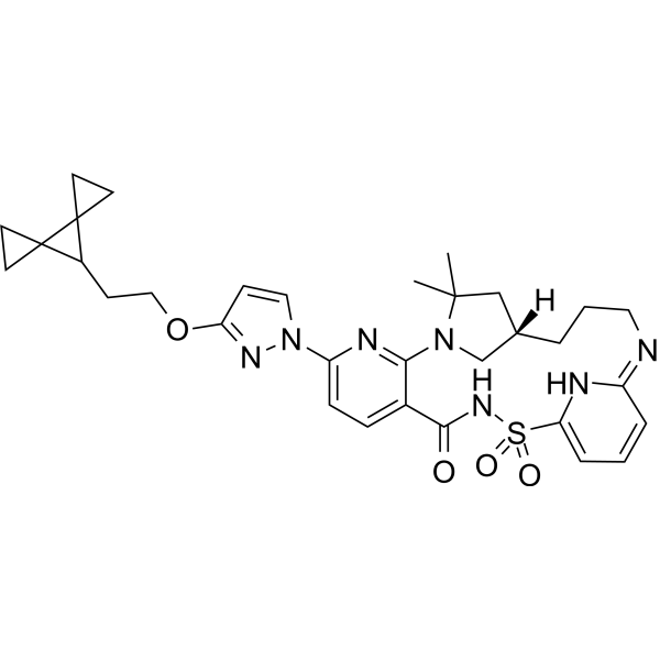
Vanzacaftor (VX-121) is an orally active noval corrector of Cystic fibrosis transmembrane conductance regulator (CFTR). Vanzacaftor improves processing and trafficking of CFTR protein as well as increases chloride transport in triple combined with Tezacaftor (HY-15448) and Deutivacaftor. Vanzacaftor-Tezacaftor-Deutivacaftor is safe and well tolerated, improving lung function, respiratory symptoms, and CFTR function with cystic fibrosis, which is promising for research in the field of cystic fibrosis diseases.
For research use only. We do not sell to patients.
- Purity : 98.72%
- CAS No.: 2374124-49-7
- Formula: C32H39N7O4S
- Molecular Weight:617.76
-
Storage:
-20°C, stored under nitrogen
* In solvent : -80°C, 6 months; -20°C, 1 month (stored under nitrogen)

To place orders, for customer services and technical support, please contact: MedChemExpress USA
Tel: 609-228-6898 E-mail: [email protected] [email protected]


-
 Biological Activity
-
Chemical Information
-
Solvent & Solubility
- Protocols
- Purity & Documentation
- References
-
 Help & FAQs


-
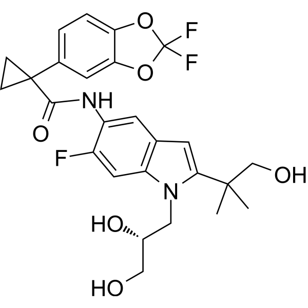
HY-15448
TezacaftorCFTR Corrector -
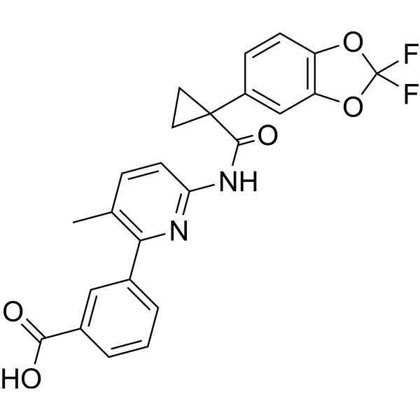
HY-13262
LumacaftorCFTR Modulator -
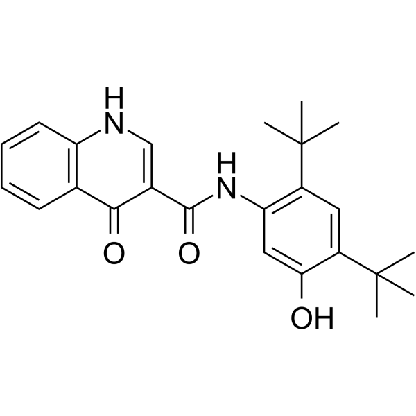
HY-13017
IvacaftorCFTR Potentiator -
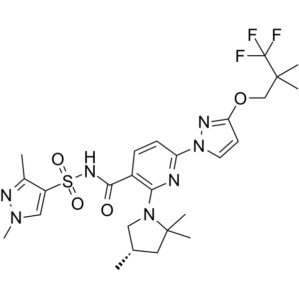
HY-111772
ElexacaftorCFTR Corrector -
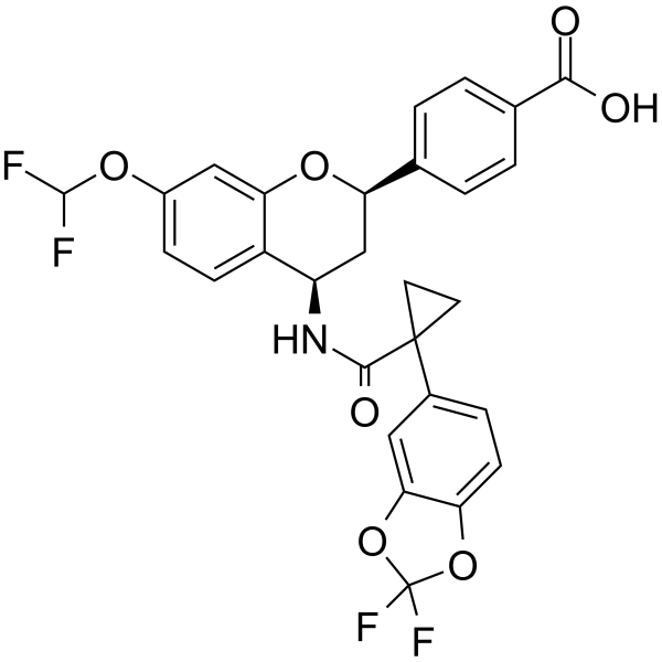
HY-111111
GalicaftorCFTR Corrector -
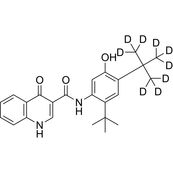
HY-13017S
Ivacaftor-d9CFTR Modulator -
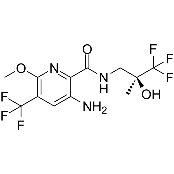
HY-109177
IcenticaftorCFTR Potentiator -
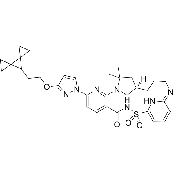
HY-145603A
(R)-VanzacaftorCFTR Modulator -
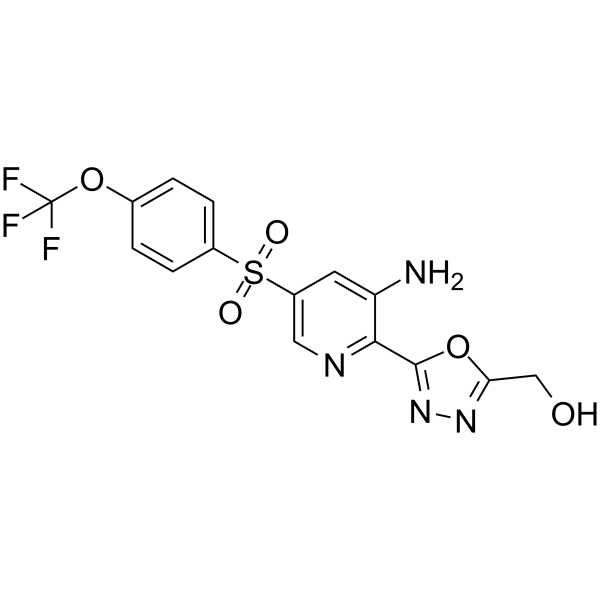
HY-109152
NavocaftorCystic Fibrosis Transmembrane Regulator -
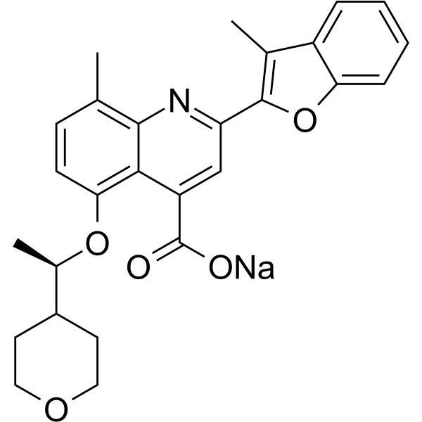
HY-109187A
Posenacaftor sodiumCFTR Modulator -
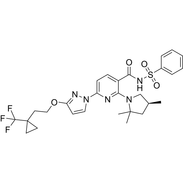
HY-126394
BamocaftorCFTR Corrector -

HY-15371
ForskolinAdenylate Cyclase Activator
Publications Citing Use of MedChemExpress (MCE) Vanzacaftor
More

Biological Activity
Description
In Vitro
Vanzacaftor (0-10 μM) stimulates K+ secretion at low micromolar concentrations in human bronchial epithelial cells (HBEs), which is inhibited by Paxilline (HY-N6778) with an EC50 of 4.4 μM [2]. Vanzacaftor (1 μM, 5 μM, 10 μM) significantly potentiates αBKCa current density, which is blocked by Paxilline (HY-N6778) in HEK cells[2]. Vanzacaftor (5 μM) reduces action potential firing frequency and alters neuronal excitability in a primary hippocampal neuron[2].
MedChemExpress (MCE) has not independently confirmed the accuracy of these methods. They are for reference only. Further protocols information, click here.
Clinical Trial
| NCT Number | Sponsor | Condition | Start Date |
Phase

|
|---|---|---|---|---|
| NCT01329991 | Plexxikon| | 2011-05 | PHASE1 |


Chemical Information
-
CAS No. 2374124-49-7
-
Appearance Solid
-
Molecular Weight 617.76
-
Formula C32H39N7O4S
-
Color Off-white to light yellow
-
SMILES
CC(C)(C[C@@](CCC/N=C1NC(S(=O)(N2)=O)=CC=C\1)([H])C3)N3C4=NC(N(C=C5)N=C5OCCC6C7(CC7)C68CC8)=CC=C4C2=O
-
Synonyms
VX-121
-
Shipping
Room temperature in continental US; may vary elsewhere.
-
Storage
-20°C, stored under nitrogen
* In solvent : -80°C, 6 months; -20°C, 1 month (stored under nitrogen)


Publications (5)
-
Journal Impact Factor
-
Most Recent
-
J Cyst Fibros
Vanzacaftor-Tezacaftor as an alternative therapeutic resource for the ETI-Resistant L467F-F508del Allele: Ex vivo prediction and exploratory clinical assessment. [Abstract]2026 Jun 4:S1569-1993(26)01638-3. PMID: 42243036 -
Am J Physiol Cell Physiol
(R)-vanzacaftor potentiates BKCa channels in the absence of CFTR correction or potentiation. [Abstract]2025 Oct 1;329(4):C1130-C1138. PMID: 40920663 -
PNAS Nexus
VX-445 (elexacaftor) inhibits chloride secretion across human bronchial epithelial cells by directly blocking KCa3.1 channels. [Abstract]2025 Jul 4;4(7):pgaf211. PMID: 40688096 -
-


Solvent & Solubility
In Vitro:
DMSO : 100 mg/mL (161.88 mM; Need ultrasonic; Hygroscopic DMSO has a significant impact on the solubility of product, please use newly opened DMSO)
Please refer to the solubility information to select the appropriate solvent. Once prepared, please aliquot and store the solution to prevent product inactivation from repeated freeze-thaw cycles.
Storage method and period of stock solution: -80°C, 6 months; -20°C, 1 month (stored under nitrogen). When stored at -80°C, please use it within 6 months. When stored at -20°C, please use it within 1 month.
Please refer to the solubility information to select the appropriate solvent. Once prepared, please aliquot and store the solution to prevent product inactivation from repeated freeze-thaw cycles.
Storage method and period of stock solution: -80°C, 6 months; -20°C, 1 month (stored under nitrogen). When stored at -80°C, please use it within 6 months. When stored at -20°C, please use it within 1 month.
Concentration (start) × Volume (start) = Concentration (final) × Volume (final)
In Vivo:
Select the appropriate dissolution method based on your experimental animal and administration route.
- For the following dissolution methods, please ensure to first prepare a clear stock solution using an In Vitro approach and then sequentially add co-solvents:
- To ensure reliable experimental results, the clarified stock solution can be appropriately stored based on storage conditions. As for the working solution for In Vivo experiments, it is recommended to prepare freshly and use it on the same day.
- The percentages shown for the solvents indicate their volumetric ratio in the final prepared solution. If precipitation or phase separation occurs during preparation, heat and/or sonication can be used to aid dissolution.
In Vivo Dissolution Calculator
Please enter the basic information of animal experiments:
-
-
-
-
Recommended: Prepare an additional quantity of animals to account for potential losses during experiments.
Please enter your animal formula composition:
-
%DMSO +
Recommended: Keep the proportion of DMSO in working solution below 2% if your animal is weak.
-
%+
-
+%Tween-80 + +
-
%Saline +
The co-solvents required include: DMSO, . All of co-solvents are available by MedChemExpress (MCE). , Tween 80. All of co-solvents are available by MedChemExpress (MCE).
Working solution concentration: 0.22 mg/mL
Method for preparing stock solution: mg drug dissolved in μL DMSO. Stock solution concentration: mg/mL. * In solvent : -80°C, 6 months; -20°C, 1 month (stored under nitrogen)
1. Take μL DMSO stock solution;
2. Add μL .
μL , mix evenly;
3. Then add μL Tween 80, mix evenly;
4. Then add μL
Please ensure that the stock solution in the first step is dissolved to a clear state, and add co-solvents in sequence. You can use ultrasonic heating (ultrasonic cleaner, recommended frequency 20-40 kHz), vortexing, etc. to assist dissolution.

Protocols
-
Fibrosis/Collagen Morphometry
Fibrosis and collagen morphometry is based on the quantitative visualization of fibrillar collagen deposition in tissue sections using histochemical stains such as Sirius Red (Picrosirius Red) or Masson's trichrome, followed by image-based or polarization-enhanced analysis to estimate collagen proportional area as a surrogate of extracellular matrix accumulation during fibrotic remodeling. Sirius Red combined with polarized light microscopy enhances detection of collagen fibers due to birefringence properties, enabling more specific visualization of collagen type I and III fibrils compared to conventional bright-field histology, while whole-section or region-restricted digital morphometry reduces field-selection bias in fibrosis assessment. Alternative quantitative approaches include second harmonic generation (SHG) and two-photon excited fluorescence microscopy, which enable label-free detection of fibrillar collagen and have been validated against histological staining and biochemica
-
Collagen: Sirius Red Staining
Sirius Red or picrosirius red staining is a histochemical method for visualizing collagen-rich extracellular matrix in tissue sections, and collagen fibers are detected as red-stained structures under bright-field microscopy with enhanced birefringence under polarized light. Picrosirius red is useful for assessing total collagen organization, distribution, and fibrosis burden, but polarized color should not be interpreted as a definitive collagen type I versus type III readout because color is affected by fiber orientation, thickness, and packing.
-
Connective Tissue: Masson's Trichrome/Collagen Trichrome Staining
Masson’s Trichrome (collagen/trichrome staining) is a histological technique that differentially stains tissue compartments using sequential acidic dyes to distinguish collagen from muscle and cytoplasmic components based on dye affinity and tissue permeability differences, enabling visualization of fibrosis and connective tissue architecture in histological sections. The classical formulation typically uses Weigert's iron hematoxylin for nuclear staining, Biebrich scarlet-acid fuchsin for cytoplasm and muscle, and aniline blue (or light green variants) for collagen, producing a characteristic blue/green collagen signal contrasted against red cytoplasm and dark nuclei. The staining principle relies on selective displacement of smaller dye molecules by larger anionic dyes in collagen-rich regions under controlled acidified conditions, which enhances collagen-specific dye retention. This property makes the method widely used for fibrosis assessment in organs such as heart, liver, lung, a


Purity & Documentation
-
Data Sheet (276 KB)
-
SDS (252 KB)
- English - EN (252 KB)
- Français - FR (252 KB)
- Deutsch - DE (252 KB)
- Norwegian - NO (252 KB)
- Español - ES (252 KB)
- Swedish - SV (252 KB)
- Italian - IT (252 KB)
- Korean - KR (252 KB)
- Portuguese - PT (252 KB)
-
Handling Instructions (2659 KB)
References
[1]. Uluer AZ, et al. Safety and efficacy of vanzacaftor-tezacaftor-deutivacaftor in adults with cystic fibrosis: randomised, double-blind, controlled, phase 2 trials[J]. Lancet Respir Med. 2023 Jun;11(6):550-562. [Content Brief]
[2]. Kolski-Andreaco A, et al. Potentiation of BKCa channels by cystic fibrosis transmembrane conductance regulator (CFTR) correctors VX-445 and VX-121[J]. J Clin Invest. 2024 Jul 2:e176328. [Content Brief]


Complete Stock Solution Preparation Table
Please refer to the solubility information to select the appropriate solvent. Once prepared, please aliquot and store the solution to prevent product inactivation from repeated freeze-thaw cycles.
Storage method and period of stock solution: -80°C, 6 months; -20°C, 1 month (stored under nitrogen). When stored at -80°C, please use it within 6 months. When stored at -20°C, please use it within 1 month.
| Optional Solvent | Concentration Solvent Mass | 1 mg | 5 mg | 10 mg | 25 mg |
|---|---|---|---|---|---|
| DMSO | 1 mM | 1.6188 mL | 8.0938 mL | 16.1875 mL | 40.4688 mL |
| 5 mM | 0.3238 mL | 1.6188 mL | 3.2375 mL | 8.0938 mL | |
| 10 mM | 0.1619 mL | 0.8094 mL | 1.6188 mL | 4.0469 mL | |
| 15 mM | 0.1079 mL | 0.5396 mL | 1.0792 mL | 2.6979 mL | |
| 20 mM | 0.0809 mL | 0.4047 mL | 0.8094 mL | 2.0234 mL | |
| 25 mM | 0.0648 mL | 0.3238 mL | 0.6475 mL | 1.6188 mL | |
| 30 mM | 0.0540 mL | 0.2698 mL | 0.5396 mL | 1.3490 mL | |
| 40 mM | 0.0405 mL | 0.2023 mL | 0.4047 mL | 1.0117 mL | |
| 50 mM | 0.0324 mL | 0.1619 mL | 0.3238 mL | 0.8094 mL | |
| 60 mM | 0.0270 mL | 0.1349 mL | 0.2698 mL | 0.6745 mL | |
| 80 mM | 0.0202 mL | 0.1012 mL | 0.2023 mL | 0.5059 mL | |
| 100 mM | 0.0162 mL | 0.0809 mL | 0.1619 mL | 0.4047 mL |


Vanzacaftor Related Classifications
HY-145603 Related Classifications
 Powered by Bioz