



VRT-532

Based on 1 Customer Validation
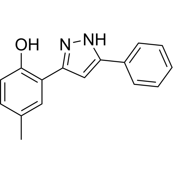
VRT-532 (CFpot-532) is a potent is a potent CFTR modulator. VRT-532 enhances channel activity in G551D-CFTR and intrinsic ATPase activity of G551D-CFTR. VRT-532 has the potential for the research of cystic fibrosis.
For research use only. We do not sell to patients.
- Purity : 98.98%
- CAS No.: 38214-71-0
- Formula: C16H14N2O
- Molecular Weight:250.30
-
Storage:Powder -20°C, 3 years ; In solvent -80°C, 6 months , -20°C, 1 month

To place orders, for customer services and technical support, please contact: MedChemExpress USA
Tel: 609-228-6898 E-mail: [email protected] [email protected]


-
 Biological Activity
-
Chemical Information
-
Solvent & Solubility
- Protocols
- Purity & Documentation
- References
-
 Help & FAQs


-
HY-109027
CavosonstatGSNOR Inhibitor -
HY-103369
PG01CFTR Potentiator -
HY-150090
SRI-41315CFTR Activator -
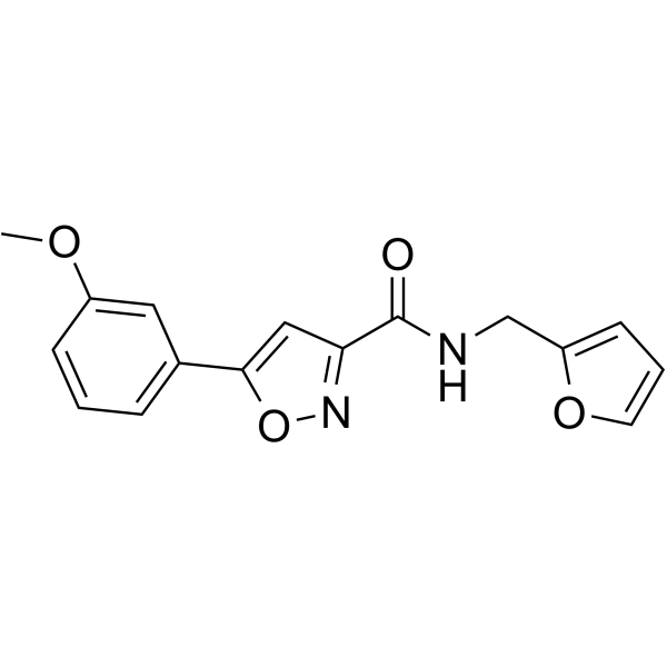
HY-147315
CFTR corrector 9CFTR Modulator -
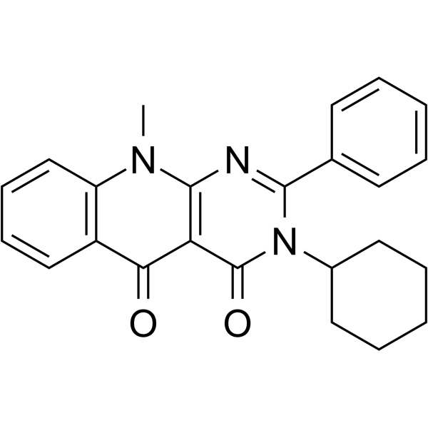
HY-150089
SRI-37240PTCs Inhibitor -
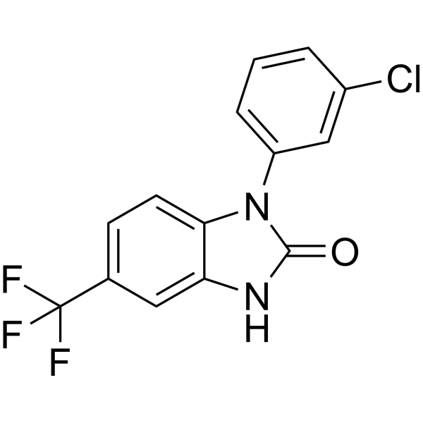
HY-119179
UCCF-853CFTR Modulator -
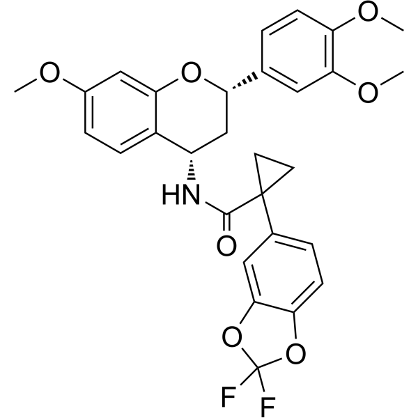
HY-147249
CFTR corrector 8CFTR Modulator -
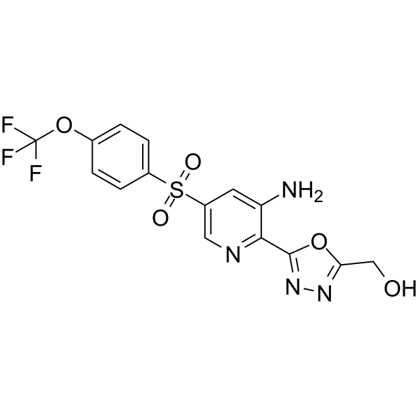
HY-109152
NavocaftorCystic Fibrosis Transmembrane Regulator -
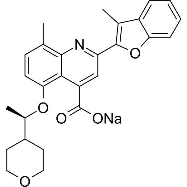
HY-109187A
Posenacaftor sodiumCFTR Modulator -
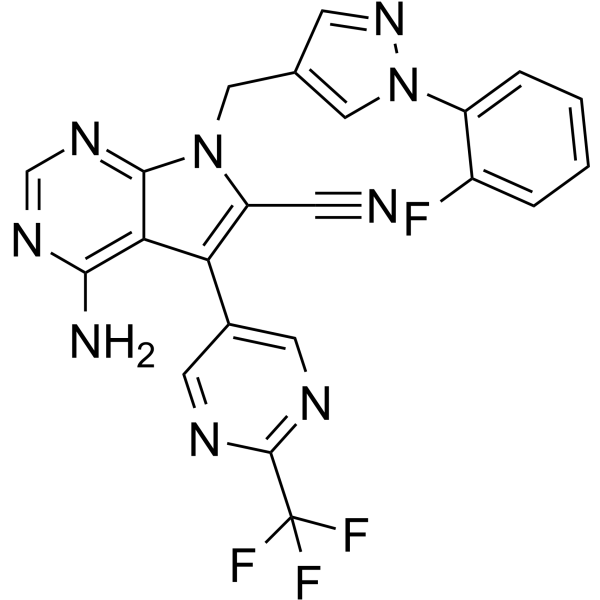
HY-136939
CFTR corrector 6CFTR Activator -

HY-109027R
Cavosonstat (Standard)Reference Standard -
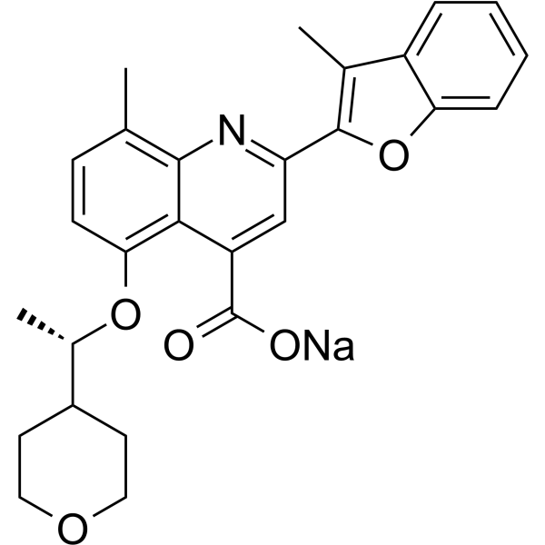
HY-109187B
(R)-Posenacaftor sodiumPosenacaftor R-Enantiomer


Biological Activity
Description
In Vitro
VRT-532 (15 µM) directly binds reconstituted G551D-CFTR and modifies its ATPase activity[1].
MedChemExpress (MCE) has not independently confirmed the accuracy of these methods. They are for reference only. Further protocols information, click here.


Chemical Information
-
CAS No. 38214-71-0
-
Appearance Solid
-
Molecular Weight 250.30
-
Formula C16H14N2O
-
Color White to off-white
-
SMILES
OC(C=CC(C)=C1)=C1C2=NNC(C3=CC=CC=C3)=C2
-
Synonyms
CFpot-532
-
Shipping
Room temperature in continental US; may vary elsewhere.
-
Storage
Powder -20°C 3 years In solvent -80°C 6 months -20°C 1 month


Solvent & Solubility
In Vitro:
DMSO : 100 mg/mL (399.52 mM; Need ultrasonic; Hygroscopic DMSO has a significant impact on the solubility of product, please use newly opened DMSO)
Please refer to the solubility information to select the appropriate solvent. Once prepared, please aliquot and store the solution to prevent product inactivation from repeated freeze-thaw cycles.
Storage method and period of stock solution: -80°C, 6 months; -20°C, 1 month. When stored at -80°C, please use it within 6 months. When stored at -20°C, please use it within 1 month.
Please refer to the solubility information to select the appropriate solvent. Once prepared, please aliquot and store the solution to prevent product inactivation from repeated freeze-thaw cycles.
Storage method and period of stock solution: -80°C, 6 months; -20°C, 1 month. When stored at -80°C, please use it within 6 months. When stored at -20°C, please use it within 1 month.
Concentration (start) × Volume (start) = Concentration (final) × Volume (final)
In Vivo:
Select the appropriate dissolution method based on your experimental animal and administration route.
- For the following dissolution methods, please ensure to first prepare a clear stock solution using an In Vitro approach and then sequentially add co-solvents:
- To ensure reliable experimental results, the clarified stock solution can be appropriately stored based on storage conditions. As for the working solution for In Vivo experiments, it is recommended to prepare freshly and use it on the same day.
- The percentages shown for the solvents indicate their volumetric ratio in the final prepared solution. If precipitation or phase separation occurs during preparation, heat and/or sonication can be used to aid dissolution.
Add each solvent one by one: 10% DMSO 40% PEG300 5% Tween-80 45% Saline
Solubility: ≥ 2.5 mg/mL (9.99 mM); Clear solution
This protocol yields a clear solution of ≥ 2.5 mg/mL (saturation unknown).
Taking 1 mL working solution as an example, add 100 μL DMSO stock solution (25.0 mg/mL) to 400 μL PEG300, and mix evenly; then add 50 μL Tween-80 and mix evenly; then add 450 μL Saline to adjust the volume to 1 mL.
Preparation of Saline: Dissolve 0.9 g sodium chloride in ddH₂O and dilute to 100 mL to obtain a clear Saline solution.
Add each solvent one by one: 10% DMSO 90% (20% SBE-β-CD in Saline)
Solubility: ≥ 2.5 mg/mL (9.99 mM); Clear solution
This protocol yields a clear solution of ≥ 2.5 mg/mL (saturation unknown).
Taking 1 mL working solution as an example, add 100 μL DMSO stock solution (25.0 mg/mL) to 900 μL 20% SBE-β-CD in Saline, and mix evenly.
Preparation of 20% SBE-β-CD in Saline (4°C, storage for one week): 2 g SBE-β-CD powder is dissolved in 10 mL Saline, completely dissolve until clear.
In Vivo Dissolution Calculator
Please enter the basic information of animal experiments:
-
-
-
-
Recommended: Prepare an additional quantity of animals to account for potential losses during experiments.
Please enter your animal formula composition:
-
%DMSO +
Recommended: Keep the proportion of DMSO in working solution below 2% if your animal is weak.
-
%+
-
+%Tween-80 + +
-
%Saline +
The co-solvents required include: DMSO, . All of co-solvents are available by MedChemExpress (MCE). , Tween 80. All of co-solvents are available by MedChemExpress (MCE).
Working solution concentration: 0.22 mg/mL
Method for preparing stock solution: mg drug dissolved in μL DMSO. Stock solution concentration: mg/mL.
1. Take μL DMSO stock solution;
2. Add μL .
μL , mix evenly;
3. Then add μL Tween 80, mix evenly;
4. Then add μL
Please ensure that the stock solution in the first step is dissolved to a clear state, and add co-solvents in sequence. You can use ultrasonic heating (ultrasonic cleaner, recommended frequency 20-40 kHz), vortexing, etc. to assist dissolution.

Protocols
-
Fibrosis/Collagen Morphometry
Fibrosis and collagen morphometry is based on the quantitative visualization of fibrillar collagen deposition in tissue sections using histochemical stains such as Sirius Red (Picrosirius Red) or Masson's trichrome, followed by image-based or polarization-enhanced analysis to estimate collagen proportional area as a surrogate of extracellular matrix accumulation during fibrotic remodeling. Sirius Red combined with polarized light microscopy enhances detection of collagen fibers due to birefringence properties, enabling more specific visualization of collagen type I and III fibrils compared to conventional bright-field histology, while whole-section or region-restricted digital morphometry reduces field-selection bias in fibrosis assessment. Alternative quantitative approaches include second harmonic generation (SHG) and two-photon excited fluorescence microscopy, which enable label-free detection of fibrillar collagen and have been validated against histological staining and biochemica
-
Collagen: Sirius Red Staining
Sirius Red or picrosirius red staining is a histochemical method for visualizing collagen-rich extracellular matrix in tissue sections, and collagen fibers are detected as red-stained structures under bright-field microscopy with enhanced birefringence under polarized light. Picrosirius red is useful for assessing total collagen organization, distribution, and fibrosis burden, but polarized color should not be interpreted as a definitive collagen type I versus type III readout because color is affected by fiber orientation, thickness, and packing.
-
Connective Tissue: Masson's Trichrome/Collagen Trichrome Staining
Masson’s Trichrome (collagen/trichrome staining) is a histological technique that differentially stains tissue compartments using sequential acidic dyes to distinguish collagen from muscle and cytoplasmic components based on dye affinity and tissue permeability differences, enabling visualization of fibrosis and connective tissue architecture in histological sections. The classical formulation typically uses Weigert's iron hematoxylin for nuclear staining, Biebrich scarlet-acid fuchsin for cytoplasm and muscle, and aniline blue (or light green variants) for collagen, producing a characteristic blue/green collagen signal contrasted against red cytoplasm and dark nuclei. The staining principle relies on selective displacement of smaller dye molecules by larger anionic dyes in collagen-rich regions under controlled acidified conditions, which enhances collagen-specific dye retention. This property makes the method widely used for fibrosis assessment in organs such as heart, liver, lung, a


Purity & Documentation
-
Data Sheet (275 KB)
-
SDS (614 KB)
- English - EN (614 KB)
- Français - FR (614 KB)
- Deutsch - DE (614 KB)
- Norwegian - NO (614 KB)
- Español - ES (614 KB)
- Swedish - SV (614 KB)
- Italian - IT (614 KB)
- Korean - KR (614 KB)
- Portuguese - PT (614 KB)
-
Handling Instructions (2659 KB)
References
[1]. Pasyk S, et al. Direct interaction of a small-molecule modulator with G551D-CFTR, a cystic fibrosis-causing mutation associated with severe disease. Biochem J. 2009 Feb 15;418(1):185-90. [Content Brief]
[2]. Wang Y, et al. Correctors promote maturation of cystic fibrosis transmembrane conductance regulator (CFTR)-processing mutants by binding to the protein. J Biol Chem. 2007 Nov 16;282(46):33247-33251. [Content Brief]


Complete Stock Solution Preparation Table
Please refer to the solubility information to select the appropriate solvent. Once prepared, please aliquot and store the solution to prevent product inactivation from repeated freeze-thaw cycles.
Storage method and period of stock solution: -80°C, 6 months; -20°C, 1 month. When stored at -80°C, please use it within 6 months. When stored at -20°C, please use it within 1 month.
| Optional Solvent | Concentration Solvent Mass | 1 mg | 5 mg | 10 mg | 25 mg |
|---|---|---|---|---|---|
| DMSO | 1 mM | 3.9952 mL | 19.9760 mL | 39.9521 mL | 99.8801 mL |
| 5 mM | 0.7990 mL | 3.9952 mL | 7.9904 mL | 19.9760 mL | |
| 10 mM | 0.3995 mL | 1.9976 mL | 3.9952 mL | 9.9880 mL | |
| 15 mM | 0.2663 mL | 1.3317 mL | 2.6635 mL | 6.6587 mL | |
| 20 mM | 0.1998 mL | 0.9988 mL | 1.9976 mL | 4.9940 mL | |
| 25 mM | 0.1598 mL | 0.7990 mL | 1.5981 mL | 3.9952 mL | |
| 30 mM | 0.1332 mL | 0.6659 mL | 1.3317 mL | 3.3293 mL | |
| 40 mM | 0.0999 mL | 0.4994 mL | 0.9988 mL | 2.4970 mL | |
| 50 mM | 0.0799 mL | 0.3995 mL | 0.7990 mL | 1.9976 mL | |
| 60 mM | 0.0666 mL | 0.3329 mL | 0.6659 mL | 1.6647 mL | |
| 80 mM | 0.0499 mL | 0.2497 mL | 0.4994 mL | 1.2485 mL | |
| 100 mM | 0.0400 mL | 0.1998 mL | 0.3995 mL | 0.9988 mL |


VRT-532 Related Classifications
HY-114725 Related Classifications
 Powered by Bioz