



YIGSR
Based on 1 publication(s) in Google Scholar
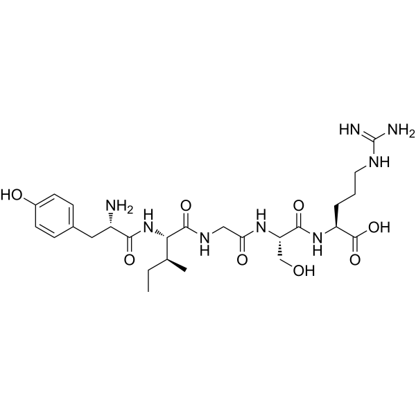
YIGSR (Laminin Fragment 929-933) is a polypeptide that inhibits tumor growth and metastasis of leukemia cells. YIGSR specifically binds to the 67kDa laminin receptor and regulates the expression of eNOS in endothelial cells. YIGSR can be used in leukemia-related research.
Nos produits utilisent uniquement pour la recherche. Nous ne vendons pas aux patients.
- Pureté : 99.38%
- CAS No.: 110590-64-2
- Formule: C26H42N8O8
- Masse moléculaire:594.66
-
Stockage:
Sealed storage, away from moisture.
Powder -80°C, 2 years , -20°C, 1 year* In solvent : -80°C, 6 months; -20°C, 1 month (sealed storage, away from moisture)

To place orders, for customer services and technical support, please contact: MedChemExpress USA
Tel: 609-228-6898 E-mail: [email protected] [email protected]


-
 Activité biologique
-
Chemical Information
-
Solvant et solubilité
- Protocole
- Pureté et documentation
- Références
-
 Help & FAQs


Publications Citing Use of MedChemExpress (MCE) YIGSR
More

Activité biologique
Description
In Vitro
YIGSR (150 μg/mL) reduces the adhesion of porcine aortic endothelial cells to laminin I to approximately 50%[1].
YIGSR (150 μg/mL; 6 h) induces the detachment of approximately 80% of porcine aortic endothelial cells coated with laminin I under shear stress[1].
YIGSR (35 μg/mL) completely abolishes the elevation of eNOS mRNA expression induced by 2-fold shear stress in primary porcine aortic endothelial cells seeded on laminin I-coated plates, with administration performed 3 days after cell seeding[1].
YIGSR (35 μg/mL) completely inhibits shear stress-induced increase in eNOS in porcine aortic endothelial cells coated with laminin I[1].
MedChemExpress (MCE) has not independently confirmed the accuracy of these methods. They are for reference only.


Chemical Information
-
CAS No. 110590-64-2
-
Appearance Solid
-
Masse moléculaire 594.66
-
Formule C26H42N8O8
-
Color White to off-white
-
Synonyms
Laminin Fragment 929-933
-
Sequence
Tyr-Ile-Gly-Ser-Arg
-
Sequence Shortening
YIGSR
-
Livraison
Room temperature in continental US; may vary elsewhere.
-
Stockage
Sealed storage, away from moisture
Powder -80°C 2 years -20°C 1 year * In solvent : -80°C, 6 months; -20°C, 1 month (sealed storage, away from moisture)


Publications (1)
-
Journal Impact Factor
-
Most Recent


Solvant et solubilité
In Vitro:
DMSO : 50 mg/mL (84.08 mM; Need ultrasonic; Hygroscopic DMSO has a significant impact on the solubility of product, please use newly opened DMSO)
Please refer to the solubility information to select the appropriate solvent. Once prepared, please aliquot and store the solution to prevent product inactivation from repeated freeze-thaw cycles.
Storage method and period of stock solution: -80°C, 6 months; -20°C, 1 month (sealed storage, away from moisture). When stored at -80°C, please use it within 6 months. When stored at -20°C, please use it within 1 month.
Please refer to the solubility information to select the appropriate solvent. Once prepared, please aliquot and store the solution to prevent product inactivation from repeated freeze-thaw cycles.
Storage method and period of stock solution: -80°C, 6 months; -20°C, 1 month (sealed storage, away from moisture). When stored at -80°C, please use it within 6 months. When stored at -20°C, please use it within 1 month.
Concentration (start) × Volume (start) = Concentration (final) × Volume (final)


Pureté et documentation
-
Fiche technique (272 KB)
-
SDS (252 KB)
- English - EN (252 KB)
- Français - FR (252 KB)
- Deutsch - DE (252 KB)
- Norwegian - NO (252 KB)
- Español - ES (252 KB)
- Swedish - SV (252 KB)
- Italian - IT (252 KB)
- Korean - KR (252 KB)
- Portuguese - PT (252 KB)
-
Instruction de manipulation (2659 KB)
Références
[1]. Gloe T, et al. The 67-kDa laminin-binding protein is involved in shear stress-dependent endothelial nitric-oxide synthase expression. J Biol Chem. 1999;274(23):15996-16002. [Content Brief]
[2]. N Yoshida, et al. The laminin-derived peptide YIGSR (Tyr–Ile–Gly–Ser–Arg) inhibits human pre-B leukaemic cell growth and dissemination to organs in SCID mice. Br J Cancer. 1999 Aug; 80(12): 1898–1904. [Content Brief]


Complete Stock Solution Preparation Table
Please refer to the solubility information to select the appropriate solvent. Once prepared, please aliquot and store the solution to prevent product inactivation from repeated freeze-thaw cycles.
Storage method and period of stock solution: -80°C, 6 months; -20°C, 1 month (sealed storage, away from moisture). When stored at -80°C, please use it within 6 months. When stored at -20°C, please use it within 1 month.
| Optional Solvent | Concentration Solvent Mass | 1 mg | 5 mg | 10 mg | 25 mg |
|---|---|---|---|---|---|
| DMSO | 1 mM | 1.6816 mL | 8.4082 mL | 16.8163 mL | 42.0408 mL |
| 5 mM | 0.3363 mL | 1.6816 mL | 3.3633 mL | 8.4082 mL | |
| 10 mM | 0.1682 mL | 0.8408 mL | 1.6816 mL | 4.2041 mL | |
| 15 mM | 0.1121 mL | 0.5605 mL | 1.1211 mL | 2.8027 mL | |
| 20 mM | 0.0841 mL | 0.4204 mL | 0.8408 mL | 2.1020 mL | |
| 25 mM | 0.0673 mL | 0.3363 mL | 0.6727 mL | 1.6816 mL | |
| 30 mM | 0.0561 mL | 0.2803 mL | 0.5605 mL | 1.4014 mL | |
| 40 mM | 0.0420 mL | 0.2102 mL | 0.4204 mL | 1.0510 mL | |
| 50 mM | 0.0336 mL | 0.1682 mL | 0.3363 mL | 0.8408 mL | |
| 60 mM | 0.0280 mL | 0.1401 mL | 0.2803 mL | 0.7007 mL | |
| 80 mM | 0.0210 mL | 0.1051 mL | 0.2102 mL | 0.5255 mL |


YIGSR Related Classifications
HY-P0132 Related Classifications
 Powered by Bioz