



α-Synuclein inhibitor 8
Based on 1 publication(s) in Google Scholar
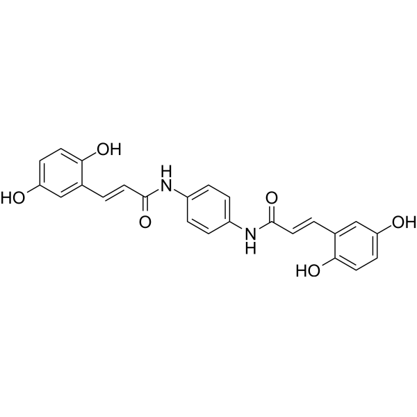
α-Synuclein inhibitor 8 is an active inhibitor of α-Synuclein with an IC50 value of 2.5 µM. α-Synuclein inhibitor 8 has highly inhibition on the aggregation and disaggregation of α-Synuclein fibers. α-Synuclein inhibitor 8 reduces the formation of inclusions in neurons that can repairs damage neurons and improves Parkinson’s disease (PD)-like symptoms. α-Synuclein inhibitor 8 has high antioxidant activity and low cytotoxicity.
For research use only. We do not sell to patients.
- CAS No.: 2883627-64-1
- Formula: C24H20N2O6
- Molecular Weight:432.43
-
Storage:
Please store the product under the recommended conditions in the Certificate of Analysis.

To place orders, for customer services and technical support, please contact: MedChemExpress USA
Tel: 609-228-6898 E-mail: [email protected] [email protected]


-
 Biological Activity
-
Chemical Information
- Protocols
- Purity & Documentation
- References
-
 Help & FAQs


Publications Citing Use of MedChemExpress (MCE) α-Synuclein inhibitor 8
MoreAll α-synuclein Isoforms
More

Biological Activity
Description
IC50 & Target
|
α-synuclein Aggregation |
In Vitro
α-Synuclein inhibitor 8 (Compound 2ee) (0-100 μM; 0-96 h) inhibits the aggregation of α-Synuclein in a dose-dependent manner[1].
α-Synuclein inhibitor 8 (100 µM; 48 h) decomposes mature fiber into soluble protein and a small amount of short insoluble fiber[1].
α-Synuclein inhibitor 8 (1-1000 µM; 0.5 h or 24 h) increases the clearance rate of reactive oxygen species in a dose-dependent manner. α-Synuclein inhibitor 8 has low cytotoxicity to H4 and SH-SY5Y cells (cell viability is higher than 80%)[1].
α-Synuclein inhibitor 8 (10 µM; 38 h) has inhibitory effect on α-Synuclein aggregation and inclusion body formation in H4 cells[1].
MedChemExpress (MCE) has not independently confirmed the accuracy of these methods. They are for reference only. Further protocols information, click here.
-
Cell Line:H4 and SH-SY5Y cells.
-
Concentration:1, 3, 10, 30, 100, 300, 500 and 1000 µM.
-
Incubation Time:24 h.
-
Result:Showed low cytotoxicity.
In Vivo
MedChemExpress (MCE) has not independently confirmed the accuracy of these methods. They are for reference only.
-
Animal Model:6-OHDA-lesioned C57 mice model of PD (8-12 weeks, 20-28 g)[1].
-
Dosage:0.9 μg/μL.
-
Administration:Intravenous injection (right striatum); single dose.
-
Result:Exhibited neuroprotective and repair effects.


Chemical Information
-
CAS No. 2883627-64-1
-
Molecular Weight 432.43
-
Formula C24H20N2O6
-
SMILES
OC1=CC=C(O)C=C1/C=C/C(NC2=CC=C(NC(/C=C/C3=CC(O)=CC=C3O)=O)C=C2)=O
-
Shipping
Room temperature in continental US; may vary elsewhere.
-
Storage
Please store the product under the recommended conditions in the Certificate of Analysis.


Publications (1)
-
Journal Impact Factor
-
Most Recent
-
J Adv Res
Fibrinogen exacerbates α-synuclein aggregation and mitochondrial dysfunction via alpha5beta3 integrin in Parkinson's disease. [Abstract]2025 May 25:S2090-1232(25)00370-4. PMID: 40425084

Protocols
-
Cell Cytotoxicity Assay
Cytotoxicity assays are usually based on the assessment of cell membrane damage, which can also be indirectly detected by measuring cell viability. Detection methods include MTT assay, CKK-8 assay, LDH assay and ATP assay, etc.
-
Mammalian live/dead viability and cytotoxicity staining
Live/dead viability and cytotoxicity staining assays are based on the simultaneous detection of intracellular esterase activity in metabolically active (viable) cells and membrane integrity loss in non-viable cells. In commonly used dual-staining approaches, membrane-permeant fluorogenic substrates are converted by intracellular esterases into fluorescent products in live cells, while impermeant DNA-binding dyes selectively enter cells with compromised plasma membranes and label nucleic acids in dead or dying cells, enabling discrimination between viable and non-viable populations by fluorescence microscopy or flow cytometry.
-
Cell Viability Determination by MTT Colorimetric Assay
The following protocol uses the MTT colorimetric assay as a classic literature-established method for assessing cell viability/metabolic activity in cultured mammalian cells. MTT[3-(4,5-dimethylthiazol-2-yl)-2,5-diphenyltetrazolium bromide] is reduced by metabolically active cells to a colored formazan product; the amount of formazan is quantified spectrophotometrically and provides an indirect measure of metabolically active viable cells. Importantly, MTT reduction reflects cellular oxidoreductase/metabolic activity rather than an absolute direct count of living cells, so changes in cellular metabolism can alter the signal independently of cell number.


Purity & Documentation
References

Calculators
Concentration (start) × Volume (start) = Concentration (final) × Volume (final)


α-Synuclein inhibitor 8 Related Classifications
HY-152552 Related Classifications
 Powered by Bioz