



1,1′-Ethylidenebis[L-tryptophan]

Based on 1 Customer Validation
1,1′-Ethylidenebis[L-tryptophan] is an orally active amino acid analog. 1,1′-Ethylidenebis[L-tryptophan] promotes the release of adrenocorticotropic hormone and corticosterone in rat plasma, and transiently inhibits CRH mRNA in the paraventricular nucleus of the hypothalamus via glucocorticoid negative feedback. 1,1′-Ethylidenebis[L-tryptophan] induces multi-tissue inflammation and fibrosis in rodents and human cells, increases the number of degranulated mast cells, and activates the IL-5 pathway in lymphocytes. 1,1′-Ethylidenebis[L-tryptophan] is activated by tryptophanyl-tRNA synthetase and replaces L-tryptophan during translation; it also interferes with tryptophan metabolism in mice via the kynurenine pathway and dynamically alters their plasma quinolinic acid levels. 1,1′-Ethylidenebis[L-tryptophan] can be used for studies on metabolism-related mechanisms.
For research use only. We do not sell to patients.
- Purity : 96.04%
- CAS No.: 132685-02-0
- Formula: C24H26N4O4
- Molecular Weight:434.49
-
Storage:Powder -20°C, 3 years ; In solvent -80°C, 6 months , -20°C, 1 month
![1,1′-Ethylidenebis[L-tryptophan] Chemical Structure](https://file.medchemexpress.com/product_pic/hy-w585843.gif "Click to View 1,1′-Ethylidenebis[L-tryptophan] Chemical Structure")

To place orders, for customer services and technical support, please contact: MedChemExpress USA
Tel: 609-228-6898 E-mail: [email protected] [email protected]


-
 Biological Activity
-
Chemical Information
-
Solvent & Solubility
- Protocols
- Purity & Documentation
- References
-
 Help & FAQs


-
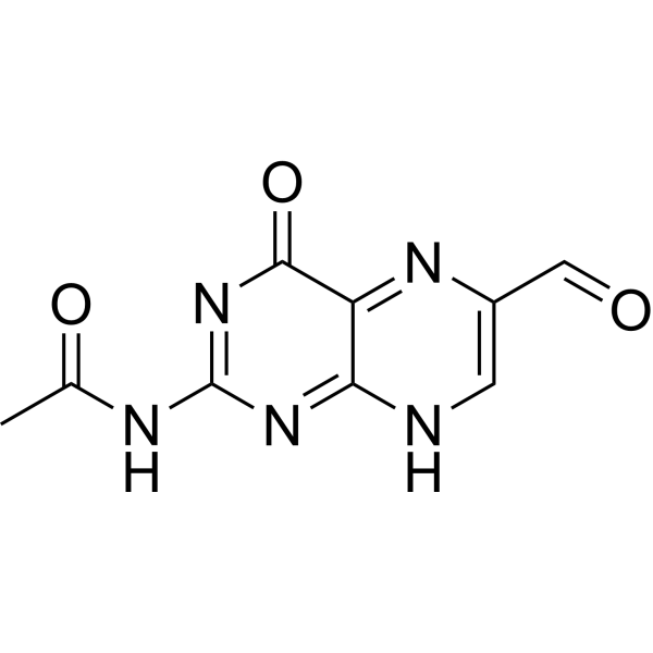
HY-124004
Acetyl-6-formylpterinMAIT Cell Antagonist -
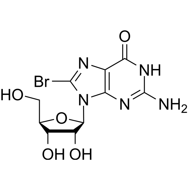
HY-W019033
8-BromoguanosinePurine Nucleoside -
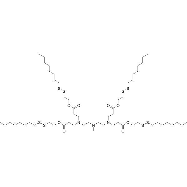
HY-W590681
113-O12BmRNA Carrier -
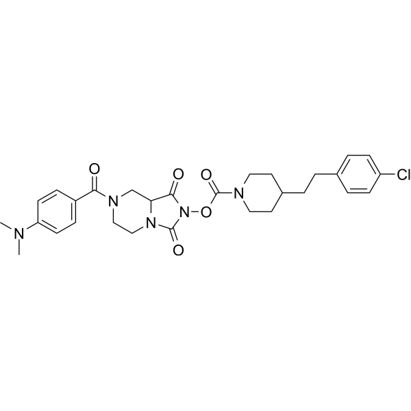
HY-116895
JJH260AIG1 Inhibitor -

HY-I0959R
5-Chlorouracil (Standard)Biochemical Reagent -
HY-I0959
5-ChlorouracilBiochemical Reagent -

HY-169478
Lipid N2-3LCationic Liposome


Biological Activity
Description
IC50 & Target
[5]|
IL-5 |
In Vitro
1,1′-Ethylidenebis[L-tryptophan] (EBT) (40-110 μM; 60 min) reduces 3H-L-tryptophan incorporation into proteins synthesized by rabbit reticulocyte lysates from BMV RNA[3].
1,1′-Ethylidenebis[L-tryptophan] (40-110 μM; 60 min) causes a small but significant reduction in 3H-L-leucine incorporation at 40 μM and 80 μM, but does not significantly reduce incorporation at 110 μM, in proteins synthesized by rabbit reticulocyte lysates from BMV RNA in lysates with 20 μM added L-tryptophan; at 110 μM, incorporation remains at 98% of control both with and without 20 μM added L-leucine[3].
1,1′-Ethylidenebis[L-tryptophan] (110 μM; 60 min) is specifically incorporated into full-length proteins synthesized by rabbit reticulocyte lysates from BMV RNA, as evidenced by discrete 14C-labeled bands at the expected BMV protein molecular weights when 110 μM 14C-labeled 1,1′-Ethylidenebis[L-tryptophan] is used[3].
MedChemExpress (MCE) has not independently confirmed the accuracy of these methods. They are for reference only. Further protocols information, click here.
In Vivo
1,1′-Ethylidenebis[L-tryptophan] leads to myofascial thickening, pancreatic fibrosis and acinar changes in female LEW/N Lewis rats, and triggers dermal and subcutaneous fibrosis in female C57BL/6 mice[3].
1,1′-Ethylidenebis[L-tryptophan] (40 μg/kg; i.p.; daily; up to 6 weeks) induces EMS-like dermal and subcutaneous inflammation and fibrosis, increases mast cell counts, thickens muscle fascia, elevates necrotic muscle fiber counts, and triggers a biphasic change in plasma quinolinic acid levels in female C57BL/6 mice[2].
MedChemExpress (MCE) has not independently confirmed the accuracy of these methods. They are for reference only.
-
Animal Model:LEW/N (female, 100 g on arrival)[1]
-
Dosage:40 mg/kg
-
Administration:p.o.; 6 days/week; 1 week, 2 weeks, 6 weeks
-
Result:Elevated plasma ACTH concentrations twice those of vehicle control rats at week 1.
Raised plasma CORT to levels matching L-Trp control rats and lowered PVN CRH mRNA expression at week 2.
Restored plasma ACTH and CORT to baseline vehicle levels while keeping reduced PVN CRH mRNA expression at week 6.
-
Animal Model:C57BL/6 (6-week-old female, 20-22 g, EMS induced via daily intraperitoneal administration of 1,1′-Ethylidenebis[L-tryptophan])[2]
-
Dosage:40 μg/kg
-
Administration:i.p.; daily; up to 6 weeks
-
Result:Induced focal skin and subcutaneous inflammation at day 3 that advanced to widespread moderate-severe inflammation with epidermal hyperplasia in partial animals at week 3.
Triggered focal dermal and subcutaneous fibrosis at day 6 that developed into full-thickness diffuse fibrosis at week 3.
Elevated mast cell numbers across skin, adipose and muscle fascia layers at day 3 and week 6 relative to saline control groups.
Widened fascia thickness markedly at week 6 compared with saline and L-tryptophan control groups.
Raised necrotic muscle fiber counts obviously at week 4 versus saline and L-tryptophan control groups.
Altered plasma quinolinic acid concentrations: decreased at day 3 and day 6, rose sharply at week 21, then dropped gradually at week 28 and week 42 relative to saline controls.


Chemical Information
-
CAS No. 132685-02-0
-
Appearance Solid
-
Molecular Weight 434.49
-
Formula C24H26N4O4
-
Color White to off-white
-
SMILES
CC(N1C2=CC=CC=C2C(C[C@H](N)C(O)=O)=C1)N3C4=CC=CC=C4C(C[C@H](N)C(O)=O)=C3
-
Shipping
Room temperature in continental US; may vary elsewhere.
-
Storage
Powder -20°C 3 years In solvent -80°C 6 months -20°C 1 month


Solvent & Solubility
In Vitro:
1 M NaOH : 50 mg/mL (115.08 mM; Need ultrasonic)
Please refer to the solubility information to select the appropriate solvent. Once prepared, please aliquot and store the solution to prevent product inactivation from repeated freeze-thaw cycles.
Storage method and period of stock solution: -80°C, 6 months; -20°C, 1 month. When stored at -80°C, please use it within 6 months. When stored at -20°C, please use it within 1 month.
Please refer to the solubility information to select the appropriate solvent. Once prepared, please aliquot and store the solution to prevent product inactivation from repeated freeze-thaw cycles.
Storage method and period of stock solution: -80°C, 6 months; -20°C, 1 month. When stored at -80°C, please use it within 6 months. When stored at -20°C, please use it within 1 month.
Concentration (start) × Volume (start) = Concentration (final) × Volume (final)

Protocols
-
Fibrosis/Collagen Morphometry
Fibrosis and collagen morphometry is based on the quantitative visualization of fibrillar collagen deposition in tissue sections using histochemical stains such as Sirius Red (Picrosirius Red) or Masson's trichrome, followed by image-based or polarization-enhanced analysis to estimate collagen proportional area as a surrogate of extracellular matrix accumulation during fibrotic remodeling. Sirius Red combined with polarized light microscopy enhances detection of collagen fibers due to birefringence properties, enabling more specific visualization of collagen type I and III fibrils compared to conventional bright-field histology, while whole-section or region-restricted digital morphometry reduces field-selection bias in fibrosis assessment. Alternative quantitative approaches include second harmonic generation (SHG) and two-photon excited fluorescence microscopy, which enable label-free detection of fibrillar collagen and have been validated against histological staining and biochemica
-
RNA extraction experimental
By lysing cells, releasing RNA, and removing impurities such as proteins and DNA, high-purity RNA products are finally obtained. The commonly used traditional method is the guanidine isothiocyanate/phenol/chloroform method (Trizol), which is suitable for a variety of animal materials including animal tissues, microorganisms, cultured cells, etc., and most plant materials.
-
Collagen: Sirius Red Staining
Sirius Red or picrosirius red staining is a histochemical method for visualizing collagen-rich extracellular matrix in tissue sections, and collagen fibers are detected as red-stained structures under bright-field microscopy with enhanced birefringence under polarized light. Picrosirius red is useful for assessing total collagen organization, distribution, and fibrosis burden, but polarized color should not be interpreted as a definitive collagen type I versus type III readout because color is affected by fiber orientation, thickness, and packing.
-
Research Protocol for Endocrine Diseases
Endocrine diseases often arise from disrupted hormone production, hormone signaling, or target-tissue responsiveness; for diabetes-focused endocrine disease models, insulin signaling regulates glucose uptake, hepatic glucose output, lipid metabolism, and β-cell compensation. Type 2 diabetes develops through interacting defects in insulin resistance, β-cell dysfunction, adipose inflammation, hepatic glucose overproduction, altered incretin signaling, and ectopic lipid metabolism. A major unresolved question is whether endocrine dysfunction is driven primarily by target-tissue insulin resistance, intrinsic β-cell failure, immune/inflammatory stress, or combined multi-organ failure that differs by disease stage.
-
Connective Tissue: Masson's Trichrome/Collagen Trichrome Staining
Masson’s Trichrome (collagen/trichrome staining) is a histological technique that differentially stains tissue compartments using sequential acidic dyes to distinguish collagen from muscle and cytoplasmic components based on dye affinity and tissue permeability differences, enabling visualization of fibrosis and connective tissue architecture in histological sections. The classical formulation typically uses Weigert's iron hematoxylin for nuclear staining, Biebrich scarlet-acid fuchsin for cytoplasm and muscle, and aniline blue (or light green variants) for collagen, producing a characteristic blue/green collagen signal contrasted against red cytoplasm and dark nuclei. The staining principle relies on selective displacement of smaller dye molecules by larger anionic dyes in collagen-rich regions under controlled acidified conditions, which enhances collagen-specific dye retention. This property makes the method widely used for fibrosis assessment in organs such as heart, liver, lung, a
-
Research Protocol for Inflammation-related Diseases
The NLRP3 inflammasome is a cytosolic innate immune signaling platform that integrates priming signals and danger-signal activation to promote caspase-1 activation, maturation of IL-1β and IL-18, and gasdermin D-mediated pyroptotic cell death. The core experimental logic is to determine whether inflammatory disease phenotypes are driven by increased NLRP3 expression, ASC-containing inflammasome assembly, caspase-1 cleavage, GSDMD cleavage, and extracellular release of IL-1β/IL-18 rather than by nonspecific cell injury alone. The pathway is strongly linked to inflammation-related disease phenotypes because monosodium urate crystals activate NALP3/NLRP3 inflammasome signaling in gout-like crystal inflammation, cholesterol crystals activate NLRP3 inflammasomes in atherogenesis models, and DSS-induced intestinal inflammation has been reported to involve NLRP3 inflammasome activity. However, experimental colitis studies also show context-dependent protective effects of NLRP3 inflammasome co


Purity & Documentation
-
Data Sheet (283 KB)
-
SDS (394 KB)
- English - EN (394 KB)
- Français - FR (394 KB)
- Deutsch - DE (394 KB)
- Norwegian - NO (394 KB)
- Español - ES (394 KB)
- Swedish - SV (394 KB)
- Italian - IT (394 KB)
- Korean - KR (394 KB)
- Portuguese - PT (394 KB)
-
Handling Instructions (2659 KB)
References
[1]. Brady LS, et al. 1,1'-Ethylidenebis[L-tryptophan], a contaminant implicated in L-tryptophan eosinophilia myalgia syndrome, suppresses mRNA expression of hypothalamic corticotropin-releasing hormone in Lewis (LEW/N) rat brain. Neuroimmunomodulation. 1994 Jan;1(1):59-65. [Content Brief]
[2]. Silver RM, et al. A murine model of the eosinophilia-myalgia syndrome induced by 1,1'-ethylidenebis (L-tryptophan). The Journal of clinical investigation. 1994 Apr;93(4):1473-80. [Content Brief]
[3]. Buss WC, et al. EBT, a tryptophan contaminant associated with eosinophilia myalgia syndrome, is incorporated into proteins during translation as an amino acid analog. Autoimmunity. 1996;25(1):33-45. [Content Brief]


Complete Stock Solution Preparation Table
Please refer to the solubility information to select the appropriate solvent. Once prepared, please aliquot and store the solution to prevent product inactivation from repeated freeze-thaw cycles.
Storage method and period of stock solution: -80°C, 6 months; -20°C, 1 month. When stored at -80°C, please use it within 6 months. When stored at -20°C, please use it within 1 month.
| Optional Solvent | Concentration Solvent Mass | 1 mg | 5 mg | 10 mg | 25 mg |
|---|---|---|---|---|---|
| 1 M NaOH | 1 mM | 2.3015 mL | 11.5077 mL | 23.0155 mL | 57.5387 mL |
| 5 mM | 0.4603 mL | 2.3015 mL | 4.6031 mL | 11.5077 mL | |
| 10 mM | 0.2302 mL | 1.1508 mL | 2.3015 mL | 5.7539 mL | |
| 15 mM | 0.1534 mL | 0.7672 mL | 1.5344 mL | 3.8359 mL | |
| 20 mM | 0.1151 mL | 0.5754 mL | 1.1508 mL | 2.8769 mL | |
| 25 mM | 0.0921 mL | 0.4603 mL | 0.9206 mL | 2.3015 mL | |
| 30 mM | 0.0767 mL | 0.3836 mL | 0.7672 mL | 1.9180 mL | |
| 40 mM | 0.0575 mL | 0.2877 mL | 0.5754 mL | 1.4385 mL | |
| 50 mM | 0.0460 mL | 0.2302 mL | 0.4603 mL | 1.1508 mL | |
| 60 mM | 0.0384 mL | 0.1918 mL | 0.3836 mL | 0.9590 mL | |
| 80 mM | 0.0288 mL | 0.1438 mL | 0.2877 mL | 0.7192 mL | |
| 100 mM | 0.0230 mL | 0.1151 mL | 0.2302 mL | 0.5754 mL |


1,1′-Ethylidenebis[L-tryptophan] Related Classifications
HY-W585843 Related Classifications
 Powered by Bioz
Keywords
- 1,1′-Ethylidenebis[L-tryptophan]
- 132685-02-0
- Amino Acid Derivatives
- Interleukin Related
- CRFR
- Aminoacyl-tRNA Synthetase
- rat hypothalamic paraventricular nuclei
- glucocorticoid negative feedback
- tryptophanyl-tRNA synthetase
- CRH mRNA
- eosinophilia myalgia syndrome
- kynurenine pathway
- quinolinic acid
- corticosterone
- adrenocorticotropic hormone
- rabbit reticulocyte lysates
- Inhibitor
- inhibitor
- inhibit