



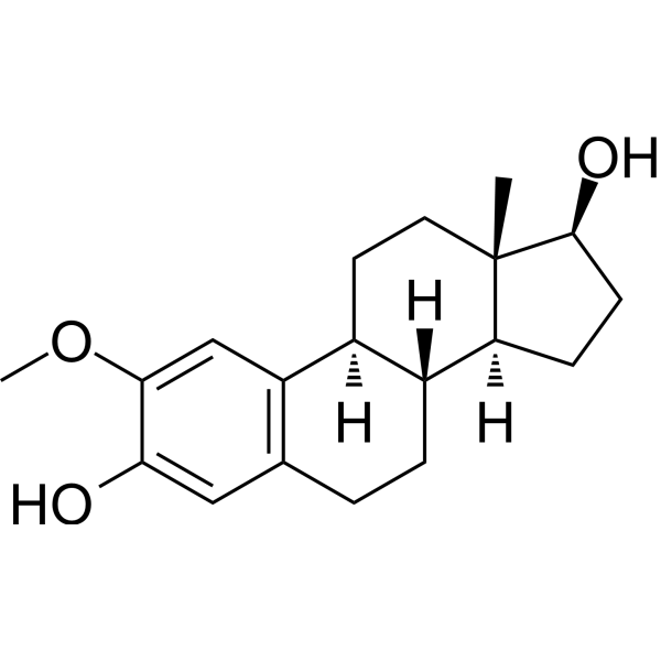
2-Methoxyestradiol
Based on 33 publication(s) in Google Scholar
2-Methoxyestradiol (2-ME2), an orally active endogenous metabolite of 17β-estradiol (E2), is an apoptosis inducer and an angiogenesis inhibitor with potent antineoplastic activity. 2-Methoxyestradiol also destablize microtubules. 2-Methoxyestradio, also a potent superoxide dismutase (SOD) inhibitor and a ROS-generating agent, induces autophagy in the transformed cell line HEK293 and the cancer cell lines U87 and HeLa.
For research use only. We do not sell to patients.
- Purity : 99.76%
- CAS No.: 362-07-2
- Formula: C19H26O3
- Molecular Weight:302.41
-
Storage:Powder -20°C, 3 years , 4°C, 2 years ; In solvent -80°C, 2 years , -20°C, 1 year

To place orders, for customer services and technical support, please contact: MedChemExpress USA
Tel: 609-228-6898 E-mail: [email protected] [email protected]


-
 Biological Activity
-
Chemical Information
-
Solvent & Solubility
- Protocols
- Purity & Documentation
- References
-
 Help & FAQs


-
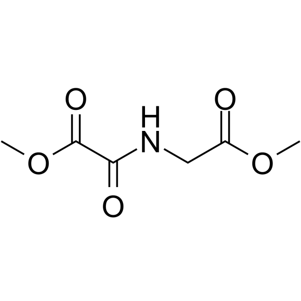
HY-15893
DMOGHIF-PH Inhibitor -
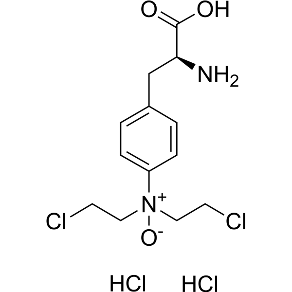
HY-10231
PX-478HIF-1α Inhibitor -

HY-100579
Ferrostatin-1Ferroptosis Inhibitor -
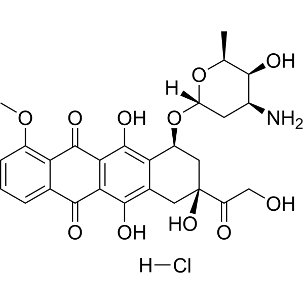
HY-15142
Doxorubicin hydrochlorideTopoisomerase Inhibitor -
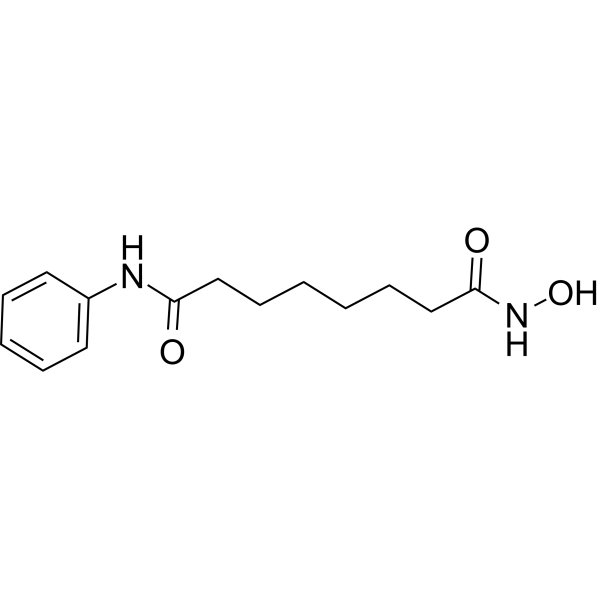
HY-10221
VorinostatHDAC Inhibitor -
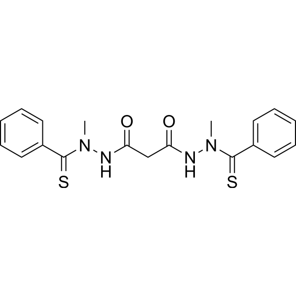
HY-12040
ElesclomolCuproptosis Inducer -
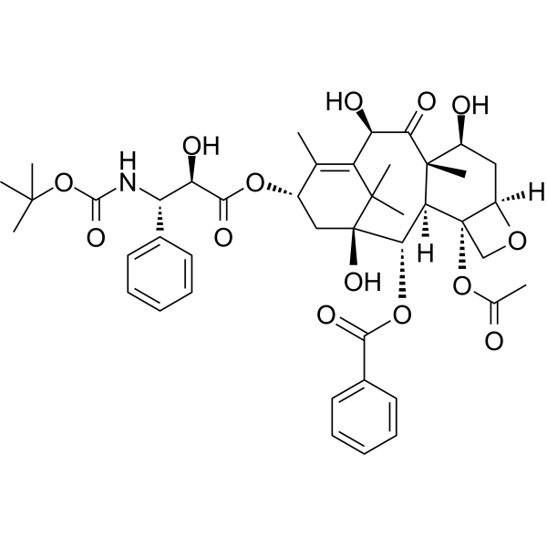
HY-B0011
DocetaxelMicrotubule Depolymerization Inhibitor -
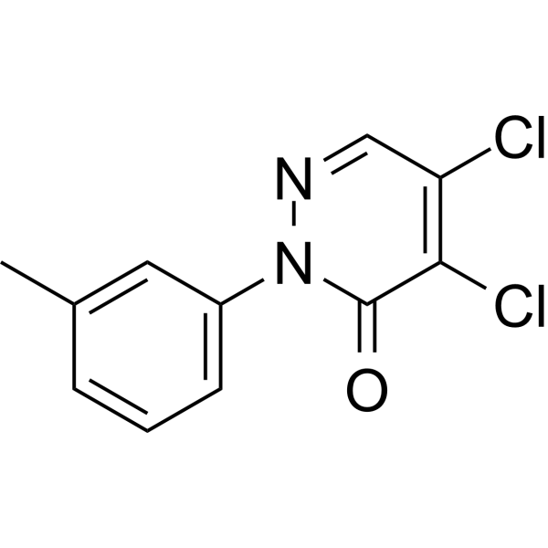
HY-115445
LCS-1SOD1 Inhibitor -
HY-13671
LW6Apoptosis Inducer -

HY-10219
RapamycinmTOR Inhibitor -
HY-12867
PT-2385HIF-2α Inhibitor -
HY-113252
2-MethoxyestroneEstrogen Metabolite
Publications Citing Use of MedChemExpress (MCE) 2-Methoxyestradiol
More- Cell Res. 2026 Mar;36(3):219-232. [Abstract]
- J Nanobiotechnology. 2023 Jul 22;21(1):233. [Abstract]
- Theranostics. 2024 May 27;14(8):3339-3357. [Abstract]
- J Exp Clin Cancer Res. 2018 Jun 28;37(1):128. [Abstract]
- Carbohydr Polym. 2025 Aug 15:362:123685. [Abstract]
- Cell Death Dis. 2025 Jun 20;16(1):462. [Abstract]
- Cancer Lett. 2016 Nov 1;382(1):44-52. [Abstract]
- Mol Ther. 2021 Dec 1;29(12):3449-3464. [Abstract]
- Acta Biomater. 2025 Aug:202:248-261. [Abstract]
- J Transl Med. 2023 Oct 26;21(1):754. [Abstract]
- Free Radic Biol Med. 2021 Jun:169:271-282. [Abstract]
- Stem Cell Res Ther. 2022 Feb 5;13(1):59. [Abstract]
- Front Immunol. 2021 May 12:12:639049. [Abstract]
- Ann Hepatol. 2023 Nov-Dec;28(6):101135. [Abstract]
- Eur J Pharmacol. 2022 Oct 15:933:175276. [Abstract]
- Cancer Biol Ther. 2016 Jun 2;17(6):625-34. [Abstract]
- Int J Mol Sci. 2025 Jul 8;26(14):6554. [Abstract]
- Front Cell Dev Biol. 2021 Feb 23:9:607142. [Abstract]
- Chem Biol Interact. 2023 Jan 5:369:110277. [Abstract]
- Pestic Biochem Physiol. 2023 Dec:197:105647. [Abstract]
- Rheumatology (Oxford). 2019 Nov 1;58(11):1966-1975. [Abstract]
- Aquaculture. 2020, 735393.
- Front Cell Neurosci. 2018 Dec 21:12:504. [Abstract]
- Am J Physiol Lung Cell Mol Physiol. 2025 Aug 1;329(2):L282-L295. [Abstract]
- Peptides. 2024 Nov:181:171299. [Abstract]
- J Immunol Res. 2022 Apr 28;2022:6324326. [Abstract]
- Biomed Res Int. 2016:2016:7948345. [Abstract]
- Thorac Cancer. 2019 Apr;10(4):676-685. [Abstract]
- Exp Ther Med. 2017 Aug;14(2):1825-1831. [Abstract]
- SSRN. 2025 Mar 17.
- Research Square Print. 2022 Aug.
- Research Square Print. 2022 May.
- Patent. US20180263995A1.
 Customer Validation & Images

-
WB
-
WB
-
WB
All Endogenous Metabolite Isoforms
More

Biological Activity
Description
IC50 & Target
|
Human Endogenous Metabolite |
Cellular Effect
|
Cell Line

|
Type | Value | Description | References |
|---|---|---|---|---|
| A549 | IC50 |
1.1 μM
Compound: 2ME2
|
Antitumour activity against human A549 cells assessed as cell growth inhibition measured after 72 hrs by SRB assay
Antitumour activity against human A549 cells assessed as cell growth inhibition measured after 72 hrs by SRB assay
|
[PMID: 31102934] |
| B16 | IC50 |
3.4 μM
Compound: 2ME2
|
Antitumour activity against mouse B16 cells assessed as cell growth inhibition measured after 72 hrs by SRB assay
Antitumour activity against mouse B16 cells assessed as cell growth inhibition measured after 72 hrs by SRB assay
|
[PMID: 31102934] |
| Cancer cell lines | GI50 |
0.08 μM
Compound: 1
|
Cytotoxicity against the cancer cell lines breast MDA-MB-435
Cytotoxicity against the cancer cell lines breast MDA-MB-435
|
[PMID: 12361402] |
| Cancer cell lines | GI50 |
0.21 μM
Compound: 1
|
Cytotoxicity against the cancer cell lines ovarian OVCAR-3
Cytotoxicity against the cancer cell lines ovarian OVCAR-3
|
[PMID: 12361402] |
| Cancer cell lines | GI50 |
0.32 μM
Compound: 1
|
Cytotoxicity against the cancer cell lines CNS SF-539
Cytotoxicity against the cancer cell lines CNS SF-539
|
[PMID: 12361402] |
| Cancer cell lines | GI50 |
0.36 μM
Compound: 1
|
Cytotoxicity against the cancer cell lines melanoma UACC-62
Cytotoxicity against the cancer cell lines melanoma UACC-62
|
[PMID: 12361402] |
| Cancer cell lines | GI50 |
0.47 μM
Compound: 1
|
Cytotoxicity against the cancer cell lines colon HCT116
Cytotoxicity against the cancer cell lines colon HCT116
|
[PMID: 12361402] |
| Cancer cell lines | GI50 |
0.7 μM
Compound: 2
|
Concentration required to inhibit the growth of HOP-6 human lung cancer cells by 50%
Concentration required to inhibit the growth of HOP-6 human lung cancer cells by 50%
|
[PMID: 16078843] |
| Cancer cell lines | GI50 |
0.7 μM
Compound: 1
|
Cytotoxicity against the cancer cell lines lung HOP-62
Cytotoxicity against the cancer cell lines lung HOP-62
|
[PMID: 12361402] |
| Cancer cell lines | GI50 |
0.95 μM
Compound: 1
|
Cytotoxicity against the cancer cell lines renal SN12C
Cytotoxicity against the cancer cell lines renal SN12C
|
[PMID: 12361402] |
| Cancer cell lines | GI50 |
1.3 μM
Compound: 1
|
Growth inhibitory concentration against human cancer cell line
Growth inhibitory concentration against human cancer cell line
|
[PMID: 15456256] |
| Cancer cell lines | GI50 |
1.8 μM
Compound: 1
|
Cytotoxicity against the cancer cell lines prostate DU-145
Cytotoxicity against the cancer cell lines prostate DU-145
|
[PMID: 12361402] |
| DU-145 | GI50 |
1.2 μM
Compound: 2-methoxyestradiol
|
Antiproliferative activity against human DU145 cells at 96 hrs by WST1 assay
Antiproliferative activity against human DU145 cells at 96 hrs by WST1 assay
|
[PMID: 22247790] |
| DU-145 | GI50 |
1.22 μM
Compound: 1
|
Antiproliferative activity against human DU145 cells after 96 hrs by WST1 assay
Antiproliferative activity against human DU145 cells after 96 hrs by WST1 assay
|
[PMID: 17696419] |
| DU-145 | GI50 |
1.22 μM
Compound: 1
|
Antiproliferative activity against human DU145 cells after 96 hrs by WST-1 assay
Antiproliferative activity against human DU145 cells after 96 hrs by WST-1 assay
|
[PMID: 20225862] |
| DU-145 | GI50 |
1.22 μM
Compound: 1
|
Antiproliferative activity against human DU145 cells
Antiproliferative activity against human DU145 cells
|
[PMID: 18260615] |
| DU-145 | GI50 |
1.8 μM
Compound: 2
|
Concentration required to inhibit the growth of DU-145 human prostate cancer cells by 50%
Concentration required to inhibit the growth of DU-145 human prostate cancer cells by 50%
|
[PMID: 16078843] |
| DU-145 | GI50 |
1.8 μM
Compound: 1
|
Growth inhibitory concentration against human prostate DU-145 cancer cell line
Growth inhibitory concentration against human prostate DU-145 cancer cell line
|
[PMID: 15456256] |
| DU-145 | GI50 |
1.8 μM
Compound: 1
|
Cytotoxicity against human DU145 cells
Cytotoxicity against human DU145 cells
|
[PMID: 18052315] |
| DU-145 | GI50 |
1.8 μM
Compound: 1
|
Cytotoxicity against prostate DU-145 cancer cell lines.
Cytotoxicity against prostate DU-145 cancer cell lines.
|
[PMID: 7783135] |
| DU-145 | GI50 |
1.8 μM
Compound: 1
|
The In vitro cytotoxicity value corresponding to 50% growth inhibition against DU-145 (prostate) cell line.
The In vitro cytotoxicity value corresponding to 50% growth inhibition against DU-145 (prostate) cell line.
|
[PMID: 9240348] |
| ECa-109 cell line | IC50 |
4.1 μM
Compound: 2ME2
|
Antitumour activity against human EC109 cells assessed as cell growth inhibition measured after 72 hrs by SRB assay
Antitumour activity against human EC109 cells assessed as cell growth inhibition measured after 72 hrs by SRB assay
|
[PMID: 31102934] |
| HCT-116 | GI50 |
0.47 μM
Compound: 1
|
Antiproliferative activity against human HCT116 cells after 96 hrs by WST-1 assay
Antiproliferative activity against human HCT116 cells after 96 hrs by WST-1 assay
|
[PMID: 20225862] |
| HCT-116 | GI50 |
0.47 μM
Compound: 2
|
Concentration required to inhibit the growth of HCT116 human colon cancer cells by 50%
Concentration required to inhibit the growth of HCT116 human colon cancer cells by 50%
|
[PMID: 16078843] |
| HCT-116 | GI50 |
0.47 μM
Compound: 1
|
Growth inhibitory concentration against human colon cancer HCT116 cell line
Growth inhibitory concentration against human colon cancer HCT116 cell line
|
[PMID: 15456256] |
| HCT-116 | GI50 |
0.47 μM
Compound: 1
|
Cytotoxicity against human HCT116 cells
Cytotoxicity against human HCT116 cells
|
[PMID: 18052315] |
| HCT-116 | GI50 |
0.47 μM
Compound: 1
|
Antiproliferative activity against human HCT116 cells
Antiproliferative activity against human HCT116 cells
|
[PMID: 18260615] |
| HCT-116 | GI50 |
0.47 μM
Compound: 1
|
Cytotoxicity against colon HCT116 cancer cell lines.
Cytotoxicity against colon HCT116 cancer cell lines.
|
[PMID: 7783135] |
| HCT-116 | GI50 |
0.47 μM
Compound: 1
|
The In vitro cytotoxicity value is the concentration of the compound corresponding to 50% growth inhibition againstHCT-116 (colon) cell line.
The In vitro cytotoxicity value is the concentration of the compound corresponding to 50% growth inhibition againstHCT-116 (colon) cell line.
|
[PMID: 9240348] |
| HEK-293T | IC50 |
4.6 μM
Compound: 2-methoxyestradiol
|
Cytotoxicity against HEK293T cells assessed as inhibition of cell growth incubated for 72 hrs by MTT assay
Cytotoxicity against HEK293T cells assessed as inhibition of cell growth incubated for 72 hrs by MTT assay
|
[PMID: 36455509] |
| HeLa | IC50 |
586 nM
Compound: 2ME2
|
Antiproliferative activity against human HeLa cells expressing tubulin 3beta by sulforhodamine B assay
Antiproliferative activity against human HeLa cells expressing tubulin 3beta by sulforhodamine B assay
|
[PMID: 20973488] |
| HeLa | IC50 |
608 nM
Compound: 2ME2
|
Antiproliferative activity against human HeLa cells by sulforhodamine B assay
Antiproliferative activity against human HeLa cells by sulforhodamine B assay
|
[PMID: 20973488] |
| HeLa | IC50 |
8.6 μM
Compound: 2-methoxyestradiol
|
Antiproliferative activity against human HeLa cells assessed as inhibition of cell growth incubated for 72 hrs by MTT assay
Antiproliferative activity against human HeLa cells assessed as inhibition of cell growth incubated for 72 hrs by MTT assay
|
[PMID: 36455509] |
| HepG2 | IC50 |
11.03 μM
Compound: 2; 2-ME2
|
Antiproliferative activity against human HepG2 cells after 48 hrs by MTT assay
Antiproliferative activity against human HepG2 cells after 48 hrs by MTT assay
|
[PMID: 28810190] |
| HepG2 | IC50 |
2.3 μM
Compound: 2-methoxyestradiol
|
Antiproliferative activity against human HepG2 cells assessed as inhibition of cell growth incubated for 72 hrs by MTT assay
Antiproliferative activity against human HepG2 cells assessed as inhibition of cell growth incubated for 72 hrs by MTT assay
|
[PMID: 36455509] |
| HL-60 | IC50 |
0.9 μM
Compound: Table 3 R4C1
|
Cytotoxicity against vincristine-resistant human HL60 cells after 72 hrs MTS assay
Cytotoxicity against vincristine-resistant human HL60 cells after 72 hrs MTS assay
|
[PMID: 20411988] |
| HL-60 | IC50 |
3.4 μM
Compound: Table 3 R4C1
|
Cytotoxicity against human HL60 cells after 72 hrs MTS assay
Cytotoxicity against human HL60 cells after 72 hrs MTS assay
|
[PMID: 20411988] |
| HOP-62 | GI50 |
0.7 μM
Compound: 1
|
Antiproliferative activity against human HOP62 cells after 96 hrs by WST-1 assay
Antiproliferative activity against human HOP62 cells after 96 hrs by WST-1 assay
|
[PMID: 20225862] |
| HOP-62 | GI50 |
0.7 μM
Compound: 1
|
Growth inhibitory concentration against human lung cancer HOP-62 cell line
Growth inhibitory concentration against human lung cancer HOP-62 cell line
|
[PMID: 15456256] |
| HOP-62 | GI50 |
0.7 μM
Compound: 1
|
Cytotoxicity against human HOP62 cells
Cytotoxicity against human HOP62 cells
|
[PMID: 18052315] |
| HOP-62 | GI50 |
0.7 μM
Compound: 1
|
Antiproliferative activity against human HOP62 cells
Antiproliferative activity against human HOP62 cells
|
[PMID: 18260615] |
| HOP-62 | GI50 |
0.7 μM
Compound: 1
|
Cytotoxicity against lung HOP-62 cancer cell lines.
Cytotoxicity against lung HOP-62 cancer cell lines.
|
[PMID: 7783135] |
| HOP-62 | GI50 |
0.7 μM
Compound: 1
|
The In vitro cytotoxicity value is the concentration corresponding to 50% growth inhibition against HOP-62 (non small cell lung) cell line.
The In vitro cytotoxicity value is the concentration corresponding to 50% growth inhibition against HOP-62 (non small cell lung) cell line.
|
[PMID: 9240348] |
| HUVEC | IC50 |
0.68 μM
Compound: 2ME2, Panzem
|
Antiangiogenic activity against HUVEC assessed as growth inhibition after 48 hrs by WST1 assay
Antiangiogenic activity against HUVEC assessed as growth inhibition after 48 hrs by WST1 assay
|
[PMID: 19782564] |
| HUVEC | IC50 |
0.68 μM
Compound: 2ME2
|
Antiangiogenic activity in HUVEC assessed as inhibition of cell proliferation
Antiangiogenic activity in HUVEC assessed as inhibition of cell proliferation
|
[PMID: 19782568] |
| HUVEC | IC50 |
0.84 μM
Compound: 1, 2ME2
|
Antiproliferative activity against human HUVECs after 48 hrs
Antiproliferative activity against human HUVECs after 48 hrs
|
[PMID: 17910916] |
| HUVEC | IC50 |
0.84 μM
Compound: 2ME2
|
Antiangiogenic activity against HUVECs by Brdu incorporation assay
Antiangiogenic activity against HUVECs by Brdu incorporation assay
|
[PMID: 19762246] |
| HUVEC | IC50 |
0.84 μM
Compound: 1
|
Growth inhibitory concentration against MDA-MB-231 cancer cell line
Growth inhibitory concentration against MDA-MB-231 cancer cell line
|
[PMID: 15456256] |
| HUVEC | IC50 |
14.17 μM
Compound: 2; 2-ME2
|
Antiproliferative activity against HUVEC after 48 hrs by MTT assay
Antiproliferative activity against HUVEC after 48 hrs by MTT assay
|
[PMID: 28810190] |
| HUVEC | IC50 |
523 nM
Compound: 1
|
Antiangiogenic activity against HUVEC cells assessed as inhibition of proliferation
Antiangiogenic activity against HUVEC cells assessed as inhibition of proliferation
|
[PMID: 18260615] |
| Ishikawa | IC50 |
8.96 μM
Compound: 2; 2-ME2
|
Antiproliferative activity against human Ishikawa cells after 48 hrs by MTT assay
Antiproliferative activity against human Ishikawa cells after 48 hrs by MTT assay
|
[PMID: 28810190] |
| K562 | IC50 |
0.8 μM
Compound: 1, 2-ME
|
Anticancer activity against human K562 cells assessed as cell growth inhibition after 5 days by MTT assay
Anticancer activity against human K562 cells assessed as cell growth inhibition after 5 days by MTT assay
|
[PMID: 25108078] |
| LNCaP | IC50 |
0.5 μM
Compound: 6
|
Antiproliferative activity against human LNCaP cell line
Antiproliferative activity against human LNCaP cell line
|
[PMID: 16650989] |
| MCF7 | GI50 |
2.35 μM
Compound: 2
|
Inhibition of the growth of human breast cancer MCF-7 cells done for 2 hr at 37 degree C with compound
Inhibition of the growth of human breast cancer MCF-7 cells done for 2 hr at 37 degree C with compound
|
[PMID: 16078843] |
| MCF7 | GI50 |
2.35 μM
Compound: 1
|
Antiproliferative activity against estrogen receptor positive human MCF7 cells
Antiproliferative activity against estrogen receptor positive human MCF7 cells
|
[PMID: 18260615] |
| MCF7 | GI50 |
2.35 μM
Compound: 40
|
Growth inhibition of human MCF7 cells
Growth inhibition of human MCF7 cells
|
[PMID: 25992880] |
| MCF7 | IC50 |
>70 μM
Compound: 2-methoxyestradiol
|
Antiproliferative activity against human MCF7 cells assessed as inhibition of cell growth incubated for 72 hrs by MTT assay
Antiproliferative activity against human MCF7 cells assessed as inhibition of cell growth incubated for 72 hrs by MTT assay
|
[PMID: 36455509] |
| MCF7 | IC50 |
0.842 μM
Compound: 2-methoxyestradiol
|
Antiproliferative activity against human MCF7 cells after 72 hrs by crystal violet biomass reduction assay
Antiproliferative activity against human MCF7 cells after 72 hrs by crystal violet biomass reduction assay
|
[PMID: 20334421] |
| MCF7 | IC50 |
2.39 μM
Compound: 1 (2-MeOE2)
|
Anti-proliferative activity against MCF-7 human breast cancer cells was determined by using MCF-7 plate assay
Anti-proliferative activity against MCF-7 human breast cancer cells was determined by using MCF-7 plate assay
|
[PMID: 15149660] |
| MCF7 | IC50 |
6.01 μM
Compound: 2; 2-ME2
|
Antiproliferative activity against human MCF7 cells after 48 hrs by MTT assay
Antiproliferative activity against human MCF7 cells after 48 hrs by MTT assay
|
[PMID: 28810190] |
| MDA-MB-231 | GI50 |
0.94 μM
Compound: 1
|
Antiproliferative activity against human MDA-MB-231 cells after 96 hrs by WST1 assay
Antiproliferative activity against human MDA-MB-231 cells after 96 hrs by WST1 assay
|
[PMID: 17696419] |
| MDA-MB-231 | GI50 |
0.94 μM
Compound: 1
|
Antiproliferative activity against human MDA-MB-231 cells after 96 hrs by WST-1 assay
Antiproliferative activity against human MDA-MB-231 cells after 96 hrs by WST-1 assay
|
[PMID: 20225862] |
| MDA-MB-231 | GI50 |
0.94 μM
Compound: 1
|
Antiproliferative activity against estrogen receptor negative human MDA-MB-231 cells
Antiproliferative activity against estrogen receptor negative human MDA-MB-231 cells
|
[PMID: 18260615] |
| MDA-MB-231 | IC50 |
0.25 μM
Compound: 2-methoxyestradiol
|
Inhibition of estrogen receptor negative MDA-MB 231 breast cancer cell proliferation at 1 uM
Inhibition of estrogen receptor negative MDA-MB 231 breast cancer cell proliferation at 1 uM
|
[PMID: 9357519] |
| MDA-MB-231 | IC50 |
0.79 μM
Compound: 2ME2, Panzem
|
Antiproliferative activity against human MDA-MB-231 cells after 48 hrs by WST1 assay
Antiproliferative activity against human MDA-MB-231 cells after 48 hrs by WST1 assay
|
[PMID: 19782564] |
| MDA-MB-231 | IC50 |
0.79 μM
Compound: 2ME2
|
Antiproliferative activity against human MDA-MB-231 cells
Antiproliferative activity against human MDA-MB-231 cells
|
[PMID: 19782568] |
| MDA-MB-231 | IC50 |
1 μM
Compound: 1, 2ME2
|
Antiproliferative activity against human MDA-MB-231 cells after 48 hrs
Antiproliferative activity against human MDA-MB-231 cells after 48 hrs
|
[PMID: 17910916] |
| MDA-MB-231 | IC50 |
1 μM
Compound: 1
|
Growth inhibitory concentration against HUVEC cancer cell line
Growth inhibitory concentration against HUVEC cancer cell line
|
[PMID: 15456256] |
| MDA-MB-231 | IC50 |
5 μM
Compound: 2-methoxyestradiol
|
Inhibition of estrogen receptor negative MDA-MB 231 breast cancer cell proliferation at 10 uM
Inhibition of estrogen receptor negative MDA-MB 231 breast cancer cell proliferation at 10 uM
|
[PMID: 9357519] |
| MDA-MB-435 | GI50 |
0.08 μM
Compound: 2
|
Concentration required to inhibit the growth of MDA-MB-435 human breast cancer cells by 50%
Concentration required to inhibit the growth of MDA-MB-435 human breast cancer cells by 50%
|
[PMID: 16078843] |
| MDA-MB-435 | GI50 |
0.08 μM
Compound: 1
|
Growth inhibitory concentration against human breast MDA-MB-435 cancer cell line
Growth inhibitory concentration against human breast MDA-MB-435 cancer cell line
|
[PMID: 15456256] |
| MDA-MB-435 | GI50 |
0.08 μM
Compound: 1
|
Cytotoxicity against human MDA-MB-435 cells
Cytotoxicity against human MDA-MB-435 cells
|
[PMID: 18052315] |
| MDA-MB-435 | GI50 |
0.08 μM
Compound: 1
|
Antiproliferative activity against human MDA-MB-435 cells
Antiproliferative activity against human MDA-MB-435 cells
|
[PMID: 18260615] |
| MDA-MB-435 | GI50 |
0.08 μM
Compound: 1
|
Cytotoxicity against breast MDA-MB-435 cancer cell lines.
Cytotoxicity against breast MDA-MB-435 cancer cell lines.
|
[PMID: 7783135] |
| MDA-MB-435 | GI50 |
0.08 μM
Compound: 1
|
In vitro cytotoxicity value corresponding to 50% growth inhibition against MDA-MB-435 (breast) cell line.
In vitro cytotoxicity value corresponding to 50% growth inhibition against MDA-MB-435 (breast) cell line.
|
[PMID: 9240348] |
| MDA-MB-435 | IC50 |
1 μM
Compound: 2ME2
|
Antiproliferative activity against human MDA-MB-435 cells by Brdu incorporation assay
Antiproliferative activity against human MDA-MB-435 cells by Brdu incorporation assay
|
[PMID: 19762246] |
| MGC-803 | IC50 |
4 μM
Compound: 2ME2
|
Antitumour activity against human MGC803 cells assessed as cell growth inhibition measured after 72 hrs by SRB assay
Antitumour activity against human MGC803 cells assessed as cell growth inhibition measured after 72 hrs by SRB assay
|
[PMID: 31102934] |
| OVCAR-3 | GI50 |
0.21 μM
Compound: 1
|
Antiproliferative activity against human OVCAR-3 cells after 96 hrs by WST-1 assay
Antiproliferative activity against human OVCAR-3 cells after 96 hrs by WST-1 assay
|
[PMID: 20225862] |
| OVCAR-3 | GI50 |
0.21 μM
Compound: 2
|
Concentration required to inhibit the growth of OVCAR-3 human ovarian cancer cells by 50%
Concentration required to inhibit the growth of OVCAR-3 human ovarian cancer cells by 50%
|
[PMID: 16078843] |
| OVCAR-3 | GI50 |
0.21 μM
Compound: 1
|
Growth inhibitory concentration against human ovarian OVCAR-3 cancer cell line
Growth inhibitory concentration against human ovarian OVCAR-3 cancer cell line
|
[PMID: 15456256] |
| OVCAR-3 | GI50 |
0.21 μM
Compound: 1
|
Cytotoxicity against human OVCAR-3 cells
Cytotoxicity against human OVCAR-3 cells
|
[PMID: 18052315] |
| OVCAR-3 | GI50 |
0.21 μM
Compound: 1
|
Antiproliferative activity against human OVCAR-3 cells
Antiproliferative activity against human OVCAR-3 cells
|
[PMID: 18260615] |
| OVCAR-3 | GI50 |
0.21 μM
Compound: 1
|
Cytotoxicity against ovarian OVCAR-3 cancer cell lines.
Cytotoxicity against ovarian OVCAR-3 cancer cell lines.
|
[PMID: 7783135] |
| OVCAR-3 | GI50 |
0.21 μM
Compound: 1
|
In vitro cytotoxicity value corresponding to 50% growth inhibition against OVCAR-3 (ovarian) cell line.
In vitro cytotoxicity value corresponding to 50% growth inhibition against OVCAR-3 (ovarian) cell line.
|
[PMID: 9240348] |
| OVCAR-3 | IC50 |
1.2 μM
Compound: 1, 2-ME
|
Anticancer activity against human OVCAR3 cells assessed as cell growth inhibition after 5 days by MTT assay
Anticancer activity against human OVCAR3 cells assessed as cell growth inhibition after 5 days by MTT assay
|
[PMID: 25108078] |
| Panel (Carcinoma cell lines) | GI50 |
1.3 μM
Compound: 1
|
Mean graph midpoint for all human cancer cell line cytotoxicity.
Mean graph midpoint for all human cancer cell line cytotoxicity.
|
[PMID: 7783135] |
| Panel (Carcinoma cell lines) | GI50 |
1.3 μM
Compound: 1
|
Mean graph midpoint for growth inhibition of all human cancer cell lines
Mean graph midpoint for growth inhibition of all human cancer cell lines
|
[PMID: 9240348] |
| PC-3 | IC50 |
12.31 μM
Compound: 2; 2-ME2
|
Antiproliferative activity against human PC3 cells after 48 hrs by MTT assay
Antiproliferative activity against human PC3 cells after 48 hrs by MTT assay
|
[PMID: 28810190] |
| PC-3 | IC50 |
2.65 μM
Compound: 2ME2
|
Antiproliferative activity against human PC3 cells by Brdu incorporation assay
Antiproliferative activity against human PC3 cells by Brdu incorporation assay
|
[PMID: 19762246] |
| PC-3 | IC50 |
82 μg/mL
Compound: 2-ME2
|
Cytotoxicity against human PC3 cells after 72 hrs by CellTiter one aqueous solution assay
Cytotoxicity against human PC3 cells after 72 hrs by CellTiter one aqueous solution assay
|
[PMID: 31195168] |
| SF-539 | GI50 |
0.32 μM
Compound: 1
|
Antiproliferative activity against human SF539 cells after 96 hrs by WST-1 assay
Antiproliferative activity against human SF539 cells after 96 hrs by WST-1 assay
|
[PMID: 20225862] |
| SF-539 | GI50 |
0.32 μM
Compound: 2
|
Concentration required to inhibit the growth of SF-539 human CNS cancer cells by 50%
Concentration required to inhibit the growth of SF-539 human CNS cancer cells by 50%
|
[PMID: 16078843] |
| SF-539 | GI50 |
0.32 μM
Compound: 1
|
Growth inhibitory concentration against human CNS SF-539 cancer cell line
Growth inhibitory concentration against human CNS SF-539 cancer cell line
|
[PMID: 15456256] |
| SF-539 | GI50 |
0.32 μM
Compound: 1
|
Cytotoxicity against human SF539 cells
Cytotoxicity against human SF539 cells
|
[PMID: 18052315] |
| SF-539 | GI50 |
0.32 μM
Compound: 1
|
Antiproliferative activity against human SF539 cells at
Antiproliferative activity against human SF539 cells at
|
[PMID: 18260615] |
| SF-539 | GI50 |
0.32 μM
Compound: 1
|
Cytotoxicity against CNS SF-539 cancer cell lines.
Cytotoxicity against CNS SF-539 cancer cell lines.
|
[PMID: 7783135] |
| SF-539 | GI50 |
0.32 μM
Compound: 1
|
The In vitro cytotoxicity corresponding to 50% growth inhibition against SF-59 (central nervous system) cell line.
The In vitro cytotoxicity corresponding to 50% growth inhibition against SF-59 (central nervous system) cell line.
|
[PMID: 9240348] |
| SK-N-SH | IC50 |
1.6 μM
Compound: 2ME2
|
Antitumour activity against human SK-N-SH cells assessed as cell growth inhibition measured after 72 hrs by SRB assay
Antitumour activity against human SK-N-SH cells assessed as cell growth inhibition measured after 72 hrs by SRB assay
|
[PMID: 31102934] |
| SK-OV-3 | IC50 |
16.5 μM
Compound: 2-methoxyestradiol
|
Antiproliferative activity against human SK-OV-3 cells assessed as inhibition of cell growth incubated for 72 hrs by MTT assay
Antiproliferative activity against human SK-OV-3 cells assessed as inhibition of cell growth incubated for 72 hrs by MTT assay
|
[PMID: 36455509] |
| SK-OV-3 | IC50 |
2268 nM
Compound: 2ME2
|
Antiproliferative activity against human SKOV3 cells expressing MDR1-6/6 by sulforhodamine B assay
Antiproliferative activity against human SKOV3 cells expressing MDR1-6/6 by sulforhodamine B assay
|
[PMID: 20973488] |
| SK-OV-3 | IC50 |
867 nM
Compound: 2ME2
|
Antiproliferative activity against human SKOV3 cells by sulforhodamine B assay
Antiproliferative activity against human SKOV3 cells by sulforhodamine B assay
|
[PMID: 20973488] |
| SMMC-7721 | IC50 |
8.4 μM
Compound: 2ME2
|
Antitumour activity against human SMMC7721 cells assessed as cell growth inhibition measured after 72 hrs by SRB assay
Antitumour activity against human SMMC7721 cells assessed as cell growth inhibition measured after 72 hrs by SRB assay
|
[PMID: 31102934] |
| SN12C | GI50 |
0.95 μM
Compound: 1
|
Antiproliferative activity against human SN12C cells after 96 hrs by WST-1 assay
Antiproliferative activity against human SN12C cells after 96 hrs by WST-1 assay
|
[PMID: 20225862] |
| SN12C | GI50 |
0.95 μM
Compound: 2
|
Concentration required to inhibit the growth of SN12C human renal cancer cells by 50%
Concentration required to inhibit the growth of SN12C human renal cancer cells by 50%
|
[PMID: 16078843] |
| SN12C | GI50 |
0.95 μM
Compound: 1
|
Growth inhibitory concentration against human renal SN12C cancer cell line
Growth inhibitory concentration against human renal SN12C cancer cell line
|
[PMID: 15456256] |
| SN12C | GI50 |
0.95 μM
Compound: 1
|
Cytotoxicity against human SN12C cells
Cytotoxicity against human SN12C cells
|
[PMID: 18052315] |
| SN12C | GI50 |
0.95 μM
Compound: 1
|
Antiproliferative activity against human SN12-C cells
Antiproliferative activity against human SN12-C cells
|
[PMID: 18260615] |
| SN12C | GI50 |
0.95 μM
Compound: 1
|
Cytotoxicity against renal SN12C cancer cell lines.
Cytotoxicity against renal SN12C cancer cell lines.
|
[PMID: 7783135] |
| SN12C | GI50 |
0.95 μM
Compound: 1
|
In vitro cytotoxicity value corresponding to 50% growth inhibition against SN12C (renal) cell line.
In vitro cytotoxicity value corresponding to 50% growth inhibition against SN12C (renal) cell line.
|
[PMID: 9240348] |
| T47D | IC50 |
25.2 μM
Compound: 2-methoxyestradiol
|
Antiproliferative activity against human T47D cells assessed as inhibition of cell growth incubated for 72 hrs by MTT assay
Antiproliferative activity against human T47D cells assessed as inhibition of cell growth incubated for 72 hrs by MTT assay
|
[PMID: 36455509] |
| U-87MG ATCC | IC50 |
8.54 μM
Compound: 2ME2
|
Antiproliferative activity against human U87MG cells by Brdu incorporation assay
Antiproliferative activity against human U87MG cells by Brdu incorporation assay
|
[PMID: 19762246] |
| U-937 | IC50 |
2.91 μM
Compound: 2-methoxyestradiol
|
Antiproliferative activity against human U937 cells by sulforhodamine B assay
Antiproliferative activity against human U937 cells by sulforhodamine B assay
|
[PMID: 20334421] |
| UACC-62 | GI50 |
0.36 μM
Compound: 1
|
Antiproliferative activity against human UACC62 cells after 96 hrs by WST-1 assay
Antiproliferative activity against human UACC62 cells after 96 hrs by WST-1 assay
|
[PMID: 20225862] |
| UACC-62 | GI50 |
0.36 μM
Compound: 2
|
Concentration required to inhibit the growth of UACC-62 human melanoma cells by 50%
Concentration required to inhibit the growth of UACC-62 human melanoma cells by 50%
|
[PMID: 16078843] |
| UACC-62 | GI50 |
0.36 μM
Compound: 1
|
Growth inhibitory concentration against human melanoma UACC-62 cancer cell line
Growth inhibitory concentration against human melanoma UACC-62 cancer cell line
|
[PMID: 15456256] |
| UACC-62 | GI50 |
0.36 μM
Compound: 1
|
Cytotoxicity against human UACC62 cells
Cytotoxicity against human UACC62 cells
|
[PMID: 18052315] |
| UACC-62 | GI50 |
0.36 μM
Compound: 1
|
Antiproliferative activity against human UACC62 cells
Antiproliferative activity against human UACC62 cells
|
[PMID: 18260615] |
| UACC-62 | GI50 |
0.36 μM
Compound: 1
|
Cytotoxicity against melanoma UACC-62 cancer cell lines.
Cytotoxicity against melanoma UACC-62 cancer cell lines.
|
[PMID: 7783135] |
| UACC-62 | GI50 |
0.36 μM
Compound: 1
|
In vitro cytotoxicity value corresponding to 50% growth inhibition against UACC-62(melanoma) cell line.
In vitro cytotoxicity value corresponding to 50% growth inhibition against UACC-62(melanoma) cell line.
|
[PMID: 9240348] |
In Vitro
2-Methoxyestradiol (2-ME) (5-100 μM) inhibits assembly of purified tubulin in a concentration-dependent manner, with maximal inhibition (60%) at 200 μM 2-Methoxyestradiol (2ME2). In living interphase MCF7 cells at the IC50 for mitotic arrest (1.2 μM), 2-Methoxyestradiol significantly suppresses the mean microtubule growth rate, duration and length, and the overall dynamicity, consistent with its effects in vitro, and without any observable depolymerization of microtubules. 2-Methoxyestradiol induces G2-M arrest and apoptosis in many actively dividing cell types while sparing quiescent cells. 2-Methoxyestradiol binds to tubulin at or near the colchicine site, it inhibits microtubule assembly, and high concentrations have been shown to depolymerize microtubules in cells[1].
2-Methoxyestradiol (2-ME) decreases the HIF-1α and HIF-2α nuclear staining in cells cultured under hypoxia. 2-Methoxyestradiol is an anti-angiogenic, anti-proliferative and pro-apoptotic agent that suppresses HIF-1α protein levels and its transcriptional activity. A significant decrease in the growth rate is found in the 10 μM 2-Methoxyestradiol-treated A549 cells in comparison with the DMSO-treated cells (66.2±7.2 and 101.2±2.3%, respectively; p=0.04) at 96 h. A significant increase in apoptosis is observed in cells treated with 10 μM 2-Methoxyestradiol in a normoxic condition in comparison with cells under lower O2 concentration (5.8±0.2%; p=0.003)[2].
MedChemExpress (MCE) has not independently confirmed the accuracy of these methods. They are for reference only. Further protocols information, click here.
In Vivo
Treatment with 2-Methoxyestradiol (60-600 mg/kg/d) results in a dose-dependent inhibition of tumor growth. The percentage of cells with strong pimonidazole-positive staining (+++) is significantly decreased in the 2-Methoxyestradiol-treated group (36.0% for 60 mg/kg/d and 0% for 200 and 600 mg/kg/d) compare with the vehicle-treated group (86.5%). This may be attributed to the dramatic inhibition of tumor growth in a dose-dependent manner following 2-Methoxyestradiol treatment[4].
MedChemExpress (MCE) has not independently confirmed the accuracy of these methods. They are for reference only.
Clinical Trial
| NCT Number | Sponsor | Condition | Start Date |
Phase
|
|---|---|---|---|---|
| NCT01329991 | Plexxikon| | 2011-05 | PHASE1 |


Chemical Information
-
CAS No. 362-07-2
-
Appearance Solid
-
Molecular Weight 302.41
-
Formula C19H26O3
-
Color White to off-white
-
SMILES
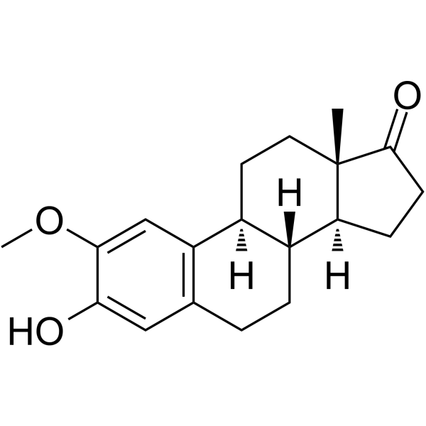
OC1=CC2=C([C@]3(CC[C@@]4([C@H](CC[C@]4([C@@]3(CC2)[H])[H])O)C)[H])C=C1OC
-
Synonyms
2-ME2; NSC-659853
-
Structure Classification
-
Shipping
Room temperature in continental US; may vary elsewhere.
-
Storage
Powder -20°C 3 years 4°C 2 years In solvent -80°C 2 years -20°C 1 year


Publications (33)
-
Journal Impact Factor
-
Most Recent
-
Cell Res
2026 Mar;36(3):219-232. PMID: 41634384 -
J Nanobiotechnology
Mesenchymal stem cells, as glioma exosomal immunosuppressive signal multipliers, enhance MDSCs immunosuppressive activity through the miR-21/SP1/DNMT1 positive feedback loop. [Abstract]2023 Jul 22;21(1):233. PMID: 37481646 -
Theranostics
Energy competition remodels the metabolic glucose landscape of psoriatic epidermal cells. [Abstract]2024 May 27;14(8):3339-3357. PMID: 38855186 -
J Exp Clin Cancer Res
Supervillin promotes epithelial-mesenchymal transition and metastasis of hepatocellular carcinoma in hypoxia via activation of the RhoA/ROCK-ERK/p38 pathway. [Abstract]2018 Jun 28;37(1):128. PMID: 29954442 -
Carbohydr Polym
Bioactive glycyrrhizic acid-astragalus polysaccharide hydrogel facilitates gastric ulcer healing via ROS scavenging and anti-apoptotic effects. [Abstract]2025 Aug 15:362:123685. PMID: 40409809 -
Cell Death Dis
ISCU-p53 axis orchestrates macrophage polarization to dictate immunotherapy response in esophageal squamous cell carcinoma. [Abstract]2025 Jun 20;16(1):462. PMID: 40541964 -
Cancer Lett
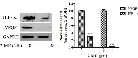
Sulfated fucoidan FP08S2 inhibits lung cancer cell growth in vivo by disrupting angiogenesis via targeting VEGFR2/VEGF and blocking VEGFR2/Erk/VEGF signaling. [Abstract]2016 Nov 1;382(1):44-52. PMID: 27569654
2-Methoxyestradiol purchased from MedChemExpress. Usage Cited in: Cancer Lett. 2016 Nov 1;382(1):44-52. [Abstract]
Downregulation of VEGF and HIF-1α by FP08S2 is dependent on Erk inactivation. VEGF and HIF-1α expression at protein level is detected after 2-ME treatment by Western blotting. 18S rRNA is used as internal control in qPCR while GAPDH is used as loading control in Western blotting.
-
Mol Ther
Exosomal miR-1246 from glioma patient body fluids drives the differentiation and activation of myeloid-derived suppressor cells. [Abstract]2021 Dec 1;29(12):3449-3464. PMID: 34217892 -
Acta Biomater
Redox-responsive dendritic copolymer-drug conjugates enhance therapeutic mitophagy through coordinated microtubule destabilization for synergistic triple-negative breast cancer therapy. [Abstract]2025 Aug:202:248-261. PMID: 40555336 -
J Transl Med
Fasting mimicking diet inhibits tumor-associated macrophage survival and pro-tumor function in hypoxia: implications for combination therapy with anti-angiogenic agent. [Abstract]2023 Oct 26;21(1):754. PMID: 37884960 -
Free Radic Biol Med
2021 Jun:169:271-282. PMID: 33895289 -
Stem Cell Res Ther
2022 Feb 5;13(1):59. PMID: 35123567 -
Front Immunol
Rapamycin Modulates the Proinflammatory Memory-Like Response of Microglia Induced by BAFF. [Abstract]2021 May 12:12:639049. PMID: 34054807 -
Ann Hepatol
Metformin ameliorates liver fibrosis induced by congestive hepatopathy via the mTOR/HIF-1α signaling pathway. [Abstract]2023 Nov-Dec;28(6):101135. PMID: 37451514 -
Eur J Pharmacol
Inhibition of JAK1/STAT3 pathway by 2-methoxyestradiol ameliorates psoriatic features in vitro and in an imiquimod-induced psoriasis-like mouse model. [Abstract]2022 Oct 15:933:175276. PMID: 36130639 -
Cancer Biol Ther
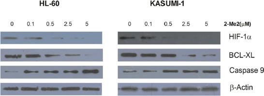
HIF-1α inhibition by 2-methoxyestradiol induces cell death via activation of the mitochondrial apoptotic pathway in acute myeloid leukemia. [Abstract]2016 Jun 2;17(6):625-34. PMID: 27082496
2-Methoxyestradiol purchased from MedChemExpress. Usage Cited in: Cancer Biol Ther. 2016 Jun 2;17(6):625-34. [Abstract]
HL-60 and Kasumi-1 cells are treated with different concentration of 2ME2 (0.1, 0.5, 2.5, or 5μM) for 24 hours or diluent control, and intracelluar protein levels are examined by Western blot.
-
Int J Mol Sci
A High-Calorie Diet Aggravates Lipopolysaccharide-Induced Pulmonary Inflammation in Juvenile Rats via Hypothalamic-Pituitary-Adrenal Axis-Related Pathways. [Abstract]2025 Jul 8;26(14):6554. PMID: 40724811 -
Front Cell Dev Biol
Hypoxia-Mediated Complement 1q Binding Protein Regulates Metastasis and Chemoresistance in Triple-Negative Breast Cancer and Modulates the PKC-NF-κB-VCAM-1 Signaling Pathway. [Abstract]2021 Feb 23:9:607142. PMID: 33708767 -
Chem Biol Interact
2023 Jan 5:369:110277. PMID: 36414027 -
Pestic Biochem Physiol
2-Methoxyestradiol ameliorates paraquat-induced pulmonary fibrosis by inhibiting the TGF-β1/Smad2/3 signaling pathway. [Abstract]2023 Dec:197:105647. PMID: 38072522 -
Rheumatology (Oxford)
Autophagy mediates 2-methoxyestradiol-inhibited scleroderma collagen synthesis and endothelial-to-mesenchymal transition induced by hypoxia. [Abstract]2019 Nov 1;58(11):1966-1975. PMID: 31049569 -
-
Front Cell Neurosci
IRAK-M Deficiency Exacerbates Ischemic Neurovascular Injuries in Experimental Stroke Mice. [Abstract]2018 Dec 21:12:504. PMID: 30622459 -
Am J Physiol Lung Cell Mol Physiol
Intrauterine inflammation-induced neonatal lung injury via succinic acid-mediated alveolar epithelial E-cadherin downregulation. [Abstract]2025 Aug 1;329(2):L282-L295. PMID: 40643013 -
Peptides
Cholecystokinin regulates atrial natriuretic peptide secretion through activation of NOX4-Sirt1-LEF1 signaling in beating rat hypoxic atria. [Abstract]2024 Nov:181:171299. PMID: 39326462 -
J Immunol Res
Combined Effects of 2-Methoxyestradiol (Hypoxia-Inducible Factor 1 α Inhibitor) and Dasatinib (A Second-Generation Tyrosine Kinase Inhibitor) on Chronic Myelocytic Leukemia Cells. [Abstract]2022 Apr 28;2022:6324326. PMID: 35528614 -
Biomed Res Int
2-Methoxyestradiol Alleviates Experimental Autoimmune Uveitis by Inhibiting Lymphocytes Proliferation and T Cell Differentiation. [Abstract]2016:2016:7948345. PMID: 27243036 -
Thorac Cancer
Mitochondrial NDUFA4L2 protein promotes the vitality of lung cancer cells by repressing oxidative stress. [Abstract]2019 Apr;10(4):676-685. PMID: 30710412 -
Exp Ther Med
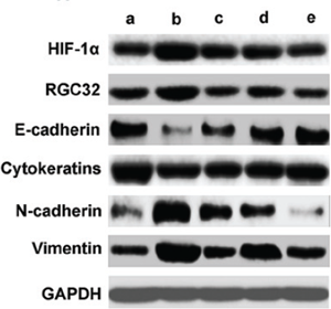
Hypoxia-inducible factor 1α participates in hypoxia-induced epithelial-mesenchymal transition via response gene to complement 32. [Abstract]2017 Aug;14(2):1825-1831. PMID: 28810656
2-Methoxyestradiol purchased from MedChemExpress. Usage Cited in: Exp Ther Med. 2017 Aug;14(2):1825-1831. [Abstract]
HIF 1α regulates the protein levels of RGC 32 and epithelial mesenchymal transition associated genes induced by hypoxia. Proteins are extracted from cells after different treatments. Protein levels of HIF 1α, RGC 32, E cadherin, cytokeratins, N cadherin and vimentin are detected by western blotting. a, no treatment cells under normoxia; b, cells under hypoxia; c, cells are pretreated with HIF 1α inhibitor for 30 min, and then incubated under hypoxia; d, cells are transfected with negative contro
-
-
-
-


Solvent & Solubility
In Vitro:
DMSO : 250 mg/mL (826.69 mM; Need ultrasonic; Hygroscopic DMSO has a significant impact on the solubility of product, please use newly opened DMSO)
H2O : < 0.1 mg/mL (insoluble)
Please refer to the solubility information to select the appropriate solvent. Once prepared, please aliquot and store the solution to prevent product inactivation from repeated freeze-thaw cycles.
Storage method and period of stock solution: -80°C, 2 years; -20°C, 1 year. When stored at -80°C, please use it within 2 years. When stored at -20°C, please use it within 1 year.
Please refer to the solubility information to select the appropriate solvent. Once prepared, please aliquot and store the solution to prevent product inactivation from repeated freeze-thaw cycles.
Storage method and period of stock solution: -80°C, 2 years; -20°C, 1 year. When stored at -80°C, please use it within 2 years. When stored at -20°C, please use it within 1 year.
Concentration (start) × Volume (start) = Concentration (final) × Volume (final)
In Vivo:
Select the appropriate dissolution method based on your experimental animal and administration route.
- For the following dissolution methods, please ensure to first prepare a clear stock solution using an In Vitro approach and then sequentially add co-solvents:
- To ensure reliable experimental results, the clarified stock solution can be appropriately stored based on storage conditions. As for the working solution for In Vivo experiments, it is recommended to prepare freshly and use it on the same day.
- The percentages shown for the solvents indicate their volumetric ratio in the final prepared solution. If precipitation or phase separation occurs during preparation, heat and/or sonication can be used to aid dissolution.
Add each solvent one by one: 10% DMSO 40% PEG300 5% Tween-80 45% Saline
Solubility: ≥ 2.08 mg/mL (6.88 mM); Clear solution
This protocol yields a clear solution of ≥ 2.08 mg/mL (saturation unknown).
Taking 1 mL working solution as an example, add 100 μL DMSO stock solution (20.8 mg/mL) to 400 μL PEG300, and mix evenly; then add 50 μL Tween-80 and mix evenly; then add 450 μL Saline to adjust the volume to 1 mL.
Preparation of Saline: Dissolve 0.9 g sodium chloride in ddH₂O and dilute to 100 mL to obtain a clear Saline solution.
Add each solvent one by one: 10% DMSO 90% (20% SBE-β-CD in Saline)
Solubility: ≥ 2.08 mg/mL (6.88 mM); Clear solution
This protocol yields a clear solution of ≥ 2.08 mg/mL (saturation unknown).
Taking 1 mL working solution as an example, add 100 μL DMSO stock solution (20.8 mg/mL) to 900 μL 20% SBE-β-CD in Saline, and mix evenly.
Preparation of 20% SBE-β-CD in Saline (4°C, storage for one week): 2 g SBE-β-CD powder is dissolved in 10 mL Saline, completely dissolve until clear.
In Vivo Dissolution Calculator
Please enter the basic information of animal experiments:
-
-
-
-
Recommended: Prepare an additional quantity of animals to account for potential losses during experiments.
Please enter your animal formula composition:
-
%DMSO +
Recommended: Keep the proportion of DMSO in working solution below 2% if your animal is weak.
-
%+
-
+%Tween-80 + +
-
%Saline +
The co-solvents required include: DMSO, . All of co-solvents are available by MedChemExpress (MCE). , Tween 80. All of co-solvents are available by MedChemExpress (MCE).
Working solution concentration: 0.22 mg/mL
Method for preparing stock solution: mg drug dissolved in μL DMSO. Stock solution concentration: mg/mL.
1. Take μL DMSO stock solution;
2. Add μL .
μL , mix evenly;
3. Then add μL Tween 80, mix evenly;
4. Then add μL
Please ensure that the stock solution in the first step is dissolved to a clear state, and add co-solvents in sequence. You can use ultrasonic heating (ultrasonic cleaner, recommended frequency 20-40 kHz), vortexing, etc. to assist dissolution.

Protocols
-
Apoptosis
Apoptosis, also called programmed cell death, is generally characterized by distinct morphological characteristics.
-
TUNEL staining for apoptotic DNA fragmentation
TUNEL staining detects DNA strand breaks by using terminal deoxynucleotidyl transferase to add labeled nucleotides to exposed 3′-OH DNA termini, generating either microscopic staining in fixed cells or tissue sections, or fluorescence/cytometric signal in cell suspensions. TUNEL positivity reflects DNA fragmentation but should not be interpreted alone as definitive apoptosis, because TUNEL can also label necrotic, autolytic, mechanically damaged, or DNA-repair-associated DNA breaks.
-
Human pluripotent stem cell endothelial-cell differentiation
Human pluripotent stem cell endothelial differentiation is based on stepwise developmental patterning: early activation of WNT/GSK3β inhibition promotes mesodermal or vascular progenitor entry, followed by endothelial specification using VEGF-related signaling, BMP4, FGF2, Notch modulation, or cAMP depending on the published protocol. Endothelial differentiation is read out by acquisition of CD31, CD34, VE-cadherin/CD144, KDR/VEGFR2, vWF, Tie2, NOS3, acetylated LDL uptake, tube/network formation, barrier function, and in vivo vessel-forming capacity where tested.
-
Vascular/Branching Fractal Analysis
Vascular/branching fractal analysis quantifies the geometric complexity of vessel trees or vascular networks from segmented 2D images, commonly by converting vessels into binary and/or skeletonized maps and estimating fractal dimension using box-counting or related approaches. Fractal dimension is interpreted as an image-derived readout of vascular branching complexity, space filling, or density, and has been applied to retinal photographs, fluorescein angiography, OCT angiography, capillary perfusion maps, and in vitro Matrigel angiogenesis networks. The assay readout is generated from vessel-positive pixels after image preprocessing, vessel segmentation, binarization, and optional skeletonization; reported outputs include fractal dimension, vessel density, branchpoint density, endpoint density, vessel length density, tortuosity, and generation-based branching metrics when VESGEN-style analysis is used. The biological interpretation is limited to quantitative vascular patterning and s
-
Annexin V plus membrane-impermeant dye apoptosis staining
Annexin V-based apoptosis assays rely on the detection of phosphatidylserine (PS) externalization from the inner leaflet of the plasma membrane to the outer leaflet, an early biochemical hallmark of apoptosis. Fluorescently labeled Annexin V binds PS in a calcium-dependent manner, enabling identification of early apoptotic cells by flow cytometry or fluorescence microscopy. When combined with a membrane-impermeant DNA-binding dye (e. g. , propidium iodide), this approach allows discrimination between viable (Annexin V−/dye−), early apoptotic (Annexin V+/dye−), and late apoptotic or necrotic (Annexin V+/dye+) cell populations by assessing membrane integrity and PS exposure.
-
ROS/oxidative-stress fluorescent staining
ROS/oxidative-stress fluorescent staining uses cell-permeant fluorogenic probes that become fluorescent after oxidation inside cells or tissues; commonly used examples include DCFH-DA/DCFDA for broad cellular oxidant detection, DHE for superoxide-related signal detection, MitoSOX for mitochondrial superoxide-related signal detection, and CellROX probes for oxidative-stress-associated fluorescence readouts. The assay detects probe oxidation rather than a single ROS species unless the probe and analysis method have been chemically validated for that species. DCFH-DA enters cells, is deacetylated by intracellular esterases to DCFH, and produces fluorescent DCF after oxidation, so the readout is used as an operational measure of total cellular oxidative stress rather than a species-specific ROS measurement. DHE and MitoSOX can report superoxide-related oxidation, but red fluorescence alone can include non-specific ethidium-like oxidation products; HPLC or optimized spectral approaches are
-
Endothelial Tube Formation Assay
Endothelial tube formation assay evaluates the ability of endothelial cells to attach, migrate, align, and organize into capillary-like networks when cultured on gelled basement membrane extract or Matrigel; the readout is the morphology and quantity of tube-like networks, which reflects an in vitro endothelial morphogenesis step related to angiogenesis. Basement membrane extract/Matrigel provides laminin-rich extracellular matrix cues that support endothelial differentiation into capillary-like structures, but it can contain biologically active growth factors, so growth-factor-reduced matrix is preferred when testing defined angiogenic stimulators or inhibitors.
-
Autophagy
Autophagy is a process in which eukaryotic cells use lysosomes to degrade their own cytoplasmic proteins and damaged organelles under the regulation of autophagy related gene (Atg). Microtubule-associated proteins light chain 3 (LC3) is recognized as autophagy marker, which transfers from cytoplasmic LC3 (LC3-I) to membrane type (LC3-II). LC3-II/I ratio could be detected by Western Blot and fluorescence microscopy.
-
Apoptosis Solutions
Apoptosis is a regulated, generally non-lytic cell-death pathway that removes unwanted, damaged, infected, or abnormal cells through coordinated morphological changes, caspase activation, DNA fragmentation, and membrane remodeling. The intrinsic apoptosis pathway is controlled mainly by mitochondrial outer membrane permeabilization, BCL-2 family proteins, cytochrome c release, apoptosome formation, caspase-9 activation, and downstream executioner caspase-3/7 activation. The extrinsic apoptosis pathway is initiated by death receptors such as Fas, TNFR, and TRAIL receptors, which recruit adaptor proteins and activate caspase-8 before engaging executioner caspases or mitochondrial amplification through BID cleavage. Apoptosis is linked to many phenotypes, including cancer cell killing, tissue homeostasis, immune regulation, neurodegeneration, infection response, and treatment-induced cytotoxicity; unresolved questions include how apoptosis interacts with necroptosis, pyroptosis, ferroptos
-
Lysosome and acidic-vesicle live-cell staining
Lysosome and acidic-vesicle live-cell staining detects acidic intracellular compartments by using membrane-permeant acidotropic probes that accumulate in low-pH vesicles, including lysosomes, late endosomes, autolysosomes, and acidic phagosomes. LysoTracker staining is commonly used as an intensity-based readout of acidic lysosomal compartment abundance or enlargement, while acridine orange produces green fluorescence in less concentrated compartments and red fluorescence after concentration-dependent accumulation in acidic vesicular organelles. Loss or reduction of acridine-orange red signal can be used as a readout of lysosomal membrane permeabilization or reduced acidic-vesicle integrity. This protocol is designed for live cultured cells and can be adapted for fluorescence microscopy, high-content imaging, plate-reader readout, or flow cytometry when the selected literature supports the readout. Because these dyes report acidotropic accumulation rather than lysosome identity alone,
-
Macroautophagy Solutions
Macroautophagy is a conserved lysosome-dependent degradation pathway in which cytoplasmic material is sequestered into double-membrane autophagosomes and delivered to lysosomes for degradation and recycling. The pathway supports cellular homeostasis during nutrient limitation, organelle stress, protein-aggregate accumulation, infection, differentiation, and tissue remodeling by coupling cargo sequestration, autophagosome maturation, lysosomal fusion, and degradation of cargo-derived macromolecules. The core molecular sequence includes initiation by nutrient- and stress-regulated autophagy machinery, autophagosome nucleation, LC3/ATG8-family conjugation to autophagosomal membranes, cargo selection through receptors such as SQSTM1/p62, autophagosome-lysosome fusion, and lysosomal degradation. LC3 was identified as a mammalian homolog of yeast Atg8 that localizes to autophagosomal membranes after processing, and p62/SQSTM1 was shown to connect ubiquitinated cargo with autophagic degradati
-
Endothelial Cell Migration/Angiogenic Sprouting Assay
Endothelial cell migration and angiogenic sprouting assays are in vitro (and partially ex vivo-adapted) functional models that quantify the ability of endothelial cells to undergo coordinated migration, extracellular matrix invasion, and multicellular organization into capillary-like sprouts in response to pro-angiogenic stimuli such as VEGF, bFGF, or conditioned microenvironments. These assays are used to model early angiogenic events including tip-cell formation, directional migration, and lumen-like sprout extension, which collectively reflect angiogenic activation and vascular morphogenesis processes observed in vivo.


Purity & Documentation
-
Data Sheet (286 KB)
-
SDS (644 KB)
- English - EN (644 KB)
- Français - FR (644 KB)
- Deutsch - DE (644 KB)
- Norwegian - NO (644 KB)
- Español - ES (644 KB)
- Swedish - SV (644 KB)
- Italian - IT (644 KB)
- Korean - KR (644 KB)
- Portuguese - PT (644 KB)
-
Handling Instructions (2659 KB)
References
[1]. Kamath K, et al. 2-Methoxyestradiol suppresses microtubule dynamics and arrests mitosis without depolymerizing microtubules. Mol Cancer Ther. 2006 Sep;5(9):2225-33. [Content Brief]
[2]. Aquino-Gálvez A, et al. Effects of 2-methoxyestradiol on apoptosis and HIF-1α and HIF-2α expression in lung cancer cells under normoxia and hypoxia. Oncol Rep. 2016 Jan;35(1):577-83. [Content Brief]
[3]. Xu L, et al. 2-Methoxyestradiol Alleviates Experimental Autoimmune Uveitis by Inhibiting Lymphocytes Proliferation and T Cell Differentiation. Biomed Res Int. 2016;2016:7948345. [Content Brief]
[4]. Kang SH, et al. Antitumor effect of 2-methoxyestradiol in a rat orthotopic brain tumor model. Cancer Res. 2006, 66(24),11991-11997. [Content Brief]
[5]. LaVallee TM, et al. 2-Methoxyestradiol inhibits proliferation and induces apoptosis independently of estrogen receptorsalpha and beta. Cancer Res. 2002 Jul 1;62(13):3691-7. [Content Brief]
[6]. Chen Y, et al. Oxidative stress induces autophagic cell death independent of apoptosis in transformed and cancer cells. Cell Death Differ. 2008;15(1):171-182. [Content Brief]


Complete Stock Solution Preparation Table
Please refer to the solubility information to select the appropriate solvent. Once prepared, please aliquot and store the solution to prevent product inactivation from repeated freeze-thaw cycles.
Storage method and period of stock solution: -80°C, 2 years; -20°C, 1 year. When stored at -80°C, please use it within 2 years. When stored at -20°C, please use it within 1 year.
| Optional Solvent | Concentration Solvent Mass | 1 mg | 5 mg | 10 mg | 25 mg |
|---|---|---|---|---|---|
| DMSO | 1 mM | 3.3068 mL | 16.5338 mL | 33.0677 mL | 82.6692 mL |
| 5 mM | 0.6614 mL | 3.3068 mL | 6.6135 mL | 16.5338 mL | |
| 10 mM | 0.3307 mL | 1.6534 mL | 3.3068 mL | 8.2669 mL | |
| 15 mM | 0.2205 mL | 1.1023 mL | 2.2045 mL | 5.5113 mL | |
| 20 mM | 0.1653 mL | 0.8267 mL | 1.6534 mL | 4.1335 mL | |
| 25 mM | 0.1323 mL | 0.6614 mL | 1.3227 mL | 3.3068 mL | |
| 30 mM | 0.1102 mL | 0.5511 mL | 1.1023 mL | 2.7556 mL | |
| 40 mM | 0.0827 mL | 0.4133 mL | 0.8267 mL | 2.0667 mL | |
| 50 mM | 0.0661 mL | 0.3307 mL | 0.6614 mL | 1.6534 mL | |
| 60 mM | 0.0551 mL | 0.2756 mL | 0.5511 mL | 1.3778 mL | |
| 80 mM | 0.0413 mL | 0.2067 mL | 0.4133 mL | 1.0334 mL | |
| 100 mM | 0.0331 mL | 0.1653 mL | 0.3307 mL | 0.8267 mL |


2-Methoxyestradiol Related Classifications
HY-12033 Related Classifications
 Powered by Bioz