



2-O-α-D-Glucopyranosyl-L-ascorbic Acid

Based on 1 Customer Validation
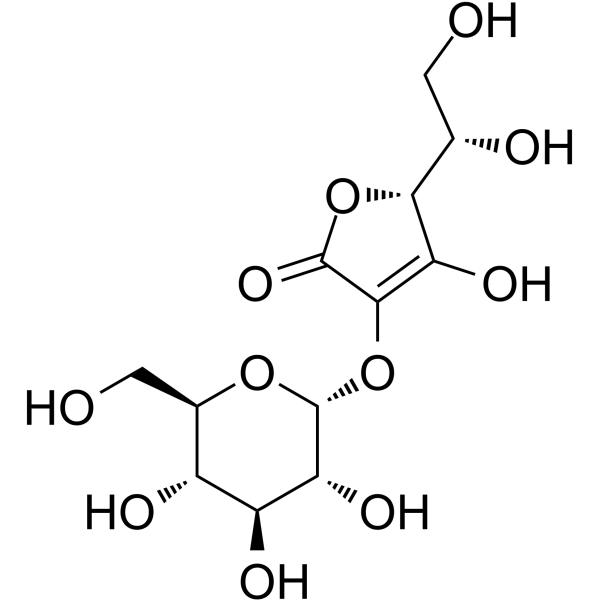
2-O-α-D-Glucopyranosyl-L-ascorbic Acid is an orally active glucoside derivative of ascorbic acid. 2-O-α-D-Glucopyranosyl-L-ascorbic Acid can be hydrolyzed by α-glucosidase to release ascorbic acid. 2-O-α-D-Glucopyranosyl-L-ascorbic Acid inhibits melanin synthesis, prevents UV-induced cell damage, and promotes collagen synthesis in skin fibroblasts. 2-O-α-D-Glucopyranosyl-L-ascorbic Acid also induces oxidative stress to inhibit tumor growth. 2-O-α-D-Glucopyranosyl-L-ascorbic Acid can be used in research related to tumors, inflammation, and other conditions.
For research use only. We do not sell to patients.
- Purity : 99.81%
- CAS No.: 129499-78-1
- Formula: C12H18O11
- Molecular Weight:338.26
-
Storage:
4°C, protect from light
* In solvent : -80°C, 6 months; -20°C, 1 month (protect from light)

To place orders, for customer services and technical support, please contact: MedChemExpress USA
Tel: 609-228-6898 E-mail: [email protected] [email protected]


-
 Biological Activity
-
Chemical Information
-
Solvent & Solubility
- Protocols
- Purity & Documentation
- References
-
 Help & FAQs


-

HY-114118
SemaglutideGLP-1R Agonist -
HY-W003607
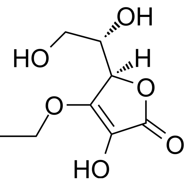
3-O-Ethyl-L-ascorbic acidVitamin C Derivative -
HY-N0684
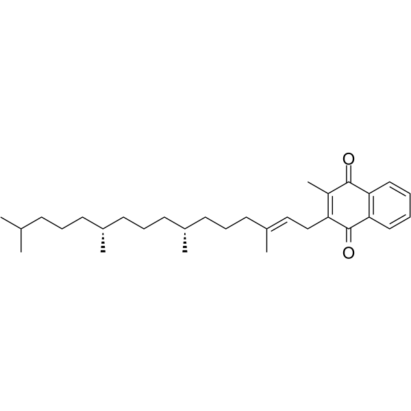
Vitamin K1Endogenous Metabolite -
HY-14649
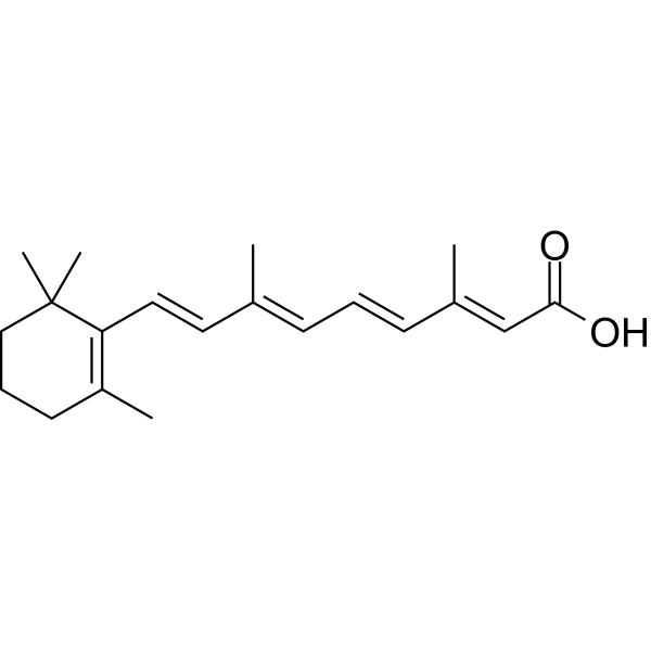
Retinoic acidRAR Agonist -
HY-B1278B
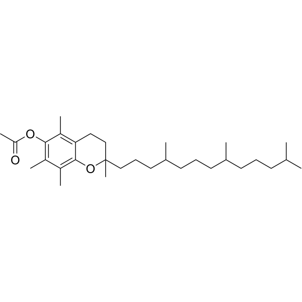
(±)-α-Tocopherol acetateVitamin E Acetate -
HY-B0987
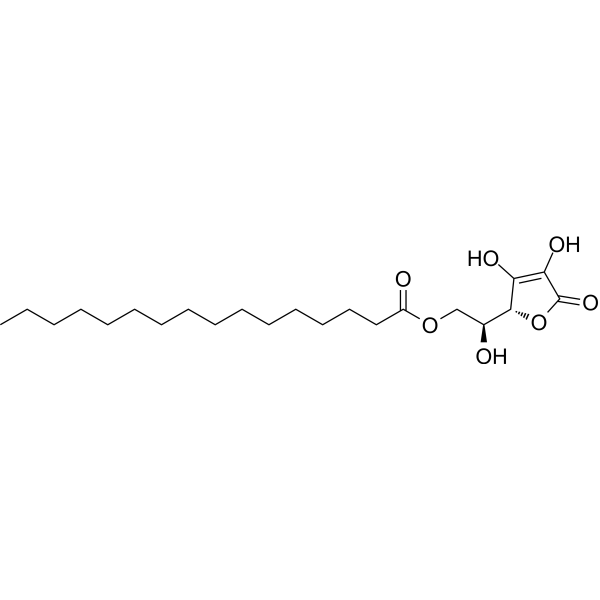
Ascorbyl palmitateEndogenous Metabolite, Antioxidant, Food Additive -
HY-16637
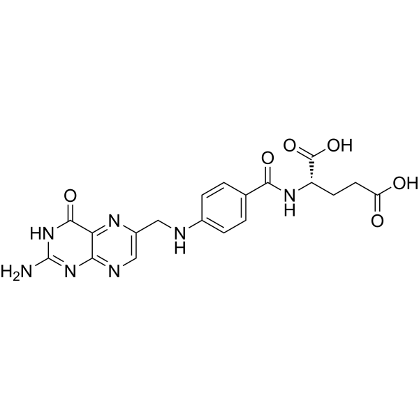
Folic acidB complex -
HY-B0456
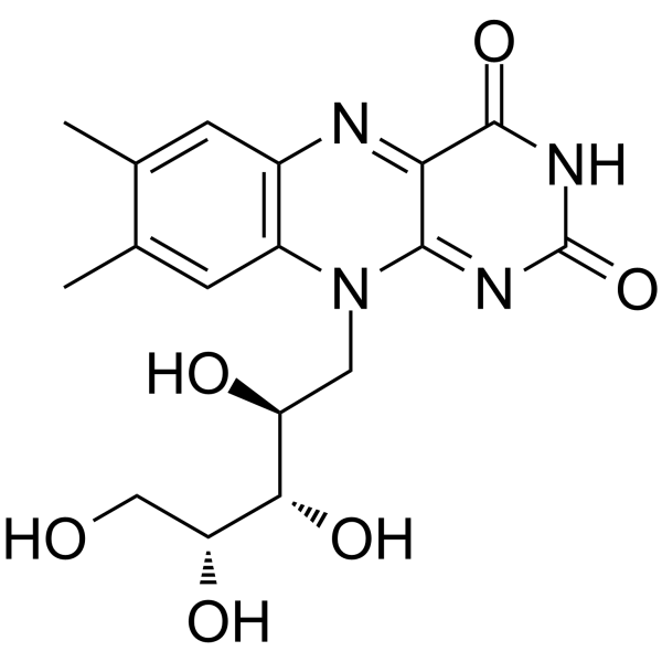
RiboflavinVitamin B2 -

HY-N6821R
2-O-α-D-Glucopyranosyl-L-ascorbic Acid (Standard)Anti-cancer Agent -

HY-16637R
Folic acid (Standard)B complex -
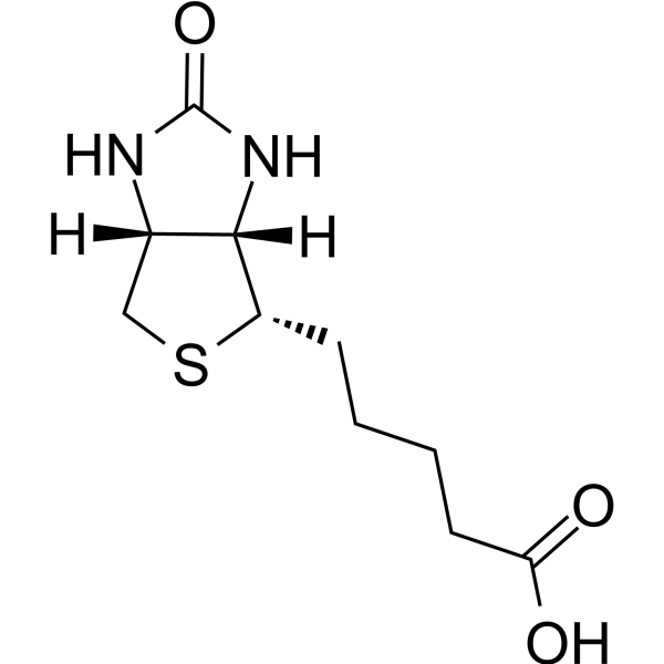
HY-B0511
BiotinVitamin B7 -
HY-B0143
NiacinB3 vitamin


Biological Activity
Description
Cellular Effect
|
Cell Line

|
Type | Value | Description | References |
|---|---|---|---|---|
| RD | EC50 |
16.3 μM
Compound: I
|
Antiviral activity against Herpes simplex virus type 1 infected in human RD cells assessed as inhibition of virus-induced cytopathic effect
Antiviral activity against Herpes simplex virus type 1 infected in human RD cells assessed as inhibition of virus-induced cytopathic effect
|
[PMID: 22390834] |
In Vitro
2-O-α-D-Glucopyranosyl-L-ascorbic acid (0.5-2 mM; 24 h) does not exert significant cytotoxic activity against murine colon carcinoma (colon-26) cells[2].
MedChemExpress (MCE) has not independently confirmed the accuracy of these methods. They are for reference only. Further protocols information, click here.
-
Cell Line:murine colon carcinoma (colon-26) cells
-
Concentration:0.5, 1, 1.5, 2 mM
-
Incubation Time:24 h
-
Result:Did not exhibit significant cytotoxic activity across all tested concentrations.
In Vivo
2-O-α-D-Glucopyranosyl-L-ascorbic Acid (19.2 mg/animal/day; p.o.; daily; 9 days) reverses weight loss, eliminates scurvy-related hemorrhage, and restores ascorbic acid levels and alkaline phosphatase activity in scorbutic male Hartley guinea pigs[1].
2-O-α-D-Glucopyranosyl-L-ascorbic Acid (equimolecular amount of 300 mg/kg of ascorbic acid; i.v.; 4 times on alternate days) significantly inhibits colon-26 tumor growth in male CDF1 mice, with associated evidence of oxidative stress induction via elevated plasma MDA levels[2].
MedChemExpress (MCE) has not independently confirmed the accuracy of these methods. They are for reference only.
-
Animal Model:Hartley guinea pigs (male, ~300 g, scurvy induced by vitamin C-deficient diet for 17 days)[1]
-
Dosage:19.2 mg/animal/day
-
Administration:p.o.; daily; 9 days
-
Result:Reversed weight loss, with animals regaining weight over the 9-day treatment period.
Eliminated subcutaneous hemorrhage.
Increased plasma ascorbic acid content, liver ascorbic acid content and brain ascorbic acid content.
Increased plasma alkaline phosphatase activity.
-
Animal Model:CDF1 (male, 5 weeks old, colon-26 tumor fragments engrafted subcutaneously)[2]
-
Dosage:Equimolecular amount of 300 mg/kg of ascorbic acid (AA)
-
Administration:i.v.; 4 times on alternate days
-
Result:Inhibited colon-26 tumor growth significantly.
Elevated plasma malondialdehyde (MDA) levels, reaching 8.5 μM at 6 hours post-administration.
Peaked plasma AA levels (hydrolyzed from AA-2G) at 90.3 μM at 15 minutes post-administration, then remained slightly above baseline (approximately 76 μM) from 1 to 6 hours.
Cleared AA-2G rapidly from plasma, with levels at 1.25 μmol/g at 15 minutes and returning to baseline within 1 hour.
Increased blood glucose levels slightly to a maximum of 85 mg/dL at 60 minutes post-administration, then returned to baseline by 90 minutes.
Decreased tumor AA levels significantly, with a maximum reduction observed at 30 minutes.


Chemical Information
-
CAS No. 129499-78-1
-
Appearance Solid
-
Molecular Weight 338.26
-
Formula C12H18O11
-
Color White to off-white
-
SMILES
OC([C@H](O1)[C@H](CO)O)=C(O[C@@H]2[C@@H]([C@H]([C@@H]([C@@H](CO)O2)O)O)O)C1=O
-
Shipping
Room temperature in continental US; may vary elsewhere.
-
Storage
4°C, protect from light
* In solvent : -80°C, 6 months; -20°C, 1 month (protect from light)


Solvent & Solubility
In Vitro:
H2O : 116.67 mg/mL (344.91 mM; Need ultrasonic)
Please refer to the solubility information to select the appropriate solvent. Once prepared, please aliquot and store the solution to prevent product inactivation from repeated freeze-thaw cycles.
Storage method and period of stock solution: -80°C, 6 months; -20°C, 1 month (protect from light). When stored at -80°C, please use it within 6 months. When stored at -20°C, please use it within 1 month.
* Note: If you choose water as the stock solution, please dilute it to the working solution, then filter and sterilize it with a 0.22 μm filter before use.
Please refer to the solubility information to select the appropriate solvent. Once prepared, please aliquot and store the solution to prevent product inactivation from repeated freeze-thaw cycles.
Storage method and period of stock solution: -80°C, 6 months; -20°C, 1 month (protect from light). When stored at -80°C, please use it within 6 months. When stored at -20°C, please use it within 1 month.
* Note: If you choose water as the stock solution, please dilute it to the working solution, then filter and sterilize it with a 0.22 μm filter before use.
Concentration (start) × Volume (start) = Concentration (final) × Volume (final)
In Vivo:
For the following dissolution methods, please prepare the working solution directly:
It is recommended to prepare fresh solutions and use them promptly within a short period of time.
The percentages shown for the solvents indicate their volumetric ratio in the final prepared solution. If precipitation or phase separation occurs during preparation, heat and/or sonication can be used to aid dissolution.
Add each solvent one by one: PBS
Solubility: 50 mg/mL (147.82 mM); Clear solution; Need ultrasonic

Protocols
-
ROS/oxidative-stress fluorescent staining
ROS/oxidative-stress fluorescent staining uses cell-permeant fluorogenic probes that become fluorescent after oxidation inside cells or tissues; commonly used examples include DCFH-DA/DCFDA for broad cellular oxidant detection, DHE for superoxide-related signal detection, MitoSOX for mitochondrial superoxide-related signal detection, and CellROX probes for oxidative-stress-associated fluorescence readouts. The assay detects probe oxidation rather than a single ROS species unless the probe and analysis method have been chemically validated for that species. DCFH-DA enters cells, is deacetylated by intracellular esterases to DCFH, and produces fluorescent DCF after oxidation, so the readout is used as an operational measure of total cellular oxidative stress rather than a species-specific ROS measurement. DHE and MitoSOX can report superoxide-related oxidation, but red fluorescence alone can include non-specific ethidium-like oxidation products; HPLC or optimized spectral approaches are
-
Patient-Derived Xenograft (PDX)
Patient-derived xenograft (PDX) models are generated by engrafting primary human tumor tissue directly into immunodeficient mice, allowing in vivo propagation of patient tumor biology without initial in vitro adaptation. These models are used to preserve key histopathological and molecular characteristics of the original tumor and enable assessment of tumor growth dynamics and therapeutic response in a living organism. The biological readout is tumor engraftment and subsequent growth in the murine host, which reflects the ability of human tumor cells to survive, vascularize, and expand in an immunocompromised microenvironment.
-
Patient-Derived Orthotopic Xenograft (PDOX)
Patient-derived orthotopic xenograft (PDOX) modeling implants fresh patient tumor tissue or patient-derived tumor cells into the anatomically corresponding organ or tissue site of immunodeficient mice, usually by surgical orthotopic implantation, to preserve patient tumor histology, local microenvironmental context, invasion, metastatic behavior, and treatment-response features better than subcutaneous implantation. PDOX readouts include tumor engraftment, orthotopic tumor growth, local invasion, metastasis, recurrence after resection, histologic similarity to the donor tumor, biomarker retention, molecular concordance, survival, and response or resistance to therapy. PDOX models are used for preclinical drug testing and individualized therapy evaluation, but engraftment success varies by tumor type and specimen quality.
-
Research Protocol for Inflammation-related Diseases
The NLRP3 inflammasome is a cytosolic innate immune signaling platform that integrates priming signals and danger-signal activation to promote caspase-1 activation, maturation of IL-1β and IL-18, and gasdermin D-mediated pyroptotic cell death. The core experimental logic is to determine whether inflammatory disease phenotypes are driven by increased NLRP3 expression, ASC-containing inflammasome assembly, caspase-1 cleavage, GSDMD cleavage, and extracellular release of IL-1β/IL-18 rather than by nonspecific cell injury alone. The pathway is strongly linked to inflammation-related disease phenotypes because monosodium urate crystals activate NALP3/NLRP3 inflammasome signaling in gout-like crystal inflammation, cholesterol crystals activate NLRP3 inflammasomes in atherogenesis models, and DSS-induced intestinal inflammation has been reported to involve NLRP3 inflammasome activity. However, experimental colitis studies also show context-dependent protective effects of NLRP3 inflammasome co
-
Subcutaneous Cell-Line-Derived Xenograft
Subcutaneous cell-line-derived xenograft (CDX) models are established by implanting cultured human cancer cell lines into immunodeficient mice, where the injected cells form localized tumors that can be monitored in vivo as a measure of tumorigenic potential, growth kinetics, and treatment response. These models are widely used in oncology research because they allow reproducible tumor formation and enable comparative assessment of tumor growth between different cell lines or genetic manipulations in a controlled in vivo microenvironment. Subcutaneous implantation of cancer cells in immunodeficient mice is a standard approach for evaluating tumor growth behavior and therapeutic response across multiple cancer types, including prostate, esophageal, pancreatic, and colon cancer models.
-
Orthotopic Cell-Line Xenograft
Orthotopic cell-line xenograft models involve implantation of human cancer cell lines into the anatomically corresponding organ of immunodeficient mice to reproduce tumor growth within a native microenvironment, enabling more clinically relevant tumor behavior compared with subcutaneous models. These models are widely used because orthotopic placement better recapitulates tumor progression, including invasion and metastatic spread, which are often underrepresented in heterotopic implantation systems. Compared with conventional xenografts, orthotopic implantation is described as more technically complex but provides improved simulation of tumor-microenvironment interactions and metastatic behavior, making it particularly valuable for translational oncology research. Surgical orthotopic implantation approaches have been emphasized as enabling faithful reproduction of clinical cancer features, including metastasis and disease progression patterns that align with the tumor’s organ of origi


Purity & Documentation
-
Data Sheet (297 KB)
-
SDS (252 KB)
- English - EN (252 KB)
- Français - FR (252 KB)
- Deutsch - DE (252 KB)
- Norwegian - NO (252 KB)
- Español - ES (252 KB)
- Swedish - SV (252 KB)
- Italian - IT (252 KB)
- Korean - KR (252 KB)
- Portuguese - PT (252 KB)
-
Handling Instructions (2659 KB)
References
[1]. Tai A, et al. Bioavailability of a series of novel acylated ascorbic acid derivatives, 6-O-acyl-2-O-alpha-D-glucopyranosyl-L-ascorbic acids, as an ascorbic acid supplement in rats and guinea pigs. Biosci Biotechnol Biochem. 2002 Aug;66(8):1628-34. [Content Brief]
[2]. Miura K, et al. 2-O-α-D-Glucopyranosyl-l-ascorbic acid as an antitumor agent for infusion therapy. Biochem Biophys Rep. 2017;10:232-236. Published 2017 Apr 22. [Content Brief]
[3]. Tai A, et al. Structural evidence for the DPPH radical-scavenging mechanism of 2-O-α-d-glucopyranosyl-l-ascorbic acid. Bioorg Med Chem. 2017 Oct 15;25(20):5303-5310. [Content Brief]


Complete Stock Solution Preparation Table
Please refer to the solubility information to select the appropriate solvent. Once prepared, please aliquot and store the solution to prevent product inactivation from repeated freeze-thaw cycles.
Storage method and period of stock solution: -80°C, 6 months; -20°C, 1 month (protect from light). When stored at -80°C, please use it within 6 months. When stored at -20°C, please use it within 1 month.
| Optional Solvent | Concentration Solvent Mass | 1 mg | 5 mg | 10 mg | 25 mg |
|---|---|---|---|---|---|
| H2O | 1 mM | 2.9563 mL | 14.7815 mL | 29.5631 mL | 73.9076 mL |
| 5 mM | 0.5913 mL | 2.9563 mL | 5.9126 mL | 14.7815 mL | |
| 10 mM | 0.2956 mL | 1.4782 mL | 2.9563 mL | 7.3908 mL | |
| 15 mM | 0.1971 mL | 0.9854 mL | 1.9709 mL | 4.9272 mL | |
| 20 mM | 0.1478 mL | 0.7391 mL | 1.4782 mL | 3.6954 mL | |
| 25 mM | 0.1183 mL | 0.5913 mL | 1.1825 mL | 2.9563 mL | |
| 30 mM | 0.0985 mL | 0.4927 mL | 0.9854 mL | 2.4636 mL | |
| 40 mM | 0.0739 mL | 0.3695 mL | 0.7391 mL | 1.8477 mL | |
| 50 mM | 0.0591 mL | 0.2956 mL | 0.5913 mL | 1.4782 mL | |
| 60 mM | 0.0493 mL | 0.2464 mL | 0.4927 mL | 1.2318 mL | |
| 80 mM | 0.0370 mL | 0.1848 mL | 0.3695 mL | 0.9238 mL | |
| 100 mM | 0.0296 mL | 0.1478 mL | 0.2956 mL | 0.7391 mL |
* Note: If you choose water as the stock solution, please dilute it to the working solution, then filter and sterilize it with a 0.22 μm filter before use.


2-O-α-D-Glucopyranosyl-L-ascorbic Acid Related Classifications
HY-N6821 Related Classifications
 Powered by Bioz