



2-Oleoylglycerol
Based on 1 publication(s) in Google Scholar
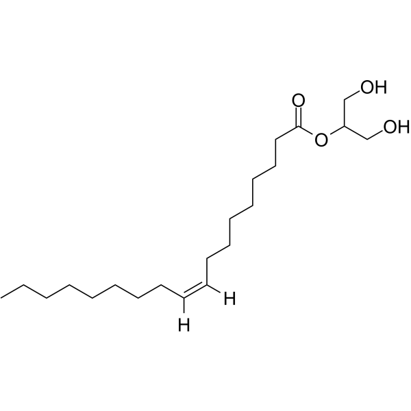
2-Oleoylglycerol (2-OG) is a lipid found in the diet. It is a GPR119 agonist, with an EC50 value of 2.5 μM in activating hGPR119 in transiently transfected COS-7 cells. 2-Oleoylglycerol enhances the inflammatory response of macrophages and promotes fibrosis by activating the GPR119/TAK1/NF-κB/TGF-β1 signaling pathway. It also stimulates glucagon-like peptide 1 (GLP-1) secretion in vivo. 2-Oleoylglycerol is expected to be used in the research of non-alcoholic steatohepatitis (NASH) .
For research use only. We do not sell to patients.
- Purity : 99.9%
- CAS No.: 3443-84-3
- Formula: C21H40O4
- Molecular Weight:356.55
-
Storage:
Solution, -20°C, 2 years

To place orders, for customer services and technical support, please contact: MedChemExpress USA
Tel: 609-228-6898 E-mail: [email protected] [email protected]


-
 Biological Activity
-
Chemical Information
- Protocols
- Purity & Documentation
- References
-
 Help & FAQs


-
HY-15291
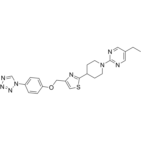
MBX-2982GPR119 Agonist -
HY-130311
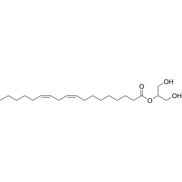
2-Linoleoyl glycerolCB1 Receptor Agonist -
HY-15565
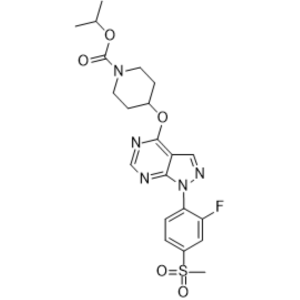
APD668GPR119 Agonist -
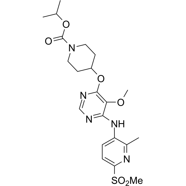
HY-15566
APD597GPR119 Agonist -
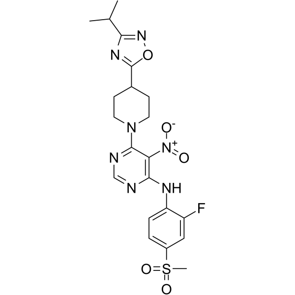
HY-15564
AR 231453GPR119 Agonist -
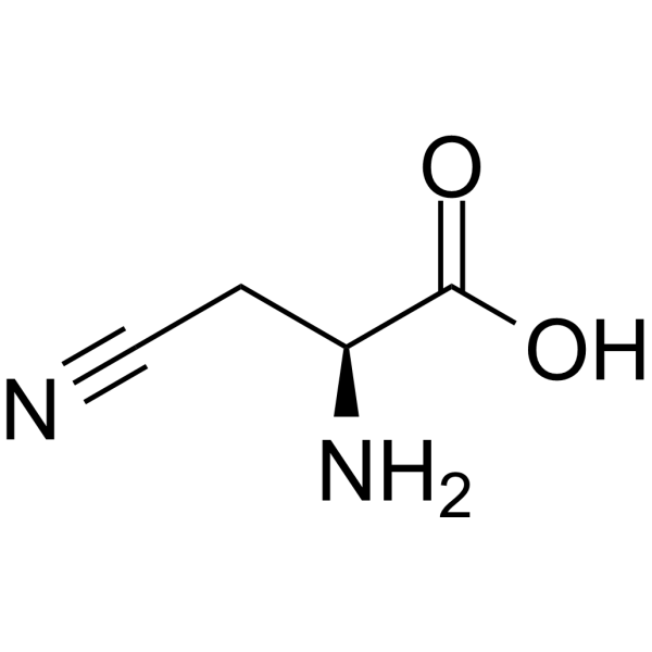
HY-125773
β-cyano-L-AlanineH2S-synthesizing Enzyme Inhibitor -
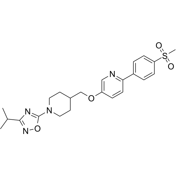
HY-12066
GSK1292263 -
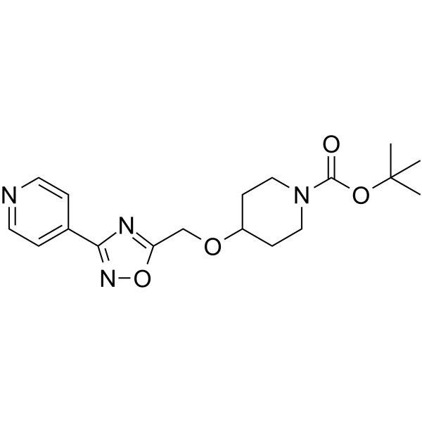
HY-16673
PSN632408GPR119 Agonist -
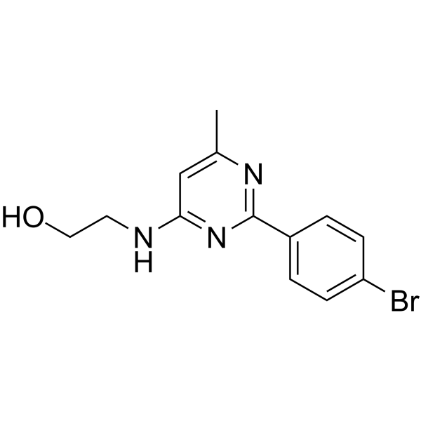
HY-107535
AS1269574GPR119 Agonist -
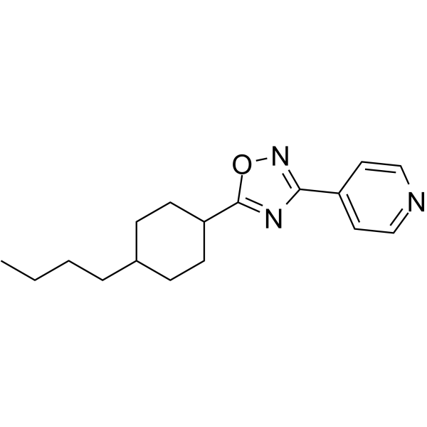
HY-108258
PSN 375963GPR119 Agonist -

HY-15565R
APD668 (Standard)GPR119 Agonist -

HY-12066R
GSK1292263 (Standard)
Publications Citing Use of MedChemExpress (MCE) 2-Oleoylglycerol
More

Biological Activity
Description
IC50 & Target
IC50: 2.5 μM (hGPR119) (in COS-7 cells)[1]
In Vitro
2-Oleoylglycerol (50 μg/mL, 24 hours) activates RAW264.7 cells via the GPR119/TAK1/NF-κB/TGF-β1 signaling pathway[3].
MedChemExpress (MCE) has not independently confirmed the accuracy of these methods. They are for reference only. Further protocols information, click here.
-
Cell Line:Mouse RAW264.7 macrophage cells
-
Concentration:50 μg/mL
-
Incubation Time:24 hours
-
Result:Increased expression of GPR119, TGF-β1, IL-1β, and TNF-α, enhancing the gene expression of HSC activation.
In Vivo
MedChemExpress (MCE) has not independently confirmed the accuracy of these methods. They are for reference only.
-
Animal Model:C57BL/6 J mice, CL-HFS-induced non-alcoholic steatohepatitis (NASH) model[3]
-
Dosage:20 μg/mouse in 0.2 mL PBS
-
Administration:Intravenous administration (I.V.), three times a week for 6 weeks
-
Result:Showed increased hepatic infiltration of inflammatory cells, increased collagen and α-SMA production; 2-OG enhanced liver resident MΦ and HSC activation.


Chemical Information
-
CAS No. 3443-84-3
-
Appearance Liquid
-
Molecular Weight 356.55
-
Formula C21H40O4
-
Color Colorless to light yellow
-
SMILES
OCC(CO)OC(CCCCCCC/C=C\CCCCCCCC)=O
-
Synonyms
2-OG
-
Structure Classification
-
Initial Source
-
Shipping
Room temperature in continental US; may vary elsewhere.
-
Storage
Solution, -20°C, 2 years


Publications (1)
-
Journal Impact Factor
-
Most Recent
-
J Adv Res
Human milk fat substitutes improve obesity-related NAFLD by enriching LPC 18:2 to activate hepatic GPR119-AMPK signaling. [Abstract]2026 Feb 2:S2090-1232(26)00108-6. PMID: 41638632

Protocols
-
Fibrosis/Collagen Morphometry
Fibrosis and collagen morphometry is based on the quantitative visualization of fibrillar collagen deposition in tissue sections using histochemical stains such as Sirius Red (Picrosirius Red) or Masson's trichrome, followed by image-based or polarization-enhanced analysis to estimate collagen proportional area as a surrogate of extracellular matrix accumulation during fibrotic remodeling. Sirius Red combined with polarized light microscopy enhances detection of collagen fibers due to birefringence properties, enabling more specific visualization of collagen type I and III fibrils compared to conventional bright-field histology, while whole-section or region-restricted digital morphometry reduces field-selection bias in fibrosis assessment. Alternative quantitative approaches include second harmonic generation (SHG) and two-photon excited fluorescence microscopy, which enable label-free detection of fibrillar collagen and have been validated against histological staining and biochemica
-
Collagen: Sirius Red Staining
Sirius Red or picrosirius red staining is a histochemical method for visualizing collagen-rich extracellular matrix in tissue sections, and collagen fibers are detected as red-stained structures under bright-field microscopy with enhanced birefringence under polarized light. Picrosirius red is useful for assessing total collagen organization, distribution, and fibrosis burden, but polarized color should not be interpreted as a definitive collagen type I versus type III readout because color is affected by fiber orientation, thickness, and packing.
-
Connective Tissue: Masson's Trichrome/Collagen Trichrome Staining
Masson’s Trichrome (collagen/trichrome staining) is a histological technique that differentially stains tissue compartments using sequential acidic dyes to distinguish collagen from muscle and cytoplasmic components based on dye affinity and tissue permeability differences, enabling visualization of fibrosis and connective tissue architecture in histological sections. The classical formulation typically uses Weigert's iron hematoxylin for nuclear staining, Biebrich scarlet-acid fuchsin for cytoplasm and muscle, and aniline blue (or light green variants) for collagen, producing a characteristic blue/green collagen signal contrasted against red cytoplasm and dark nuclei. The staining principle relies on selective displacement of smaller dye molecules by larger anionic dyes in collagen-rich regions under controlled acidified conditions, which enhances collagen-specific dye retention. This property makes the method widely used for fibrosis assessment in organs such as heart, liver, lung, a
-
Research Protocol for Inflammation-related Diseases
The NLRP3 inflammasome is a cytosolic innate immune signaling platform that integrates priming signals and danger-signal activation to promote caspase-1 activation, maturation of IL-1β and IL-18, and gasdermin D-mediated pyroptotic cell death. The core experimental logic is to determine whether inflammatory disease phenotypes are driven by increased NLRP3 expression, ASC-containing inflammasome assembly, caspase-1 cleavage, GSDMD cleavage, and extracellular release of IL-1β/IL-18 rather than by nonspecific cell injury alone. The pathway is strongly linked to inflammation-related disease phenotypes because monosodium urate crystals activate NALP3/NLRP3 inflammasome signaling in gout-like crystal inflammation, cholesterol crystals activate NLRP3 inflammasomes in atherogenesis models, and DSS-induced intestinal inflammation has been reported to involve NLRP3 inflammasome activity. However, experimental colitis studies also show context-dependent protective effects of NLRP3 inflammasome co


Purity & Documentation
-
Data Sheet (270 KB)
-
SDS (393 KB)
- English - EN (393 KB)
- Français - FR (393 KB)
- Deutsch - DE (393 KB)
- Norwegian - NO (393 KB)
- Español - ES (393 KB)
- Swedish - SV (393 KB)
- Italian - IT (393 KB)
- Korean - KR (393 KB)
- Portuguese - PT (393 KB)
-
Handling Instructions (2659 KB)
References
[1]. Hansen KB, et, al. 2-Oleoyl glycerol is a GPR119 agonist and signals GLP-1 release in humans. J Clin Endocrinol Metab. 2011 Sep;96(9):E1409-17. [Content Brief]
[2]. Ghafouri N, et, al. Inhibition of monoacylglycerol lipase and fatty acid amide hydrolase by analogues of 2-arachidonoylglycerol. Br J Pharmacol. 2004 Nov;143(6):774-84. [Content Brief]
[3]. Yang M, et al. Western diet contributes to the pathogenesis of non-alcoholic steatohepatitis in male mice via remodeling gut microbiota and increasing production of 2-oleoylglycerol. Nat Commun. 2023 Jan 16;14(1):228. [Content Brief]

Calculators
Concentration (start) × Volume (start) = Concentration (final) × Volume (final)


2-Oleoylglycerol Related Classifications
HY-W011121 Related Classifications
 Powered by Bioz