



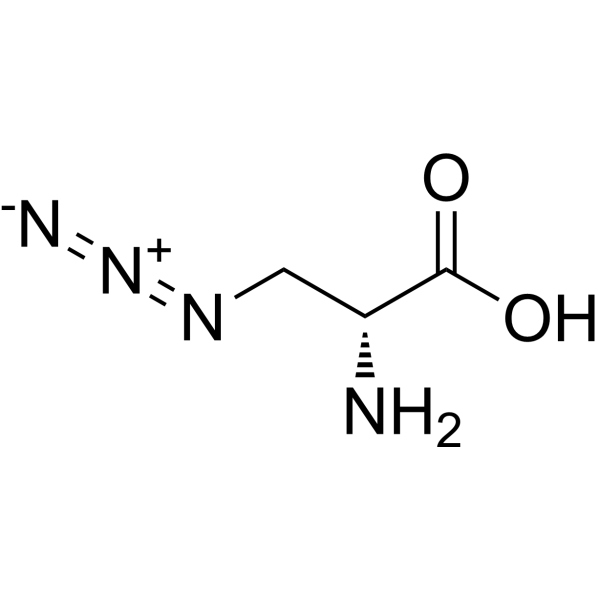
3-Azido-D-alanine
Based on 1 publication(s) in Google Scholar
3-Azido-D-alanine is an orally active, selective metabolic tracer applicable for click chemistry labeling. 3-Azido-D-alanine can be selectively incorporated into bacterial cell wall peptidoglycans, introducing azido groups to enable copper-free click chemistry-based modification, labeling and single-cell tracing, while it cannot be metabolically incorporated by mammalian cells. 3-Azido-D-alanine can be used in studies related to bacterial infection, intracranial infection, otitis media, intestinal inflammation and tissue damage, as well as sepsis.
For research use only. We do not sell to patients.
- Purity : 99.92%
- CAS No.: 105928-88-9
- Formula: C3H6N4O2
- Molecular Weight:130.11
-
Storage:Powder -20°C, 3 years ; In solvent -80°C, 6 months , -20°C, 1 month

To place orders, for customer services and technical support, please contact: MedChemExpress USA
Tel: 609-228-6898 E-mail: [email protected] [email protected]


-
 Biological Activity
-
Chemical Information
-
Solvent & Solubility
- Protocols
- Purity & Documentation
- References
-
 Help & FAQs


Publications Citing Use of MedChemExpress (MCE) 3-Azido-D-alanine
More

Biological Activity
Description
In Vitro
3-Azido-D-alanine (1 h) selectively modifies the cell walls of E. coli (ATCC 25922) and MRSA (ATCC 43300) with azide groups, enabling efficient conjugation to sulfo-DBCO-ICG via copper-free click chemistry, as detected by CLSM and flow cytometry[1].
3-Azido-D-alanine (30 min) modification enables targeted photothermal lysis of E. coli (ATCC 25922) and MRSA (ATCC 43300) via sulfo-DBCO-ICG, resulting in significant ATP release that correlates linearly with bacterial CFU counts for quantitative detection[1].
3-Azido-D-alanine (15 min) modification enables potent photothermal killing of E. coli (ATCC 25922) and MRSA (ATCC 43300) via sulfo-DBCO-ICG[1].
3-Azido-D-alanine (0.5 mM; 2 h) efficiently and specifically labels the peptidoglycan cell walls of both Escherichia coli and Staphylococcus aureus via metabolic incorporation, enabling fluorescent detection after click conjugation with Cy3-DBCO[3].
3-Azido-D-alanine (0.5 mM; 2 h) efficiently metabolically labels a range of authentic rat gut bacteria, including Bacillus subtilis, Lactobacillus murinus, and Bacillus licheniformis, as well as mixed fecal bacterial populations, with measurable fluorescence intensity increases after click conjugation with Cy3-DBCO[3].
3-Azido-D-alanine (3.84 mM; 60 min) optimally labels the peptidoglycan of Staphylococcus aureus (ATCC 25923) with azide groups for bioorthogonal fluorescence detection[5].
3-Azido-D-alanine (3.84 mM; 12 h) does not significantly reduce the viability of Staphylococcus aureus (ATCC 25923), with a survival rate above 90%[5].
3-Azido-D-alanine (3.84 mM; 60 min) enables sensitive capture and detection of Staphylococcus aureus (ATCC 25923) in whole blood, with a detection limit of 4 CFU/mL and capture efficiencies of 75.0% to 83.5% for low-concentration bacteria[5].
3-Azido-D-alanine (3.84 mM; 60 min) enables selective fluorescence labeling of Staphylococcus aureus (ATCC 25923) in mixed cultures with Escherichia coli (ATCC 10798) for Gram-type identification[5].
3-Azido-D-alanine (0-8 mM) is nontoxic to Lactobacillus reuteri at concentrations up to 8 mM, and 4 mM achieves near-saturation metabolic labeling of bacterial surface azide groups at a rate of 78.6 %[6].
MedChemExpress (MCE) has not independently confirmed the accuracy of these methods. They are for reference only. Further protocols information, click here.
In Vivo
3-Azido-D-alanine (10 mg/kg; p.o.; single dose) enables specific in vivo metabolic labeling of gut bacteria, revealing that simulated microgravity induces a significant increase in total gut bacterial content (approximately 6-fold higher than controls) and altered spatial distribution, which correlates with intestinal inflammation and tissue damage[3].
3-Azido-D-alanine hydrochloride (150 mg per 50 mg of MSP@PAA-Alkyne; p.o.) enables efficient labeling and spatiotemporal tracking of viable oral microbiota in healthy C57BL/6J mice, with labeled bacteria detectable in the gastrointestinal tract for up to 36 hours post-gavage[4].
MedChemExpress (MCE) has not independently confirmed the accuracy of these methods. They are for reference only.
-
Animal Model:Sprague-Dawley (SD) rats (simulated microgravity via tail suspension at 30° angle for 28 days)[3]
-
Dosage:10 mg/kg
-
Administration:p.o.; single dose
-
Result:Showed notably enhanced fluorescence signals across all imaging time points, with maximum intensity at 6 hours.
Revealed intestinal fluorescence intensity in simulated microgravity rats was approximately 6-fold higher than control rats.
Demonstrated fluorescence concentrated primarily in the ileum, with simulated microgravity rats showing increased gut bacterial load in the ileum compared to controls, and bacterial distribution diffused toward the jejunum and ileum.
Confirmed higher bacterial accumulation in simulated microgravity rat jejunum, ileum, and colon, correlated with pathological changes including villus rupture, crypt loss, colonic basal layer detachment, elevated IL-6 and TNF-α levels in intestinal tissues, and increased plasma endotoxin levels.
-
Animal Model:C57BL/6J (four-week-old)[4]
-
Dosage:150 mg per 50 mg of MSP@PAA-Alkyne (incorporated into MSP@PAA-ADA); 0.1 mg/mL (MSP@PAA-ADA for in vitro incubation); 200 μL (labeled bacterial solution); 200 μL of 0.1 mM (Cy5ADA for sequential labeling)
-
Administration:p.o. (single dose for labeled bacterial solution; single dose 3 hours post initial gavage for Cy5ADA)
-
Result:Detected large fluorescent signal in the mouse gastrointestinal tract at 3 hours post-gavage, which increased and centralized by 9 hours, then attenuated at 16 hours, and continued to weaken through 24 and 36 hours.
Retained detectable fluorescent signal in mice at 36 hours post-gavage.
Recovered diverse viable oral bacteria (including Gram-positive and Gram-negative bacilli, cocci, and streptococci) that retained both MSP@PAA-ADA binding and Cy5ADA fluorescence via magnetic separation of cecum contents.


Chemical Information
-
CAS No. 105928-88-9
-
Appearance Solid
-
Molecular Weight 130.11
-
Formula C3H6N4O2
-
Color White to off-white
-
SMILES
OC([C@H](N)CN=[N+]=[N-])=O
-
Shipping
Room temperature in continental US; may vary elsewhere.
-
Storage
Powder -20°C 3 years In solvent -80°C 6 months -20°C 1 month


Publications (1)
-
Journal Impact Factor
-
Most Recent
-
Nat Commun
Engineered BCG selectively triggers trained immunity in tumor-associated macrophages and sensitizes glioblastoma to radiotherapy in mice. [Abstract]2026 Apr 20;17(1):5465. PMID: 42009673


Solvent & Solubility
In Vitro:
H2O : 33.33 mg/mL (256.17 mM; Need ultrasonic)
DMSO : 1.25 mg/mL (9.61 mM; Need ultrasonic; Hygroscopic DMSO has a significant impact on the solubility of product, please use newly opened DMSO)
Please refer to the solubility information to select the appropriate solvent. Once prepared, please aliquot and store the solution to prevent product inactivation from repeated freeze-thaw cycles.
Storage method and period of stock solution: -80°C, 6 months; -20°C, 1 month. When stored at -80°C, please use it within 6 months. When stored at -20°C, please use it within 1 month.
* Note: If you choose water as the stock solution, please dilute it to the working solution, then filter and sterilize it with a 0.22 μm filter before use.
Please refer to the solubility information to select the appropriate solvent. Once prepared, please aliquot and store the solution to prevent product inactivation from repeated freeze-thaw cycles.
Storage method and period of stock solution: -80°C, 6 months; -20°C, 1 month. When stored at -80°C, please use it within 6 months. When stored at -20°C, please use it within 1 month.
* Note: If you choose water as the stock solution, please dilute it to the working solution, then filter and sterilize it with a 0.22 μm filter before use.
Concentration (start) × Volume (start) = Concentration (final) × Volume (final)

Protocols
-
EdU Incorporation Assay (Click Chemistry-Based DNA Synthesis Measurement)
The EdU incorporation assay measures DNA synthesis by adding the thymidine analog 5-ethynyl-2′-deoxyuridine to cells or tissues, where it is incorporated into newly synthesized DNA during S phase. Incorporated EdU is detected by copper-catalyzed azide-alkyne cycloaddition, in which a fluorescent azide covalently reacts with the ethynyl group on EdU, allowing S-phase cells to be detected by fluorescence microscopy, flow cytometry, or high-content imaging. EdU detection does not require DNA denaturation or anti-BrdU antibody access, which preserves sample structure and improves compatibility with immunostaining and multiparameter cytometry compared with BrdU-based detection. EdU can be cytotoxic in a cell-type- and exposure-dependent manner, so pulse duration, concentration, and continuous-labeling designs should be validated for each cell type.
-
Research Protocol for Infectious Diseases
Infectious-disease experiments test how pathogens interact with host barriers, innate immune receptors, inflammatory signaling, pathogen replication, and tissue injury; pattern-recognition receptors such as TLRs, RIG-I-like receptors, NOD-like receptors, and inflammasomes detect microbial molecules and activate NF-κB, interferon, and cytokine responses. The central hypothesis is that infection severity reflects the balance between pathogen burden and host response: protective inflammation restricts pathogen growth, whereas excessive or mislocalized inflammation contributes to tissue damage and disease phenotype. Unresolved questions include which host pathways are protective versus pathogenic, why some infection models fail to translate to human disease, and which combined readouts best predict clinically relevant infection outcomes.
-
LPS-Induced Endotoxemia/Systemic Inflammation
Lipopolysaccharide (LPS)-induced endotoxemia is a widely used in vivo model of acute systemic inflammation in which LPS, a Gram-negative bacterial endotoxin, activates innate immune signaling primarily through TLR4, leading to rapid and transient induction of pro-inflammatory cytokines such as TNF-α, IL-6, and IL-1β in circulation and tissues. This cytokine surge is commonly used as a measurable readout of systemic inflammatory activation and immune dysregulation, and is typically assessed within hours after intraperitoneal LPS administration in mouse models of endotoxemia. The model captures key features of systemic inflammatory response syndrome, including cytokine release, immune cell activation, and downstream tissue responses, and has been used to evaluate anti-inflammatory interventions such as cytokine modulation, lipid mediators, and immune cell-targeting therapies.
-
DSS-Induced Colitis
Dextran sulfate sodium (DSS)-induced colitis is generated by administering DSS in mouse drinking water, producing epithelial injury, barrier disruption, weight loss, diarrhea, fecal blood, colon shortening, histologic mucosal damage, and inflammatory mediator changes; the model is mainly used to study acute or chronic intestinal inflammation resembling selected features of ulcerative colitis. DSS injury is interpreted through clinical and tissue readouts rather than a single molecular endpoint: daily body weight, stool consistency, and bleeding are combined into a disease activity index, while colon length, histology, cytokines, myeloperoxidase activity, intestinal permeability, and tight-junction markers provide complementary measures of inflammation and barrier damage.
-
TNBS-Induced Colitis
TNBS-induced colitis is produced by intrarectal delivery of 2,4,6-trinitrobenzene sulfonic acid in ethanol, where ethanol disrupts the mucosal barrier and TNBS haptenates colonic proteins, generating immune-mediated colonic inflammation with weight loss, diarrhea, ulceration, transmural injury, inflammatory-cell infiltration, and cytokine responses. The model is used as an experimental intestinal inflammation model with Crohn’s disease–like features, especially when Th1-type responses, IL-12–dependent inflammation, chronic relapsing inflammation, or fibrosis-related endpoints are studied.
-
Research Protocol for Inflammation-related Diseases
The NLRP3 inflammasome is a cytosolic innate immune signaling platform that integrates priming signals and danger-signal activation to promote caspase-1 activation, maturation of IL-1β and IL-18, and gasdermin D-mediated pyroptotic cell death. The core experimental logic is to determine whether inflammatory disease phenotypes are driven by increased NLRP3 expression, ASC-containing inflammasome assembly, caspase-1 cleavage, GSDMD cleavage, and extracellular release of IL-1β/IL-18 rather than by nonspecific cell injury alone. The pathway is strongly linked to inflammation-related disease phenotypes because monosodium urate crystals activate NALP3/NLRP3 inflammasome signaling in gout-like crystal inflammation, cholesterol crystals activate NLRP3 inflammasomes in atherogenesis models, and DSS-induced intestinal inflammation has been reported to involve NLRP3 inflammasome activity. However, experimental colitis studies also show context-dependent protective effects of NLRP3 inflammasome co


Purity & Documentation
-
Data Sheet (286 KB)
-
SDS (644 KB)
- English - EN (644 KB)
- Français - FR (644 KB)
- Deutsch - DE (644 KB)
- Norwegian - NO (644 KB)
- Español - ES (644 KB)
- Swedish - SV (644 KB)
- Italian - IT (644 KB)
- Korean - KR (644 KB)
- Portuguese - PT (644 KB)
-
Handling Instructions (2659 KB)
References
[1]. Zhang L, et al. Bacteria Wear ICG Clothes for Rapid Detection of Intracranial Infection in Patients After Neurosurgery and Photothermal Antibacterial Therapy Against Mutans. Frontiers in bioengineering and biotechnology. 2022;10:932915. [Content Brief]
[3]. Wu Z, et al. Imaging of Gut Bacterial Macroscopic Changes in Simulated Microgravity-Exposed Rats via Metabolic Labeling. Analytical chemistry. 2024 Dec 10;96(49):19758-19767. [Content Brief]
[5]. Zhao J, et al. Sensitive NIR Fluorescence Identification of Bacteria in Whole Blood with Bioorthogonal Nanoprobes for Early Sepsis Diagnosis. Analytical chemistry. 2023 Jan 17;95(2):955-965. [Content Brief]
[6]. Liu Z, et al. Hierarchical arming of probiotics for improved viability to synergistically alleviate ulcerative colitis. Biomaterials. 2026 Apr;327:123791. [Content Brief]


Complete Stock Solution Preparation Table
Please refer to the solubility information to select the appropriate solvent. Once prepared, please aliquot and store the solution to prevent product inactivation from repeated freeze-thaw cycles.
Storage method and period of stock solution: -80°C, 6 months; -20°C, 1 month. When stored at -80°C, please use it within 6 months. When stored at -20°C, please use it within 1 month.
| Optional Solvent | Concentration Solvent Mass | 1 mg | 5 mg | 10 mg | 25 mg |
|---|---|---|---|---|---|
| DMSO / H2O | 1 mM | 7.6858 mL | 38.4290 mL | 76.8580 mL | 192.1451 mL |
| 5 mM | 1.5372 mL | 7.6858 mL | 15.3716 mL | 38.4290 mL | |
| H2O | 10 mM | 0.7686 mL | 3.8429 mL | 7.6858 mL | 19.2145 mL |
| 15 mM | 0.5124 mL | 2.5619 mL | 5.1239 mL | 12.8097 mL | |
| 20 mM | 0.3843 mL | 1.9215 mL | 3.8429 mL | 9.6073 mL | |
| 25 mM | 0.3074 mL | 1.5372 mL | 3.0743 mL | 7.6858 mL | |
| 30 mM | 0.2562 mL | 1.2810 mL | 2.5619 mL | 6.4048 mL | |
| 40 mM | 0.1921 mL | 0.9607 mL | 1.9215 mL | 4.8036 mL | |
| 50 mM | 0.1537 mL | 0.7686 mL | 1.5372 mL | 3.8429 mL | |
| 60 mM | 0.1281 mL | 0.6405 mL | 1.2810 mL | 3.2024 mL | |
| 80 mM | 0.0961 mL | 0.4804 mL | 0.9607 mL | 2.4018 mL | |
| 100 mM | 0.0769 mL | 0.3843 mL | 0.7686 mL | 1.9215 mL |
* Note: If you choose water as the stock solution, please dilute it to the working solution, then filter and sterilize it with a 0.22 μm filter before use.


3-Azido-D-alanine Related Classifications
HY-151642 Related Classifications
 Powered by Bioz
Keywords
- 3-Azido-D-alanine
- 105928-88-9
- Biochemical Assay Reagents
- Bacterial
- magnetosome production
- Staphylococcus aureus (ATCC 25923)
- copper-free click chemistry
- bacterial cell wall peptidoglycan
- E. coli (ATCC 25922)
- gut bacteria
- MRSA (ATCC 43300)
- C57BL/6J mice
- bacterial infection
- intestinal inflammation
- Inhibitor
- inhibitor
- inhibit