



4-Hydroperoxy Cyclophosphamide-d4

Based on 1 Customer Validation
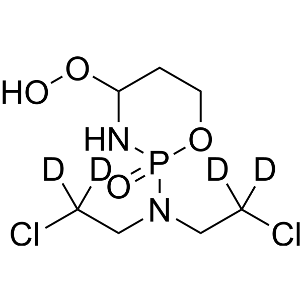
4-Hydroperoxy Cyclophosphamide-d4 is the deuterium labeled 4-Hydroperoxy cyclophosphamide. 4-Hydroperoxy cyclophosphamide is the active metabolite form of the proagent Cyclophosphamide. 4-Hydroperoxy cyclophosphamide crosslinks DNA and induces T cell apoptosis independent of death receptor activation, but activates mitochondrial death pathways through production of reactive oxygen species (ROS). 4-Hydroperoxy cyclophosphamide has the potential for lymphomas and autoimmune disorders.
Nur für Forschungszwecke. Wir verkaufen nicht an Patienten.
- Reinheit : 95.0%
- CAS. Nr.: 1246816-71-6
- Formel: C7H11D4Cl2N2O4P
- Molecular Weight:297.11
-
Speicherung:
-80°C, stored under nitrogen

To place orders, for customer services and technical support, please contact: MedChemExpress USA
Tel: 609-228-6898 E-mail: [email protected] [email protected]


-
 Biologische Aktivität
-
Chemical Information
-
Lösungsmittel & Löslichkeit
- Protokoll
- Reinheit & Dokumentation
- Verweise
-
 Help & FAQs




Biologische Aktivität
Beschreibung
In Vitro
Stable heavy isotopes of hydrogen, carbon, and other elements have been incorporated into drug molecules, largely as tracers for quantitation during the drug development process. Deuteration has gained attention because of its potential to affect the pharmacokinetic and metabolic profiles of drugs[1].
MedChemExpress (MCE) has not independently confirmed the accuracy of these methods. They are for reference only. Further protocols information, click here.
Anwendung
1. This compound can be used as a tracer
2. This compound can be used as an internal standard for quantitative analysis by NMR, GC-MS, or LC-MS.


Chemical Information
-
CAS. Nr. 1246816-71-6
-
Unlabeled CAS 39800-16-3
-
Appearance Solid
-
Molecular Weight 297.11
-
Formel C7H11D4Cl2N2O4P
-
Color Off-white to light yellow
-
SMILES
N(CC(Cl)([2H])[2H])(CC(Cl)([2H])[2H])P1(=O)NC(OO)CCO1
-
Versand
Shipping with dry ice.
-
Speicherung
-80°C, stored under nitrogen


Lösungsmittel & Löslichkeit
In Vitro:
DMSO : 50 mg/mL (168.29 mM; Need ultrasonic; Hygroscopic DMSO has a significant impact on the solubility of product, please use newly opened DMSO)
Please refer to the solubility information to select the appropriate solvent. Once prepared, please aliquot and store the solution to prevent product inactivation from repeated freeze-thaw cycles.
Storage method and period of stock solution: -80°C, 6 months (stored under nitrogen). When stored at -80°C, please use it within 6 months.
Please refer to the solubility information to select the appropriate solvent. Once prepared, please aliquot and store the solution to prevent product inactivation from repeated freeze-thaw cycles.
Storage method and period of stock solution: -80°C, 6 months (stored under nitrogen). When stored at -80°C, please use it within 6 months.
Konzentration (Stammlösung) × Volumen (Stammlösung) = Konzentration (Ziellösung) × Volumen (Ziellösung)

Protokoll
-
Detection of Apoptosis
Apoptosis, also called programmed cell death, is generally characterized by distinct morphological characteristics.
-
TUNEL staining for apoptotic DNA fragmentation
TUNEL staining detects DNA strand breaks by using terminal deoxynucleotidyl transferase to add labeled nucleotides to exposed 3′-OH DNA termini, generating either microscopic staining in fixed cells or tissue sections, or fluorescence/cytometric signal in cell suspensions. TUNEL positivity reflects DNA fragmentation but should not be interpreted alone as definitive apoptosis, because TUNEL can also label necrotic, autolytic, mechanically damaged, or DNA-repair-associated DNA breaks.
-
Annexin V plus membrane-impermeant dye apoptosis staining
Annexin V-based apoptosis assays rely on the detection of phosphatidylserine (PS) externalization from the inner leaflet of the plasma membrane to the outer leaflet, an early biochemical hallmark of apoptosis. Fluorescently labeled Annexin V binds PS in a calcium-dependent manner, enabling identification of early apoptotic cells by flow cytometry or fluorescence microscopy. When combined with a membrane-impermeant DNA-binding dye (e. g. , propidium iodide), this approach allows discrimination between viable (Annexin V−/dye−), early apoptotic (Annexin V+/dye−), and late apoptotic or necrotic (Annexin V+/dye+) cell populations by assessing membrane integrity and PS exposure.
-
ROS/oxidative-stress fluorescent staining
ROS/oxidative-stress fluorescent staining uses cell-permeant fluorogenic probes that become fluorescent after oxidation inside cells or tissues; commonly used examples include DCFH-DA/DCFDA for broad cellular oxidant detection, DHE for superoxide-related signal detection, MitoSOX for mitochondrial superoxide-related signal detection, and CellROX probes for oxidative-stress-associated fluorescence readouts. The assay detects probe oxidation rather than a single ROS species unless the probe and analysis method have been chemically validated for that species. DCFH-DA enters cells, is deacetylated by intracellular esterases to DCFH, and produces fluorescent DCF after oxidation, so the readout is used as an operational measure of total cellular oxidative stress rather than a species-specific ROS measurement. DHE and MitoSOX can report superoxide-related oxidation, but red fluorescence alone can include non-specific ethidium-like oxidation products; HPLC or optimized spectral approaches are
-
Apoptosis Solutions
Apoptosis is a regulated, generally non-lytic cell-death pathway that removes unwanted, damaged, infected, or abnormal cells through coordinated morphological changes, caspase activation, DNA fragmentation, and membrane remodeling. The intrinsic apoptosis pathway is controlled mainly by mitochondrial outer membrane permeabilization, BCL-2 family proteins, cytochrome c release, apoptosome formation, caspase-9 activation, and downstream executioner caspase-3/7 activation. The extrinsic apoptosis pathway is initiated by death receptors such as Fas, TNFR, and TRAIL receptors, which recruit adaptor proteins and activate caspase-8 before engaging executioner caspases or mitochondrial amplification through BID cleavage. Apoptosis is linked to many phenotypes, including cancer cell killing, tissue homeostasis, immune regulation, neurodegeneration, infection response, and treatment-induced cytotoxicity; unresolved questions include how apoptosis interacts with necroptosis, pyroptosis, ferroptos
-
Research Protocol for Cancer Immunology
Cancer immunology studies how the immune system recognizes, suppresses, edits, or fails to eliminate malignant cells through tumor antigen release, antigen presentation, T-cell priming, immune trafficking, tumor-cell killing, and feedback inhibition in the tumor microenvironment. The cancer-immunity cycle links tumor antigenicity, dendritic-cell priming, CD8+ T-cell infiltration, cytotoxic function, and immune-checkpoint regulation to tumor rejection or immune escape. Immune-checkpoint pathways such as PD-1/PD-L1 and CTLA-4 suppress antitumor T-cell activity and can be therapeutically blocked, but many tumors remain resistant because of poor antigen presentation, weak T-cell infiltration, suppressive myeloid cells, regulatory T cells, and tumor-intrinsic immune-exclusion programs. Unresolved questions include which immune-cell states predict response, how tumor-intrinsic pathways exclude immune cells, how myeloid suppression limits checkpoint blockade, and which combination strategies


Reinheit & Dokumentation
-
Data Sheet (277 KB)
-
SDS (393 KB)
- English - EN (393 KB)
- Français - FR (393 KB)
- Deutsch - DE (393 KB)
- Norwegian - NO (393 KB)
- Español - ES (393 KB)
- Swedish - SV (393 KB)
- Italian - IT (393 KB)
- Korean - KR (393 KB)
- Portuguese - PT (393 KB)
-
Handling Instructions (2659 KB)
Verweise
[1]. Russak EM, et al. Impact of Deuterium Substitution on the Pharmacokinetics of Pharmaceuticals. Ann Pharmacother. 2019;53(2):211-216. [Content Brief]
[2]. 4-hydroperoxy-cyclophosphamide mediates caspase-independent T-cell apoptosis involving oxidative stress-induced nuclear relocation of mitochondrial apoptogenic factors AIF and EndoG [Content Brief]


Complete Stock Solution Preparation Table
Please refer to the solubility information to select the appropriate solvent. Once prepared, please aliquot and store the solution to prevent product inactivation from repeated freeze-thaw cycles.
Storage method and period of stock solution: -80°C, 6 months (stored under nitrogen). When stored at -80°C, please use it within 6 months.
| Optional Solvent | Concentration Solvent Mass | 1 mg | 5 mg | 10 mg | 25 mg |
|---|---|---|---|---|---|
| DMSO | 1 mM | 3.3658 mL | 16.8288 mL | 33.6576 mL | 84.1439 mL |
| 5 mM | 0.6732 mL | 3.3658 mL | 6.7315 mL | 16.8288 mL | |
| 10 mM | 0.3366 mL | 1.6829 mL | 3.3658 mL | 8.4144 mL | |
| 15 mM | 0.2244 mL | 1.1219 mL | 2.2438 mL | 5.6096 mL | |
| 20 mM | 0.1683 mL | 0.8414 mL | 1.6829 mL | 4.2072 mL | |
| 25 mM | 0.1346 mL | 0.6732 mL | 1.3463 mL | 3.3658 mL | |
| 30 mM | 0.1122 mL | 0.5610 mL | 1.1219 mL | 2.8048 mL | |
| 40 mM | 0.0841 mL | 0.4207 mL | 0.8414 mL | 2.1036 mL | |
| 50 mM | 0.0673 mL | 0.3366 mL | 0.6732 mL | 1.6829 mL | |
| 60 mM | 0.0561 mL | 0.2805 mL | 0.5610 mL | 1.4024 mL | |
| 80 mM | 0.0421 mL | 0.2104 mL | 0.4207 mL | 1.0518 mL | |
| 100 mM | 0.0337 mL | 0.1683 mL | 0.3366 mL | 0.8414 mL |


4-Hydroperoxy Cyclophosphamide-d4 Related Classifications
HY-117433S Related Classifications
 Powered by Bioz