



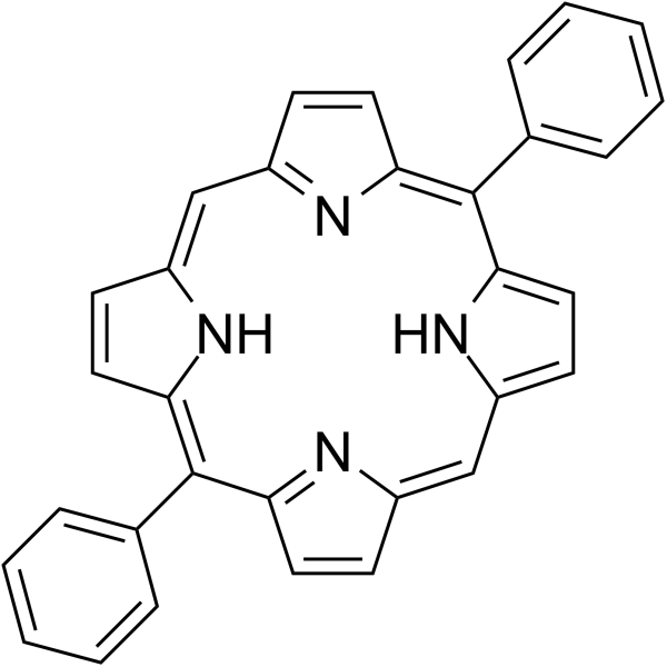
5,15-Diphenylporphyrin

Based on 1 Customer Validation
5,15-Diphenylporphyrin (5,15-DPP) is an inhibitor of the oncogenic transcription factor STAT3. 5,15-Diphenylporphyrin specifically binds to the SH2 domain of STAT3, blocking the pTyr-SH2 interaction, thereby inhibiting the dimerization, nuclear translocation and DNA binding activity of STAT3, and ultimately inhibiting the expression of cancer cell-related genes. 5,15-Diphenylporphyrin can be used in research fields related to the development of anticancer drugs
For research use only. We do not sell to patients.
- Purity : 95.0%
- CAS No.: 22112-89-6
- Formula: C32H22N4
- Molecular Weight:462.54
-
Storage:
4°C, protect from light
* In solvent : -80°C, 6 months; -20°C, 1 month (protect from light)

To place orders, for customer services and technical support, please contact: MedChemExpress USA
Tel: 609-228-6898 E-mail: [email protected] [email protected]


-
 Biological Activity
-
Chemical Information
-
Solvent & Solubility
- Protocols
- Purity & Documentation
- References
-
 Help & FAQs


-
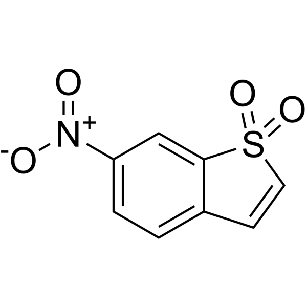
HY-13818
StatticSTAT3 Inhibitor -
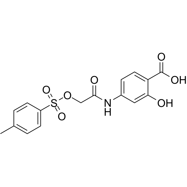
HY-15146
NSC 74859STAT3 Inhibitor -
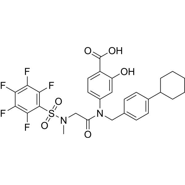
HY-100493
BP-1-102STAT3 Inhibitor -

HY-18739
Phorbol 12-myristate 13-acetatePKC Activator -
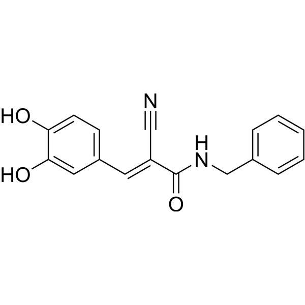
HY-12000
AG490EGFR Inhibitor -
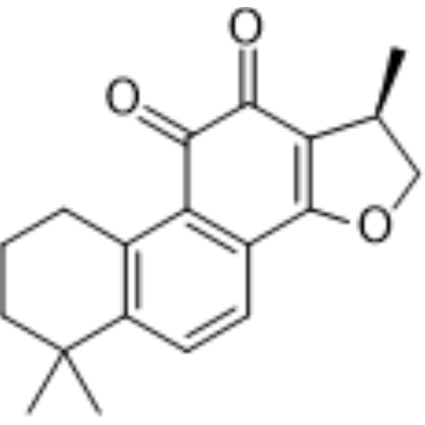
HY-N0174
CryptotanshinoneAutophagy Inducer -
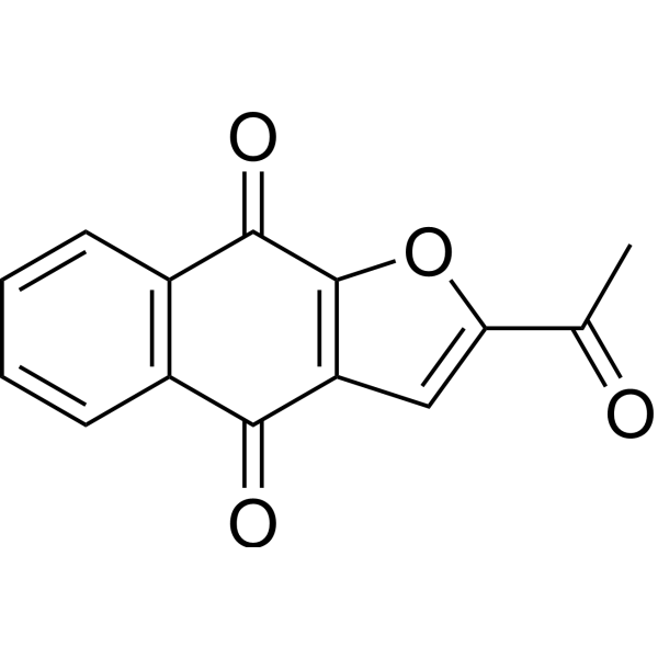
HY-13919
NapabucasinSTAT3 Inhibitor -
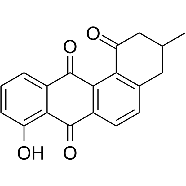
HY-18061
OchromycinoneSTAT3 Inhibitor -
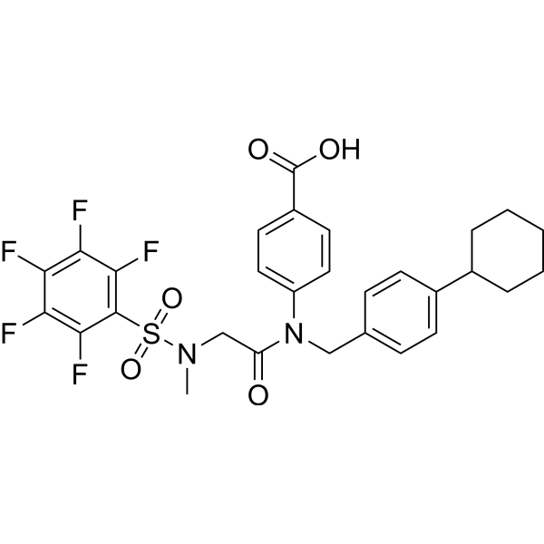
HY-16975
SH-4-54STAT3/5 Inhibitor -

HY-13818R
Stattic (Standard)STAT3 Inhibitor -
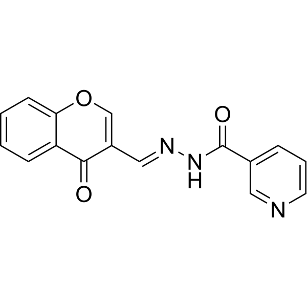
HY-101853
STAT5-IN-1STAT5 Inhibitor -
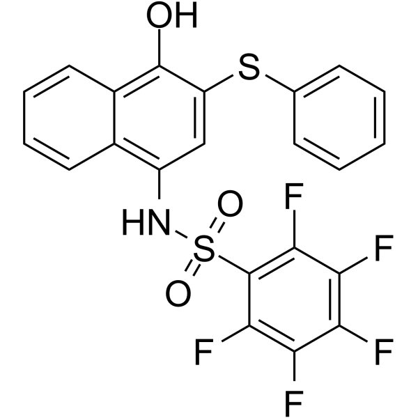
HY-121725
MM-206STAT3 Inhibitor


Biological Activity
Description
Cellular Effect
|
Cell Line

|
Type | Value | Description | References |
|---|---|---|---|---|
| HCT-116 | IC50 |
2.63 ng/mL
Compound: 8
|
Cytotoxicity against human HCT116 cell line
Cytotoxicity against human HCT116 cell line
|
[PMID: 16722648] |
| HCT-116 | IC50 |
5.66 nM
Compound: 8
|
Cytotoxicity against human HCT116 cell line
Cytotoxicity against human HCT116 cell line
|
[PMID: 16722648] |
| HCT-116 | IC50 |
5.66 nM
Compound: 3
|
Phototoxicity against human HCT116 cells after 24 hrs irradiated with tungsten-halogen white light for 2 hrs followed by incubation in drug free medium for 24 hrs by MTT assay
Phototoxicity against human HCT116 cells after 24 hrs irradiated with tungsten-halogen white light for 2 hrs followed by incubation in drug free medium for 24 hrs by MTT assay
|
[PMID: 19211252] |
| Peritoneal macrophage | CC50 |
>100 μM
Compound: 4a
|
Cytotoxicity against Swiss mouse peritoneal macrophages at 20 uM after 5 days by MTT assay
Cytotoxicity against Swiss mouse peritoneal macrophages at 20 uM after 5 days by MTT assay
|
[PMID: 23851117] |
In Vitro
5,15-Diphenylporphyrin (50 μM; 24 h) inhibits STAT3 dimerization, nuclear translocation, DNA binding activity, c-myc promoter binding, and c-myc protein expression in MDA-MB-468 cells and HEK293 cells, and is selective for STAT3 over STAT1 and Grb2 in an AlphaScreen-based assay[1].
MedChemExpress (MCE) has not independently confirmed the accuracy of these methods. They are for reference only. Further protocols information, click here.


Chemical Information
-
CAS No. 22112-89-6
-
Appearance Solid
-
Molecular Weight 462.54
-
Formula C32H22N4
-
Color Brown to black
-
SMILES
C1(/C2=CC=CC=C2)=C3C=CC(/C=C4C=C/C(N/4)=C(C5=CC=CC=C5)/C(C=C/6)=NC6=C/C7=CC=C1N7)=N/3
-
Synonyms
5,15-DPP
-
Shipping
Room temperature in continental US; may vary elsewhere.
-
Storage
4°C, protect from light
* In solvent : -80°C, 6 months; -20°C, 1 month (protect from light)


Solvent & Solubility
In Vitro:
DMF : 5 mg/mL (10.81 mM; ultrasonic and warming and heat to 60°C)
DMSO : 2 mg/mL (4.32 mM; ultrasonic and warming and heat to 60°C; Hygroscopic DMSO has a significant impact on the solubility of product, please use newly opened DMSO)
Please refer to the solubility information to select the appropriate solvent. Once prepared, please aliquot and store the solution to prevent product inactivation from repeated freeze-thaw cycles.
Storage method and period of stock solution: -80°C, 6 months; -20°C, 1 month (protect from light). When stored at -80°C, please use it within 6 months. When stored at -20°C, please use it within 1 month.
Please refer to the solubility information to select the appropriate solvent. Once prepared, please aliquot and store the solution to prevent product inactivation from repeated freeze-thaw cycles.
Storage method and period of stock solution: -80°C, 6 months; -20°C, 1 month (protect from light). When stored at -80°C, please use it within 6 months. When stored at -20°C, please use it within 1 month.
Concentration (start) × Volume (start) = Concentration (final) × Volume (final)

Protocols
-
Nuclear Protein Extraction (High-Salt/Hypotonic Fractionation)
The high-salt/hypotonic fractionation method for nuclear protein extraction is based on the differential solubility of cellular components. Cytoplasmic proteins are extracted first using a hypotonic buffer that causes cell swelling and membrane rupture, followed by centrifugation to separate the cytoplasmic supernatant from the nuclear pellet. The nuclear pellet is then subjected to high-salt extraction (e. g. , 0. 4 M (NH4)2SO4 or 1 M NaCl) to solubilize tightly bound nuclear matrix proteins, including transcription factors, histones, and structural proteins associated with chromatin and the nuclear scaffold. This approach allows for the isolation of both soluble cytoplasmic proteins and salt-resistant nuclear proteins while minimizing cross-contamination.
-
Protocol for Electrophoretic Mobility Shift Assay (EMSA)
Electrophoretic mobility shift assay detects protein-nucleic acid binding by incubating a labeled DNA or RNA probe with purified protein or cell extract, then separating free probe from slower-migrating protein-probe complexes on a native gel. For cancer cells, primary neurons, mouse tumor samples, intestinal organoids, inflammatory macrophages, or drug-treated samples, EMSA can measure transcription-factor DNA binding or RNA-binding protein activity in extracts, but it does not directly measure transcription, protein expression, or chromatin occupancy in intact cells. Specificity is judged by competition with unlabeled wild-type probe, failure of mutated or unrelated competitors to compete, and antibody supershift or disruption when the binding protein identity must be confirmed.
-
Cytoplasmic-Nuclear Fractionated Protein Extraction
Cytoplasmic-nuclear fractionated protein extraction separates soluble cytoplasmic proteins from nuclear-enriched proteins by mild plasma-membrane permeabilization, differential centrifugation, washing of nuclei, and extraction of nuclear proteins for downstream immunoblotting or related molecular analysis. The readout is the relative abundance of a protein in cytoplasmic and nuclear fractions, commonly assessed by western blotting together with compartment markers such as tubulin or pyruvate kinase for cytoplasm and lamin, nucleoporin, hnRNP, H2AX, or Lamin B for nuclear fractions.
-
RNA extraction experimental
By lysing cells, releasing RNA, and removing impurities such as proteins and DNA, high-purity RNA products are finally obtained. The commonly used traditional method is the guanidine isothiocyanate/phenol/chloroform method (Trizol), which is suitable for a variety of animal materials including animal tissues, microorganisms, cultured cells, etc., and most plant materials.


Purity & Documentation
-
Data Sheet (280 KB)
-
SDS (394 KB)
- English - EN (394 KB)
- Français - FR (394 KB)
- Deutsch - DE (394 KB)
- Norwegian - NO (394 KB)
- Español - ES (394 KB)
- Swedish - SV (394 KB)
- Italian - IT (394 KB)
- Korean - KR (394 KB)
- Portuguese - PT (394 KB)
-
Handling Instructions (2659 KB)
References


Complete Stock Solution Preparation Table
Please refer to the solubility information to select the appropriate solvent. Once prepared, please aliquot and store the solution to prevent product inactivation from repeated freeze-thaw cycles.
Storage method and period of stock solution: -80°C, 6 months; -20°C, 1 month (protect from light). When stored at -80°C, please use it within 6 months. When stored at -20°C, please use it within 1 month.
| Optional Solvent | Concentration Solvent Mass | 1 mg | 5 mg | 10 mg | 25 mg |
|---|---|---|---|---|---|
| DMSO / DMF | 1 mM | 2.1620 mL | 10.8099 mL | 21.6198 mL | 54.0494 mL |
| DMF | 5 mM | 0.4324 mL | 2.1620 mL | 4.3240 mL | 10.8099 mL |
| 10 mM | 0.2162 mL | 1.0810 mL | 2.1620 mL | 5.4049 mL |


5,15-Diphenylporphyrin Related Classifications
HY-W035137 Related Classifications
 Powered by Bioz