



5-HMSiR-Hoechst

Based on 1 Customer Validation
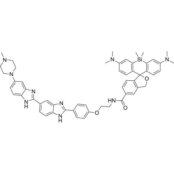
5-HMSiR-Hoechst is a DNA probe. 5-HMSiR-Hoechst comprises Hoechst 33258 and spontaneously blinking far-red hydroxymethyl silicon-rhodamine (HMSiR). 5-HMSiR-Hoechst can stain living cells. Ex/Em=640 nm/675 nm.
For research use only. We do not sell to patients.
- Purity : 98.68%
- CAS No.: 2996108-30-4
- Formula: C54H57N9O3Si
- Molecular Weight:908.17
-
Storage:
-20°C, protect from light
* In solvent : -80°C, 6 months; -20°C, 1 month (protect from light)

To place orders, for customer services and technical support, please contact: MedChemExpress USA
Tel: 609-228-6898 E-mail: [email protected] [email protected]


-
 Biological Activity
-
Chemical Information
- Protocols
- Purity & Documentation
- References
-
 Help & FAQs




Biological Activity
Description
In Vitro
Guidelines (Following is our recommended protocol. This protocol only provides a guideline, and should be modified according to your specific needs).
1. Stock solution preparation: Prepare a 1 mM stock solution with anhydrous DMSO and store it at -20℃.
2. Working solution preparation: Dilute the stock solution with phenol red-free DMEM/HDMEM (containing 10% FBS, 10 mM HEPES) to a final concentration of 0.1-1 μM (0.5 μM is commonly used), and prepare it fresh before use.
3. Staining: Aspirate the old medium, add complete medium containing 0.5 μM 5-HMSiR-Hoechst, and incubate at 37℃, 5% CO2 in the dark for 30-60 min.
4. Fluorescence detection: Wash twice with pre-warmed PBS, replace with fresh medium, and then perform imaging detection. The excitation wavelength is 640 nm, and the emission wavelength is 675 nm.
MedChemExpress (MCE) has not independently confirmed the accuracy of these methods. They are for reference only. Further protocols information, click here.


Chemical Information
-
CAS No. 2996108-30-4
-
Appearance Solid
-
Molecular Weight 908.17
-
Formula C54H57N9O3Si
-
Color Green to dark green
-
SMILES
CN1CCN(CC1)C2=CC=C3NC(C4=CC=C5NC(C6=CC=C(C=C6)OCCNC(C7=CC8=C(C9(C%10=C([Si](C%11=C9C=CC(N(C)C)=C%11)(C)C)C=C(C=C%10)N(C)C)OC8)C=C7)=O)=NC5=C4)=NC3=C2
-
Shipping
Room temperature in continental US; may vary elsewhere.
-
Storage
-20°C, protect from light
* In solvent : -80°C, 6 months; -20°C, 1 month (protect from light)

Protocols
-
Nuclear DNA counterstaining and nuclear morphology staining
Nuclear DNA counterstaining uses DNA-binding fluorescent dyes to visualize nuclei and chromatin so that nuclei can be located, counted, segmented, and evaluated for morphology; Hoechst 33342, DAPI, propidium iodide, and DRAQ5 are commonly reported nuclear stains, while live-cell DNA labeling is better supported for Hoechst dyes and DRAQ5 than for propidium iodide in intact viable cells. Nuclear morphology staining can detect apoptosis-associated nuclear changes, including chromatin condensation, nuclear shrinkage, nuclear fragmentation, reduced nuclear area/perimeter/axis length, and increased nuclear fluorescence intensity; these morphology readouts have been compared with apoptosis markers such as TUNEL and caspase-3 immunofluorescence.


Purity & Documentation
-
Data Sheet (269 KB)
-
SDS (252 KB)
- English - EN (252 KB)
- Français - FR (252 KB)
- Deutsch - DE (252 KB)
- Norwegian - NO (252 KB)
- Español - ES (252 KB)
- Swedish - SV (252 KB)
- Italian - IT (252 KB)
- Korean - KR (252 KB)
- Portuguese - PT (252 KB)
-
Handling Instructions (2659 KB)
References

Calculators
Concentration (start) × Volume (start) = Concentration (final) × Volume (final)


5-HMSiR-Hoechst Related Classifications
HY-D2222 Related Classifications
 Powered by Bioz