



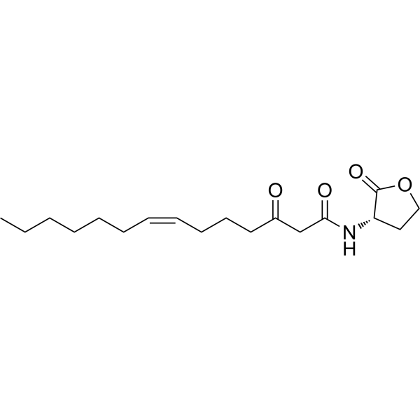
Δ7(Z)-C14-HSL
Δ7(Z)-C14-HSL (Compound 12) is an immunosuppressive agent that can inhibit the proliferation of mouse splenic cells with an IC50 of 17 μM. Δ7(Z)-C14-HSL can be used for further studying its potential as a molecular mechanism in TNF-R-driven immune diseases, especially autoimmune diseases such as psoriasis, rheumatoid arthritis, and type 1 diabetes.
For research use only. We do not sell to patients.
- CAS No.: 482598-46-9
- Formula: C18H29NO4
- Molecular Weight:323.43
-
Storage:
Please store the product under the recommended conditions in the Certificate of Analysis.

To place orders, for customer services and technical support, please contact: MedChemExpress USA
Tel: 609-228-6898 E-mail: [email protected] [email protected]


-
 Biological Activity
-
Chemical Information
- Protocols
- Purity & Documentation
- References
-
 Help & FAQs


-
HY-B1402B
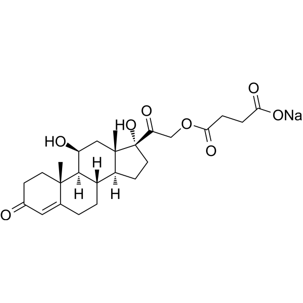
Hydrocortisone hemisuccinate sodiumGlucocorticoid -
HY-108700
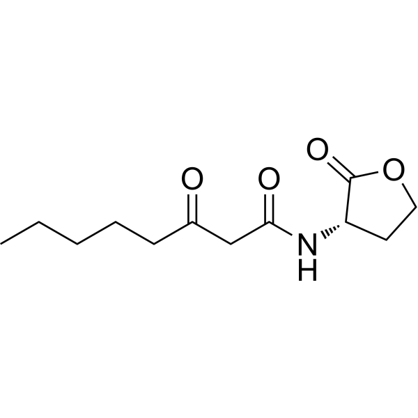
N-3-Oxo-octanoyl-L-homoserine lactoneQuorum-sensing Signal -
HY-132237
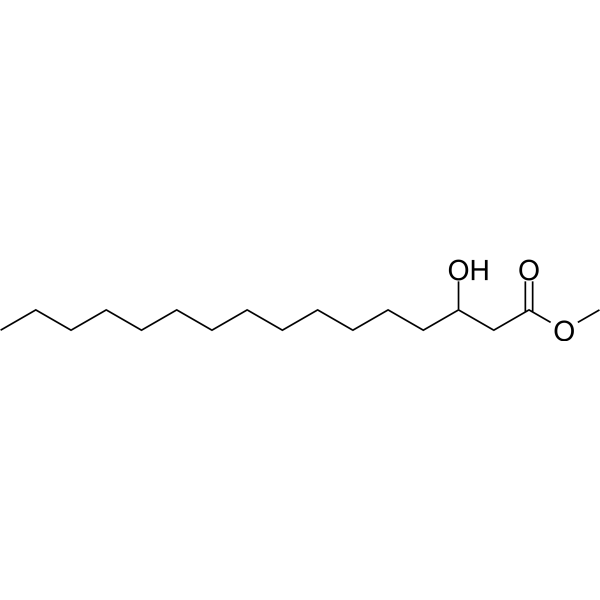
Methyl 3-hydroxyhexadecanoateBiochemical Assay Reagent -
HY-115399A
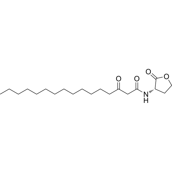
N-3-Oxo-hexadecanoyl-L-Homoserine lactoneN-acyl-homoserine lactone -
HY-W045071
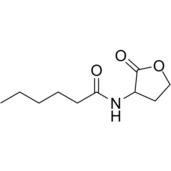
N-Hexanoyl-DL-homoserine lactoneQuorum Sensing Signal -
HY-134560
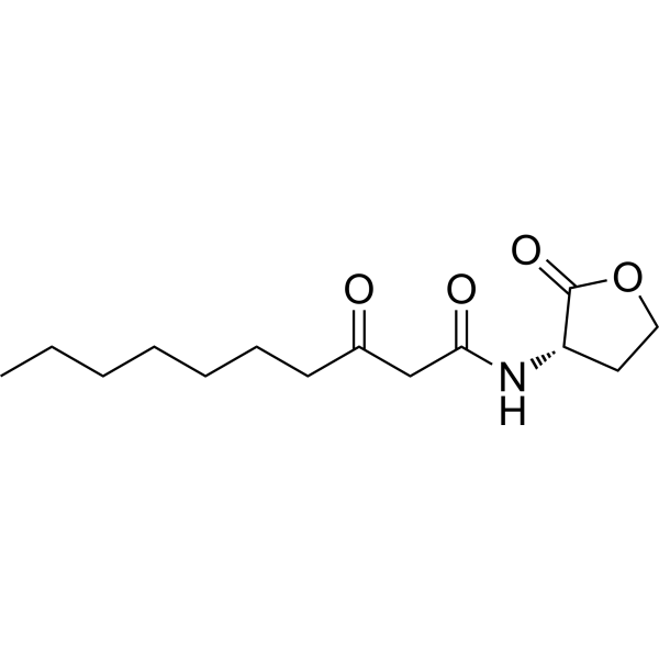
N-(3-Oxodecanoyl)-L-homoserine lactoneSignaling Molecule -
HY-118040
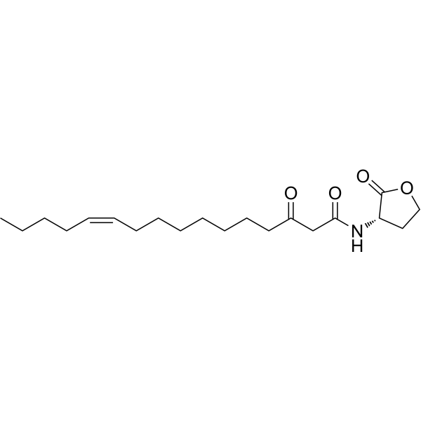
3-Oxo-C16:1N-acyl L-homoserine Lactone -
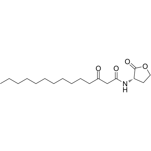
HY-116536
N-3-Oxo-tetradecanoyl-L-homoserine lactoneRhizobacterial Inducer -
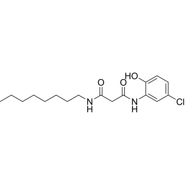
HY-116460
N'-(5-Chloro-2-hydroxyphenyl)-N-octylpropanediamideOrganic -
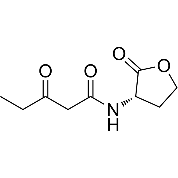
HY-141641
N-(3-Oxopentanoyl)-L-homoserine lactoneAutoinducer -
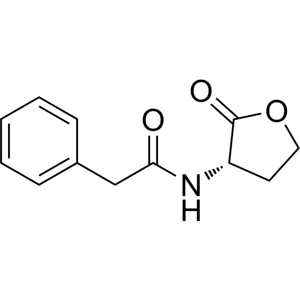
HY-137407
N-Phenylacetyl-L-homoserine lactoneSignaling Molecule -

HY-W408239
N-Pentadecanoyl-L-homoserine lactoneBacterial Regulator


Biological Activity
Description


Chemical Information
-
CAS No. 482598-46-9
-
Molecular Weight 323.43
-
Formula C18H29NO4
-
SMILES
CCCCCC/C=C\CCCC(CC(N[C@@H]1C(OCC1)=O)=O)=O
-
Synonyms
N-(3-Oxo-7Z-tetradecenoyl)-L-homoserine lactone
-
Shipping
Room temperature in continental US; may vary elsewhere.
-
Storage
Please store the product under the recommended conditions in the Certificate of Analysis.

Protocols
-
Collagen-Induced Arthritis
Collagen-induced arthritis (CIA) is an autoimmune murine model of rheumatoid arthritis in which immunization with type II collagen (CII) emulsified in an adjuvant induces a T cell- and autoantibody-driven inflammatory arthritis characterized by synovial hyperplasia, immune cell infiltration, and joint destruction. The model typically relies on genetically susceptible mouse strains (e. g. , DBA/1) and reproduces key features of human rheumatoid arthritis, including anti-collagen immune responses and progressive joint inflammation. Disease onset generally occurs within ~3-4 weeks after immunization, depending on antigen/adjuvant combinations and protocol variation. The immunopathology is driven by adaptive immune activation against CII, leading to systemic and local joint inflammation mediated by pro-inflammatory cytokines and effector immune cells, making CIA a standard preclinical platform for evaluating immunomodulatory and anti-arthritic interventions.
-
Cell Cytotoxicity Assay
Cytotoxicity assays are usually based on the assessment of cell membrane damage, which can also be indirectly detected by measuring cell viability. Detection methods include MTT assay, CKK-8 assay, LDH assay and ATP assay, etc.
-
Imiquimod-Induced Psoriasiform Dermatitis
Imiquimod (IMQ)-induced psoriasiform dermatitis is a widely used murine model in which topical application of IMQ, a Toll-like receptor 7 (TLR7) agonist, triggers innate immune activation in the skin and induces a psoriasis-like inflammatory cascade characterized by epidermal hyperplasia, immune cell infiltration, and cytokine production dominated by the IL-23/IL-17 axis. This inflammatory response is mediated through activation of dendritic cells and downstream induction of IL-23, IL-17A, IL-22, and related pro-inflammatory mediators, recapitulating key features of human plaque psoriasis and enabling mechanistic and therapeutic studies. The model is commonly induced using Aldara (5% IMQ cream) applied topically to murine skin, resulting in rapid onset of erythema, scaling, and thickening that can be quantified as disease severity indices and validated histologically.
-
Research Protocol for Metabolic Diseases
AMP-activated protein kinase, AMPK, is a conserved cellular energy sensor that responds to reduced cellular energy status and coordinates metabolism by increasing ATP-generating catabolic pathways while suppressing ATP-consuming anabolic processes. In metabolic disease research, the AMPK pathway is experimentally relevant because it regulates hepatic lipid synthesis, fatty acid oxidation, glucose production, skeletal-muscle glucose disposal, mTORC1-linked biosynthesis, autophagy, mitochondrial homeostasis, and whole-body energy balance. The central pathway logic is that energy stress, metformin, exercise-like stimulation, or direct AMPK activators increase AMPKα Thr172 phosphorylation and downstream substrate phosphorylation, including ACC and RAPTOR. Phosphorylation of ACC suppresses lipogenesis and supports fatty acid oxidation, whereas phosphorylation of RAPTOR suppresses mTORC1 signaling and links cellular energy status to growth and protein synthesis control. The pathway is linked
-
Cell Viability Determination by MTT Colorimetric Assay
The following protocol uses the MTT colorimetric assay as a classic literature-established method for assessing cell viability/metabolic activity in cultured mammalian cells. MTT[3-(4,5-dimethylthiazol-2-yl)-2,5-diphenyltetrazolium bromide] is reduced by metabolically active cells to a colored formazan product; the amount of formazan is quantified spectrophotometrically and provides an indirect measure of metabolically active viable cells. Importantly, MTT reduction reflects cellular oxidoreductase/metabolic activity rather than an absolute direct count of living cells, so changes in cellular metabolism can alter the signal independently of cell number.


Purity & Documentation
References

Calculators
Concentration (start) × Volume (start) = Concentration (final) × Volume (final)


Δ7(Z)-C14-HSL Related Classifications
HY-145492 Related Classifications
 Powered by Bioz