



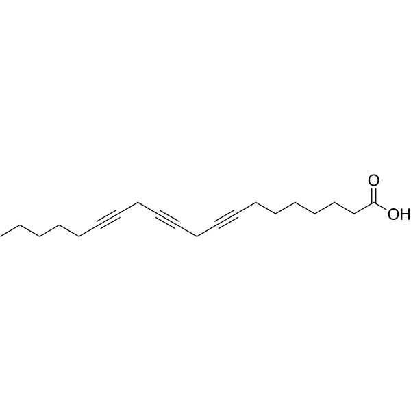
8,11,14-Eicosatriynoic acid
8,11,14-Eicosatriynoic Acid, as an inhibitor of prostaglandin, leukotriene biosynthesis, and arachidonic acid-induced platelet aggregation, blocks human 12-lipoxygenase (12-LO), cyclooxygenase (COX)and 5-lipoxygenase (5-LO) with IC50 values of 0.46 μM, 14 μMand 25 μM, respectively. In addition, 8,11,14-Eicosatriynoic Acid inhibits the action of slow-reacting substances of allergic reactions, with IC50 value of 10 μM. Lipoxygenase is widely found in fungi, plants and animals. 12-LO involves in many important disease states and may play a role in oxidative glutamate toxicity. COX enzymes play complex roles in human physiology and pathology involving the neuronal, immune, renal, cardiovascular, gastrointestinal and reproductive systems. COX enzymes are blocked by aspirin and a variety of other NSAIDs, which makes them clinically important. 5-LO involves in cancer pathology. It is expressed by a variety of cancer cells, including colon, lung, breast, and prostate cancers, and promotes cancer cell growth and neovascularization. 8,11,14-Eicosatriynoic acid is a click chemistry reagent, it contains an Alkyne group and can undergo copper-catalyzed azide-alkyne cycloaddition (CuAAc) with molecules containing Azide groups.
For research use only. We do not sell to patients.
- CAS No.: 34262-64-1
- Formula: C20H28O2
- Molecular Weight:300.44
-
Storage:
Please store the product under the recommended conditions in the Certificate of Analysis.

To place orders, for customer services and technical support, please contact: MedChemExpress USA
Tel: 609-228-6898 E-mail: [email protected] [email protected]


-
 Biological Activity
-
Chemical Information
- Protocols
- Purity & Documentation
- References
-
 Help & FAQs


-
HY-124252
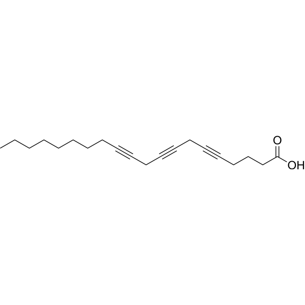
5,8,11-Eicosatriynoic acidLipoxygenase Inhibitor -
HY-113465
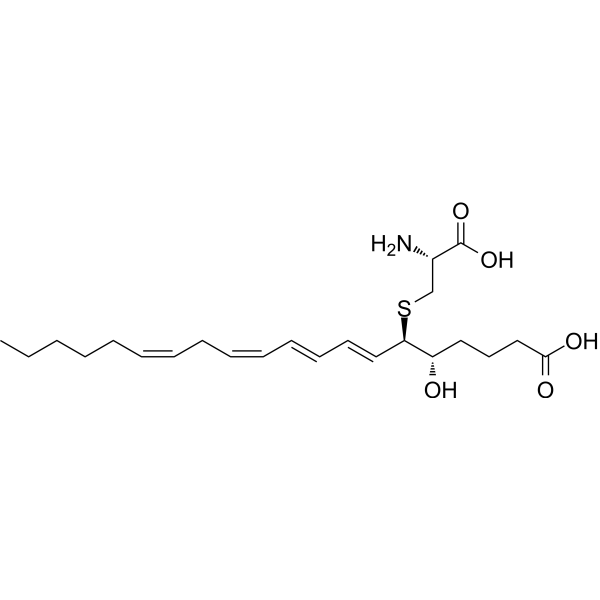
Leukotriene E4Cysteinyl Leukotriene Pathway Marker -
HY-N8276
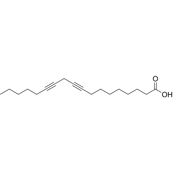
Ro 3-1314Lipoxygenase Inhibitor -

HY-108396
γ-Linolenic acid ethyl esterPolyunsaturated Fatty Acid Ester -
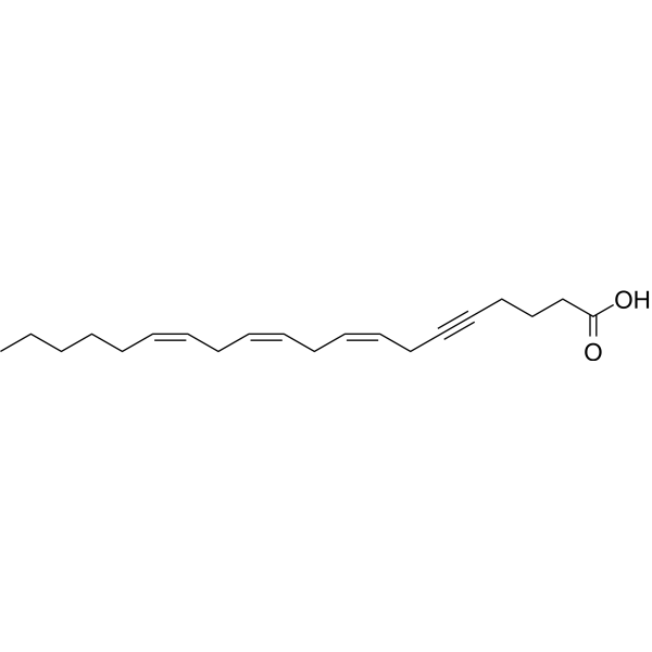
HY-131895
5,6-Dehydroarachidonic acid5-LOX Inhibitor -
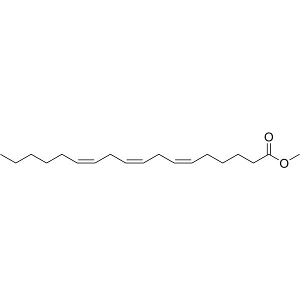
HY-W009276
γ-Linolenic Acid methyl esterGLA Derivative -
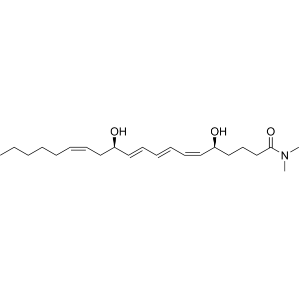
HY-139112
Leukotriene B4 dimethyl amideLeukotriene -

HY-W747567
Leukotriene E4 methyl esterLeukotriene -

HY-W009276R
γ-Linolenic Acid methyl ester (Standard)GLA Derivative -

HY-108396R
γ-Linolenic acid ethyl ester (Standard)Reference Standard


Biological Activity
Description


Chemical Information
-
CAS No. 34262-64-1
-
Molecular Weight 300.44
-
Formula C20H28O2
-
SMILES
CCCCCC#CCC#CCC#CCCCCCCC(O)=O
-
Synonyms
8,11,14-Icosatriynoic acid
-
Shipping
Room temperature in continental US; may vary elsewhere.
-
Storage
Please store the product under the recommended conditions in the Certificate of Analysis.

Protocols
-
EdU Incorporation Assay (Click Chemistry-Based DNA Synthesis Measurement)
The EdU incorporation assay measures DNA synthesis by adding the thymidine analog 5-ethynyl-2′-deoxyuridine to cells or tissues, where it is incorporated into newly synthesized DNA during S phase. Incorporated EdU is detected by copper-catalyzed azide-alkyne cycloaddition, in which a fluorescent azide covalently reacts with the ethynyl group on EdU, allowing S-phase cells to be detected by fluorescence microscopy, flow cytometry, or high-content imaging. EdU detection does not require DNA denaturation or anti-BrdU antibody access, which preserves sample structure and improves compatibility with immunostaining and multiparameter cytometry compared with BrdU-based detection. EdU can be cytotoxic in a cell-type- and exposure-dependent manner, so pulse duration, concentration, and continuous-labeling designs should be validated for each cell type.
-
Cell Cytotoxicity Assay
Cytotoxicity assays are usually based on the assessment of cell membrane damage, which can also be indirectly detected by measuring cell viability. Detection methods include MTT assay, CKK-8 assay, LDH assay and ATP assay, etc.
-
Protocol for Cell Counting and Cell Density Analysis
Cell counting and cell-density analysis estimate the number of cells in a known volume or field area. Manual hemocytometer counting uses a chamber of defined geometry to convert counted cells into cells/mL, while automated counters and image-analysis workflows detect cell objects from optical, brightfield, fluorescence, impedance, or digital-image features. Trypan blue viability counting is based on dye exclusion: viable cells with intact membranes exclude dye, while non-viable cells with compromised membranes stain blue. The readout is total cell density, viable-cell density, dead-cell density, and percent viability. Cell density can also be estimated from microscopy images by counting objects per image area, from flow cytometry using calibrated volume or reference particles, or from in situ microscopy in bioreactors after calibration against reference methods such as hemocytometer or flow cytometry.
-
Somatic Cell Culture
A method of simulating the in vivo environment in vitro to maintain the cell growth, differentation and main functions.
-
CCK-8/WST-8 Cell Proliferation Assay
The CCK-8/WST-8 assay is based on the reduction of the water-soluble tetrazolium salt WST-8 to a water-soluble formazan product by cellular dehydrogenases in metabolically active cells, where the generated formazan amount is proportional to the number of living cells and is quantified by measuring absorbance in the visible range, providing a colorimetric readout for cell viability and proliferation assessment. This class of tetrazolium-based assays improves upon earlier MTT-based systems by producing a water-soluble formazan, eliminating the need for organic solubilization steps and enabling direct spectrophotometric measurement in culture medium.
-
Protocol for Hematoxylin-Eosin (H&E) Staining
Hematoxylin-eosin staining is a routine histological method that stains nuclei mainly blue-purple with hemalum and stains cytoplasm, extracellular matrix, and many stromal components pink with eosin, allowing tissue architecture, cell morphology, necrosis, inflammation, fibrosis, tumor growth pattern, and treatment-associated injury to be evaluated by light microscopy. In cancer cells, primary neurons, mouse tumor models, intestinal organoids, inflammatory macrophage preparations, and drug-screening tissues, H&E is a morphology assay rather than a molecular assay; it should be interpreted with complementary molecular or immunostaining assays when the biological question concerns specific proteins, RNA levels, ferroptosis, mitophagy, or immune phenotypes.
-
Cell Counting-Based Growth Curve Assay
Cell counting-based growth curve assays quantify cell proliferation by directly measuring changes in viable cell number over time using manual or automated counting methods such as hemocytometer-based counting or instrument-assisted cell enumeration, enabling construction of growth curves that reflect population expansion dynamics in response to culture conditions. A widely used approach is trypan blue exclusion with hemocytometer counting, where membrane-compromised (non-viable) cells take up the dye, allowing discrimination between viable and non-viable cells while simultaneously enabling total cell number quantification. Repeated sampling across time points allows estimation of proliferation rate, growth phases, and comparative growth kinetics between experimental conditions.
-
MTT Cell Proliferation Assay
The MTT assay is a colorimetric endpoint assay for estimating viable cell number, cell growth, cytotoxicity, or cell activation in cultured mammalian cells. Living cells reduce the yellow tetrazolium salt MTT into purple/blue formazan, while dead cells do not generate the same signal; the resulting color can be quantified with a multiwell spectrophotometer. MTT reduction is commonly interpreted as a readout of metabolic activity that often correlates with viable cell number, but it should not be treated as a direct cell-counting method unless the assay is optimized for the cell type and experimental condition. Studies show that MTT reduction can involve mitochondrial and non-mitochondrial reducing systems, and formazan may accumulate in intracellular lipid droplets rather than simply marking mitochondria.
-
Cell Viability Determination by MTT Colorimetric Assay
The following protocol uses the MTT colorimetric assay as a classic literature-established method for assessing cell viability/metabolic activity in cultured mammalian cells. MTT[3-(4,5-dimethylthiazol-2-yl)-2,5-diphenyltetrazolium bromide] is reduced by metabolically active cells to a colored formazan product; the amount of formazan is quantified spectrophotometrically and provides an indirect measure of metabolically active viable cells. Importantly, MTT reduction reflects cellular oxidoreductase/metabolic activity rather than an absolute direct count of living cells, so changes in cellular metabolism can alter the signal independently of cell number.


Purity & Documentation
-
Data Sheet (262 KB)
-
SDS (252 KB)
- Français - FR (252 KB)
- Deutsch - DE (252 KB)
- Norwegian - NO (252 KB)
- Español - ES (252 KB)
- Swedish - SV (252 KB)
- Italian - IT (252 KB)
- Korean - KR (252 KB)
- Portuguese - PT (252 KB)
-
Handling Instructions (2659 KB)
References

Calculators
Concentration (start) × Volume (start) = Concentration (final) × Volume (final)


8,11,14-Eicosatriynoic acid Related Classifications
HY-131384 Related Classifications
 Powered by Bioz