



Amoxapine
Based on 4 publication(s) in Google Scholar
Amoxapine (CL-67772) is a norepinephrine reuptake blocker and a 5-HT2/5-HT3 antagonist. Amoxapine can be used for the research of depression. Amoxapine has antibacterial activity. Amoxapine can enhance the killing effect of macrophages on mycobacterium by inducing autophagy, while protecting the cells from death.
For research use only. We do not sell to patients.
- Purity : 99.96%
- CAS No.: 14028-44-5
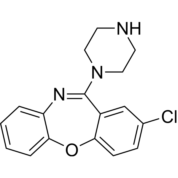
- Formula: C17H16ClN3O
- Molecular Weight:313.79
-
Storage:Powder -20°C, 3 years , 4°C, 2 years ; In solvent -80°C, 2 years , -20°C, 1 year

To place orders, for customer services and technical support, please contact: MedChemExpress USA
Tel: 609-228-6898 E-mail: [email protected] [email protected]


-
 Biological Activity
-
Chemical Information
-
Solvent & Solubility
- Protocols
- Purity & Documentation
- References
-
 Help & FAQs


-

HY-B0015
PaclitaxelAntitumor Agent -
HY-B0725
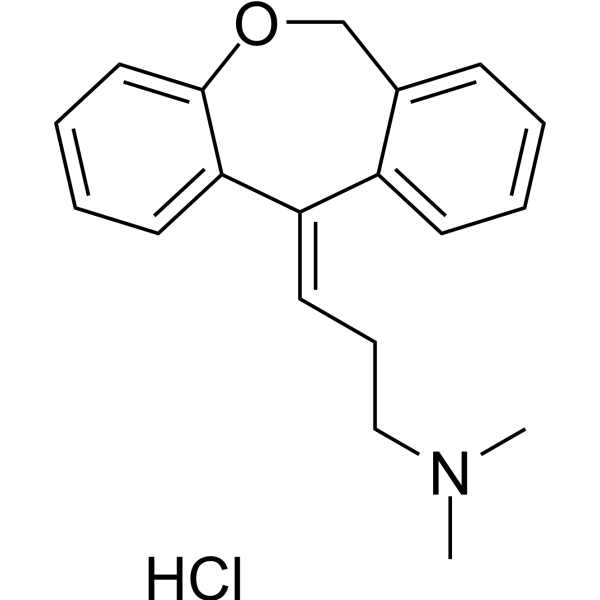
Doxepin HydrochlorideAntidepressant -
HY-14539
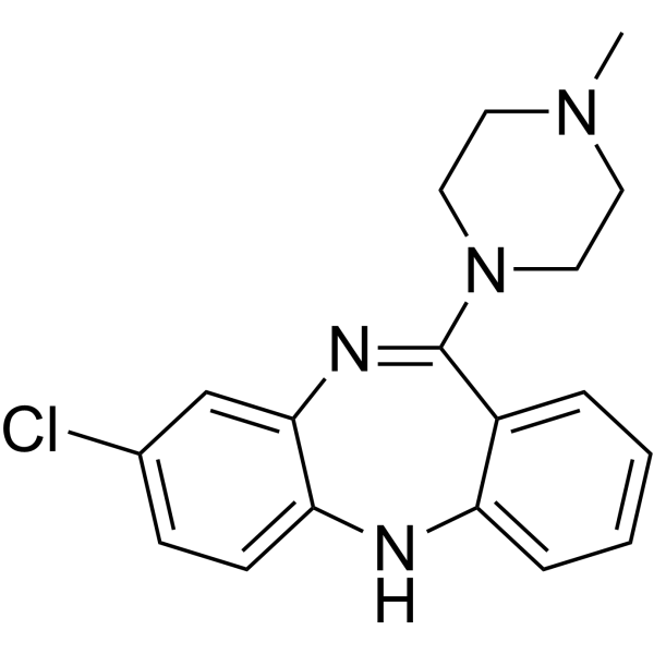
ClozapineAntipsychotic Agent -
HY-66005
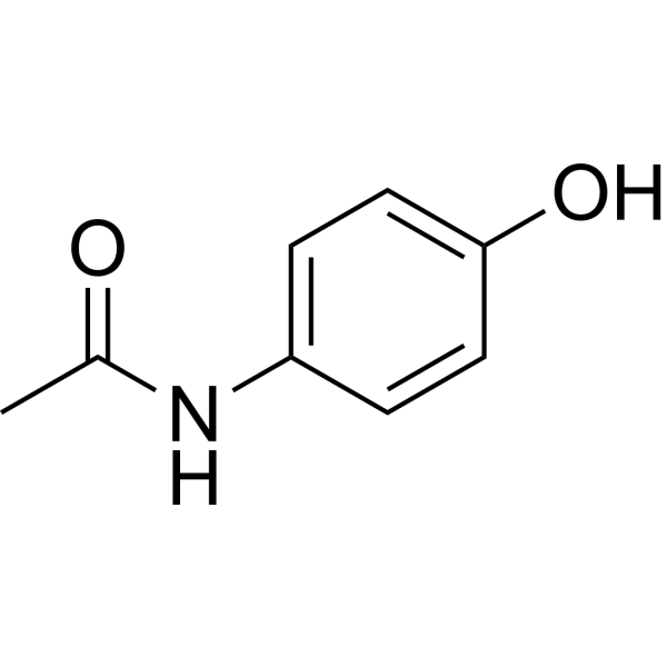
AcetaminophenCOX-2 Inhibitor -
HY-N0219
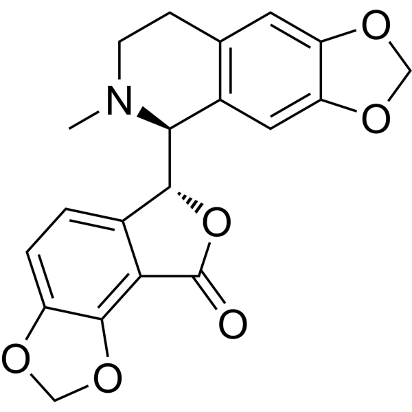
BicucullineGABAA Receptor Antagonist -
HY-17386
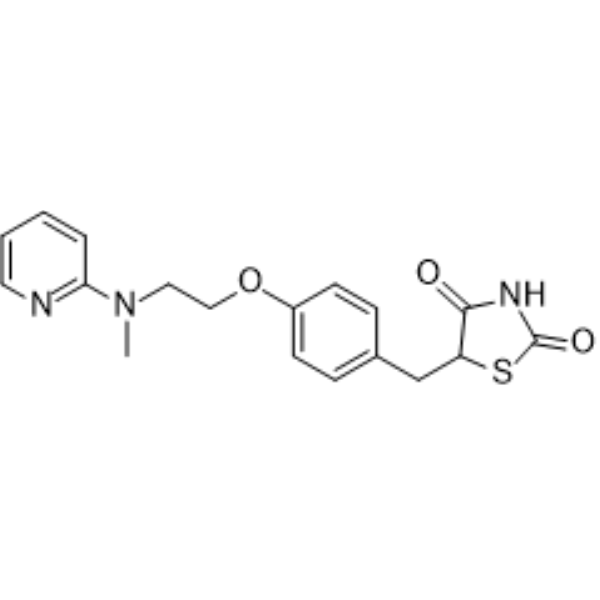
RosiglitazonePPARγ Agonist -
HY-50935
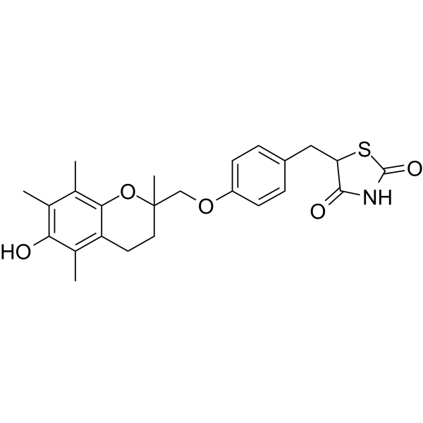
TroglitazonePPARγ Agonist -
HY-16569
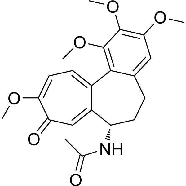
ColchicineTubulin Inhibitor -
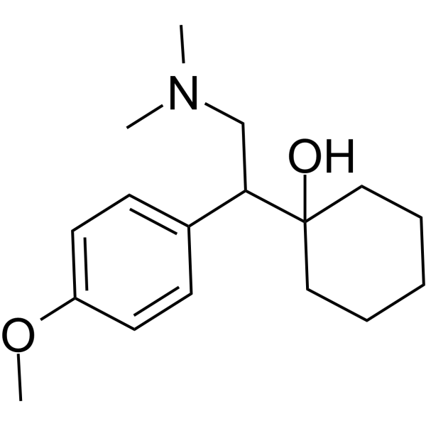
HY-B0196
VenlafaxineSerotonin/Norepinephrine Reuptake Inhibitor -
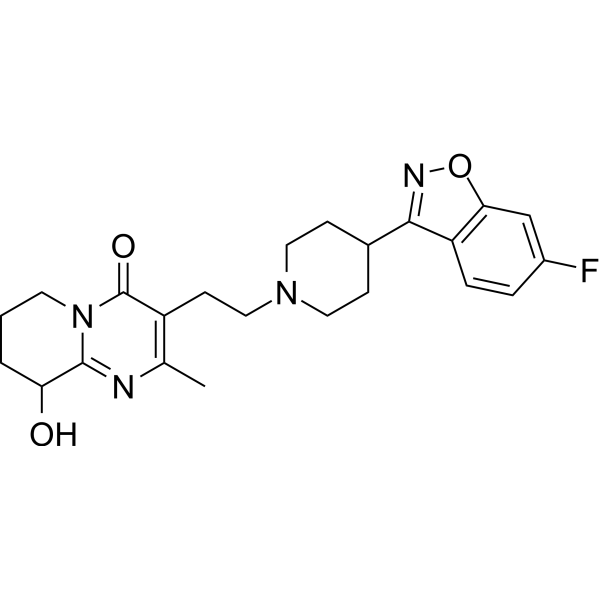
HY-A0019
PaliperidoneDopamine D2/5-HT2A Antagonist -
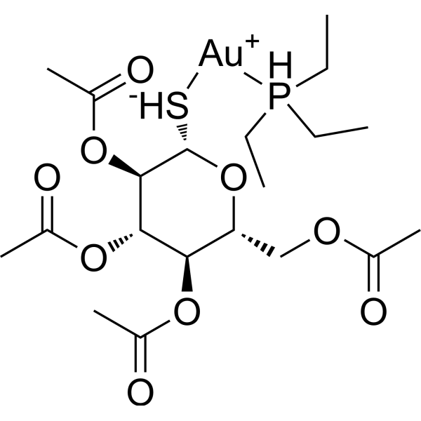
HY-B1123
AuranofinBacterial Inhibitor -

HY-P7077
IL-2 Protein, Mouse
Publications Citing Use of MedChemExpress (MCE) Amoxapine
More Customer Validation & Images

-
WB


Biological Activity
Description
IC50 & Target
5-HT Receptor and Serotonin Transporter[3]
Cellular Effect
|
Cell Line

|
Type | Value | Description | References |
|---|---|---|---|---|
| RAW264.7 | CC50 |
107.1 μM
Compound: Loxapine
|
Cytotoxicity against mouse RAW264.7 cells assessed as reduction in number of cells by DAPI staining based high-content image analysis
Cytotoxicity against mouse RAW264.7 cells assessed as reduction in number of cells by DAPI staining based high-content image analysis
|
[PMID: 36439974] |
In Vitro
Amoxapine (10 μM; 3-48 h) reduces the cytotoxicity induced by mycobacterium bovis (BCG) and inhibits the survival of BCG in macrophages. The minimum inhibitory concentration (MIC90) for BCG is 53 μM[1].
Amoxapine (10 μM; 24 h) induces autophagy in mycobacteria-infected macrophages in a mTOR-dependent manner[1].
MedChemExpress (MCE) has not independently confirmed the accuracy of these methods. They are for reference only. Further protocols information, click here.
-
Cell Line:BCG treated RAW 264.7 cells
-
Concentration:10 μM
-
Incubation Time:3 h
-
Result:Reduced the percentage of dead cells.
-
Cell Line:BCG treated RAW 264.7 cells
-
Concentration:10 μM
-
Incubation Time:24 h
-
Result:Significantly enhanced levels of LC3B-II protein and reduced levels of p62.
Significantly reduced the ratio of phosphorylation at Ser 2448 of mTOR to total mTOR.
In Vivo
Amoxapine (10 mg/kg; Intraperitoneal injection; 28 days) can affect the electrophysiology of rat atrial[2].
MedChemExpress (MCE) has not independently confirmed the accuracy of these methods. They are for reference only.
-
Animal Model:Mtb strain H37Rv treated BALB/c and C57BL/6 mice[1]
-
Dosage:3 and 5 mg/kg
-
Administration:Oral administration (p.o.); 2 weeks
-
Result:Significantly reduced bacterial burdens in the lungs.
Enhanced host defense against Mtb in vivo.
-
Animal Model:Sprague-Dawley rats (200-300g)[2]
-
Dosage:10 mg/kg
-
Administration:Intraperitoneal injection (i.p.); 28 days
-
Result:Reduced atrial rate, contractile force and df/dtmax prolonged the sinus node recovery time and decreased atrial excitability.
Decreased amplitude and Vmax of the upstroke, prolonged the duration of the action potential (APD) and effective refractory period (ERP) and reduced the resting membrane potential.
Inhibited the slow action potentials and contractions induced by isoprenaline in Kdepolarized atria.


Chemical Information
-
CAS No. 14028-44-5
-
Appearance Solid
-
Molecular Weight 313.79
-
Formula C17H16ClN3O
-
Color White to light yellow
-
SMILES
ClC1=CC=C(OC2=CC=CC=C2N=C3N4CCNCC4)C3=C1
-
Synonyms
CL-67772
-
Shipping
Room temperature in continental US; may vary elsewhere.
-
Storage
Powder -20°C 3 years 4°C 2 years In solvent -80°C 2 years -20°C 1 year


Publications (4)
-
Journal Impact Factor
-
Most Recent
-
Nat Commun
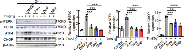
Maprotiline restores ER homeostasis and rescues neurodegeneration via Histamine Receptor H1 inhibition in retinal ganglion cells. [Abstract]2022 Nov 10;13(1):6796. PMID: 36357388
Amoxapine purchased from MedChemExpress. Usage Cited in: Nat Commun. 2022 Nov 10;13(1):6796. [Abstract]
10 µM Amoxapine (Amo), Desloratadine (Desl) and Maprotiline (Map) are incubated with HEK293T cells for 24 h and significantly inhibits PERK phosphorylation and expression of ATF4 and CHOP .
-
J Med Chem
Exploring Simple Drug Scaffolds from the Generated Database Chemical Space Reveals a Chiral Bicyclic Azepane with Potent Neuropharmacology. [Abstract]2025 Apr 24. PMID: 40274264 -
PLoS Negl Trop Dis
Identification of anti-flaviviral drugs with mosquitocidal and anti-Zika virus activity in Aedes aegypti. [Abstract]2019 Aug 20;13(8):e0007681. PMID: 31430351 -


Solvent & Solubility
In Vitro:
DMSO : 8.33 mg/mL (26.55 mM; Need ultrasonic; Hygroscopic DMSO has a significant impact on the solubility of product, please use newly opened DMSO)
Please refer to the solubility information to select the appropriate solvent. Once prepared, please aliquot and store the solution to prevent product inactivation from repeated freeze-thaw cycles.
Storage method and period of stock solution: -80°C, 2 years; -20°C, 1 year. When stored at -80°C, please use it within 2 years. When stored at -20°C, please use it within 1 year.
Please refer to the solubility information to select the appropriate solvent. Once prepared, please aliquot and store the solution to prevent product inactivation from repeated freeze-thaw cycles.
Storage method and period of stock solution: -80°C, 2 years; -20°C, 1 year. When stored at -80°C, please use it within 2 years. When stored at -20°C, please use it within 1 year.
Concentration (start) × Volume (start) = Concentration (final) × Volume (final)
In Vivo:
Select the appropriate dissolution method based on your experimental animal and administration route.
- For the following dissolution methods, please ensure to first prepare a clear stock solution using an In Vitro approach and then sequentially add co-solvents:
- To ensure reliable experimental results, the clarified stock solution can be appropriately stored based on storage conditions. As for the working solution for In Vivo experiments, it is recommended to prepare freshly and use it on the same day.
- The percentages shown for the solvents indicate their volumetric ratio in the final prepared solution. If precipitation or phase separation occurs during preparation, heat and/or sonication can be used to aid dissolution.
Add each solvent one by one: 10% DMSO 40% PEG300 5% Tween-80 45% Saline
Solubility: ≥ 1.56 mg/mL (4.97 mM); Clear solution
This protocol yields a clear solution of ≥ 1.56 mg/mL (saturation unknown).
Taking 1 mL working solution as an example, add 100 μL DMSO stock solution (15.6 mg/mL) to 400 μL PEG300, and mix evenly; then add 50 μL Tween-80 and mix evenly; then add 450 μL Saline to adjust the volume to 1 mL.
Preparation of Saline: Dissolve 0.9 g sodium chloride in ddH₂O and dilute to 100 mL to obtain a clear Saline solution.
Add each solvent one by one: 10% DMSO 90% (20% SBE-β-CD in Saline)
Solubility: ≥ 1.56 mg/mL (4.97 mM); Clear solution
This protocol yields a clear solution of ≥ 1.56 mg/mL (saturation unknown).
Taking 1 mL working solution as an example, add 100 μL DMSO stock solution (15.6 mg/mL) to 900 μL 20% SBE-β-CD in Saline, and mix evenly.
Preparation of 20% SBE-β-CD in Saline (4°C, storage for one week): 2 g SBE-β-CD powder is dissolved in 10 mL Saline, completely dissolve until clear.
In Vivo Dissolution Calculator
Please enter the basic information of animal experiments:
-
-
-
-
Recommended: Prepare an additional quantity of animals to account for potential losses during experiments.
Please enter your animal formula composition:
-
%DMSO +
Recommended: Keep the proportion of DMSO in working solution below 2% if your animal is weak.
-
%+
-
+%Tween-80 + +
-
%Saline +
The co-solvents required include: DMSO, . All of co-solvents are available by MedChemExpress (MCE). , Tween 80. All of co-solvents are available by MedChemExpress (MCE).
Working solution concentration: 0.22 mg/mL
Method for preparing stock solution: mg drug dissolved in μL DMSO. Stock solution concentration: mg/mL.
1. Take μL DMSO stock solution;
2. Add μL .
μL , mix evenly;
3. Then add μL Tween 80, mix evenly;
4. Then add μL
Please ensure that the stock solution in the first step is dissolved to a clear state, and add co-solvents in sequence. You can use ultrasonic heating (ultrasonic cleaner, recommended frequency 20-40 kHz), vortexing, etc. to assist dissolution.

Protocols
-
Research Protocol for Infectious Diseases
Infectious-disease experiments test how pathogens interact with host barriers, innate immune receptors, inflammatory signaling, pathogen replication, and tissue injury; pattern-recognition receptors such as TLRs, RIG-I-like receptors, NOD-like receptors, and inflammasomes detect microbial molecules and activate NF-κB, interferon, and cytokine responses. The central hypothesis is that infection severity reflects the balance between pathogen burden and host response: protective inflammation restricts pathogen growth, whereas excessive or mislocalized inflammation contributes to tissue damage and disease phenotype. Unresolved questions include which host pathways are protective versus pathogenic, why some infection models fail to translate to human disease, and which combined readouts best predict clinically relevant infection outcomes.
-
Autophagy
Autophagy is a process in which eukaryotic cells use lysosomes to degrade their own cytoplasmic proteins and damaged organelles under the regulation of autophagy related gene (Atg). Microtubule-associated proteins light chain 3 (LC3) is recognized as autophagy marker, which transfers from cytoplasmic LC3 (LC3-I) to membrane type (LC3-II). LC3-II/I ratio could be detected by Western Blot and fluorescence microscopy.
-
Lysosome and acidic-vesicle live-cell staining
Lysosome and acidic-vesicle live-cell staining detects acidic intracellular compartments by using membrane-permeant acidotropic probes that accumulate in low-pH vesicles, including lysosomes, late endosomes, autolysosomes, and acidic phagosomes. LysoTracker staining is commonly used as an intensity-based readout of acidic lysosomal compartment abundance or enlargement, while acridine orange produces green fluorescence in less concentrated compartments and red fluorescence after concentration-dependent accumulation in acidic vesicular organelles. Loss or reduction of acridine-orange red signal can be used as a readout of lysosomal membrane permeabilization or reduced acidic-vesicle integrity. This protocol is designed for live cultured cells and can be adapted for fluorescence microscopy, high-content imaging, plate-reader readout, or flow cytometry when the selected literature supports the readout. Because these dyes report acidotropic accumulation rather than lysosome identity alone,
-
Bacterial live/dead nucleic-acid viability staining
The LIVE/DEAD bacterial viability staining method is based on differential permeability of nucleic-acid-binding fluorescent dyes, most commonly SYTO 9 and propidium iodide (PI), which enables discrimination of bacterial populations with intact versus compromised cytoplasmic membranes. SYTO 9 penetrates both intact and damaged bacterial membranes and binds nucleic acids to produce green fluorescence, whereas propidium iodide penetrates only cells with compromised membranes and fluoresces red while also reducing SYTO 9 signal through competitive binding and fluorescence interactions. The resulting fluorescence pattern is interpreted as a proxy for membrane integrity, which is widely used as an indicator of bacterial viability in microscopy, flow cytometry, and spectroscopic platforms. However, mechanistic studies show that SYTO 9 and PI interactions involve displacement and fluorescence resonance energy transfer effects, which can influence signal interpretation depending on dye ratios a
-
Macroautophagy Solutions
Macroautophagy is a conserved lysosome-dependent degradation pathway in which cytoplasmic material is sequestered into double-membrane autophagosomes and delivered to lysosomes for degradation and recycling. The pathway supports cellular homeostasis during nutrient limitation, organelle stress, protein-aggregate accumulation, infection, differentiation, and tissue remodeling by coupling cargo sequestration, autophagosome maturation, lysosomal fusion, and degradation of cargo-derived macromolecules. The core molecular sequence includes initiation by nutrient- and stress-regulated autophagy machinery, autophagosome nucleation, LC3/ATG8-family conjugation to autophagosomal membranes, cargo selection through receptors such as SQSTM1/p62, autophagosome-lysosome fusion, and lysosomal degradation. LC3 was identified as a mammalian homolog of yeast Atg8 that localizes to autophagosomal membranes after processing, and p62/SQSTM1 was shown to connect ubiquitinated cargo with autophagic degradati


Purity & Documentation
-
Data Sheet (285 KB)
-
SDS (393 KB)
- English - EN (393 KB)
- Français - FR (393 KB)
- Deutsch - DE (393 KB)
- Norwegian - NO (393 KB)
- Español - ES (393 KB)
- Swedish - SV (393 KB)
- Italian - IT (393 KB)
- Korean - KR (393 KB)
- Portuguese - PT (393 KB)
-
Handling Instructions (2659 KB)
References
[1]. Wang J, et al. FDA-Approved Amoxapine Effectively Promotes Macrophage Control of Mycobacteria by Inducing Autophagy. Microbiol Spectr. 2022 Oct 26;10(5):e0250922. [Content Brief]
[2]. Delgado C, Manzanares J, Tamargo J, Valenzuela C. Electrophysiological effects of amoxapine in untreated and in amoxapine-pretreated rat atria. Br J Pharmacol. 1986 Feb;87(2):317-25. [Content Brief]
[3]. Maudhuit C, et al. Effects of acute and chronic treatment with amoxapine and cericlamine on the sleep-wakefulness cycle in the rat. Neuropharmacology. 1994 Aug;33(8):1017-25. [Content Brief]


Complete Stock Solution Preparation Table
Please refer to the solubility information to select the appropriate solvent. Once prepared, please aliquot and store the solution to prevent product inactivation from repeated freeze-thaw cycles.
Storage method and period of stock solution: -80°C, 2 years; -20°C, 1 year. When stored at -80°C, please use it within 2 years. When stored at -20°C, please use it within 1 year.
| Optional Solvent | Concentration Solvent Mass | 1 mg | 5 mg | 10 mg | 25 mg |
|---|---|---|---|---|---|
| DMSO | 1 mM | 3.1869 mL | 15.9343 mL | 31.8687 mL | 79.6716 mL |
| 5 mM | 0.6374 mL | 3.1869 mL | 6.3737 mL | 15.9343 mL | |
| 10 mM | 0.3187 mL | 1.5934 mL | 3.1869 mL | 7.9672 mL | |
| 15 mM | 0.2125 mL | 1.0623 mL | 2.1246 mL | 5.3114 mL | |
| 20 mM | 0.1593 mL | 0.7967 mL | 1.5934 mL | 3.9836 mL | |
| 25 mM | 0.1275 mL | 0.6374 mL | 1.2747 mL | 3.1869 mL |


Amoxapine Related Classifications
HY-B0991 Related Classifications
 Powered by Bioz