



Cholesteryl linoleate

Based on 1 Customer Validation
Cholesteryl linoleate is a cholesteryl ester with antibacterial activity that is present in low-density lipoprotein (LDL) particles, human nasal fluid, and respiratory epithelial secretions. Cholesteryl linoleate is oxidized by 12/15-lipoxygenase in macrophages and participates in selective uptake and efflux via LDL receptor-related protein. Cholesteryl linoleate does not induce endothelial adhesion molecule expression, nor does it induce monocyte binding to endothelial cells. Cholesteryl linoleate liposomal formulations inhibit the growth of multiple bacteria at physiological nasal fluid concentrations and act synergistically with antimicrobial peptides. Cholesteryl linoleate is useful for research related to atherosclerosis and bacterial infections.
For research use only. We do not sell to patients.
- Purity : 99.45%
- CAS No.: 604-33-1
- Formula: C45H76O2
- Molecular Weight:649.10
-
Storage:Powder -20°C, 3 years ; In solvent -80°C, 6 months , -20°C, 1 month

To place orders, for customer services and technical support, please contact: MedChemExpress USA
Tel: 609-228-6898 E-mail: [email protected] [email protected]


-
 Biological Activity
-
Chemical Information
-
Solvent & Solubility
- Protocols
- Purity & Documentation
- References
-
 Help & FAQs


-
HY-113217
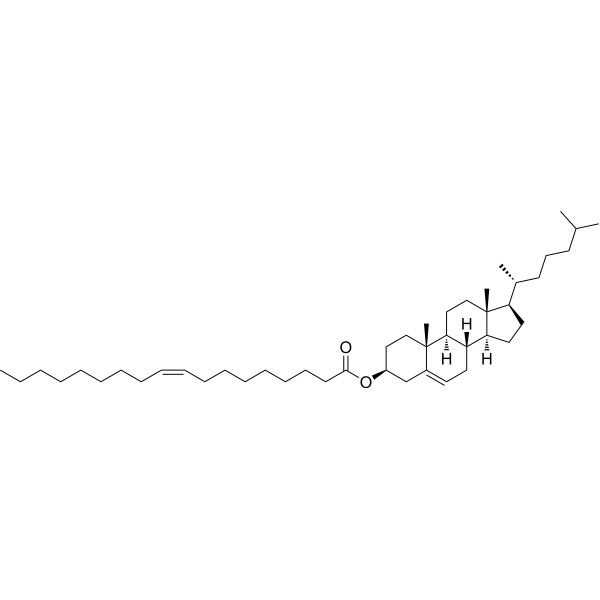
Cholesteryl oleateEndogenous Metabolite -
HY-N0322
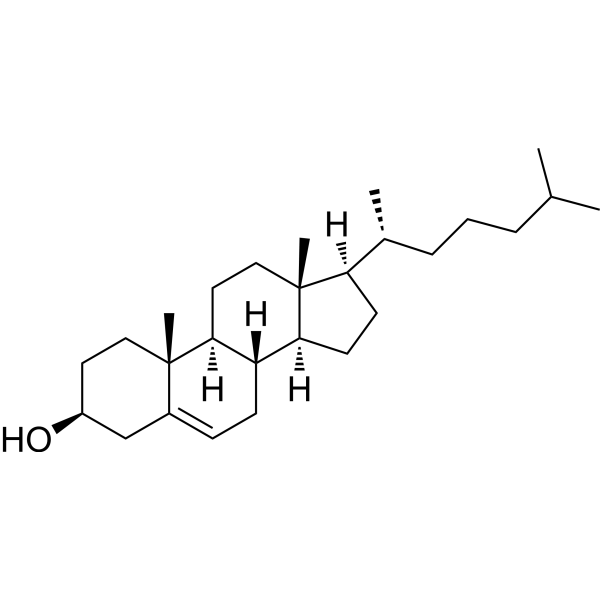
Cholesterol (from animal)ERRα Agonist -
HY-113369
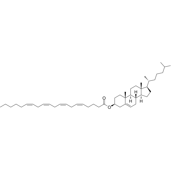
Cholesteryl arachidonateEndogenous Metabolite -
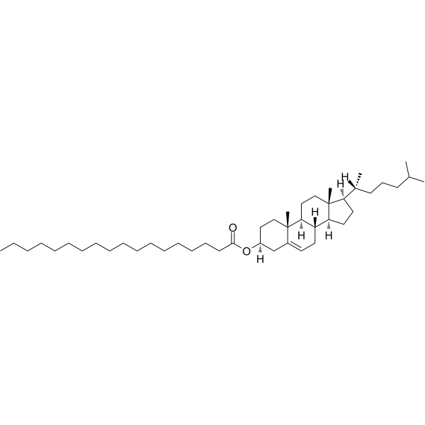
HY-W010695
Cholesteryl stearateBiochemical Assay Reagent -
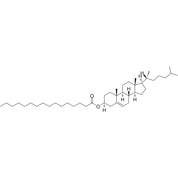
HY-W010708
Cholesteryl palmitate -
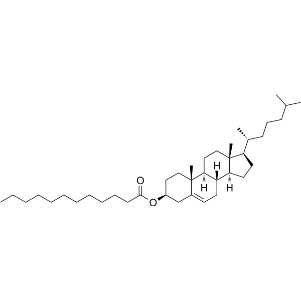
HY-W127400
Cholesteryl dodecanoateBiochemical Assay Reagent -
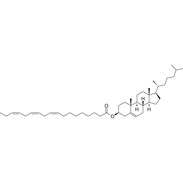
HY-125910
Cholesteryl LinolenateEndogenous Metabolite -
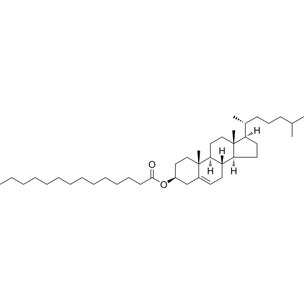
HY-N2338
Cholesterol myristateApoptosis Inhibitor -
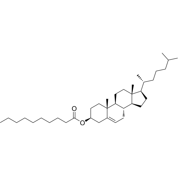
HY-W127351
Cholesterol decanoateBiochemical Assay Reagent -
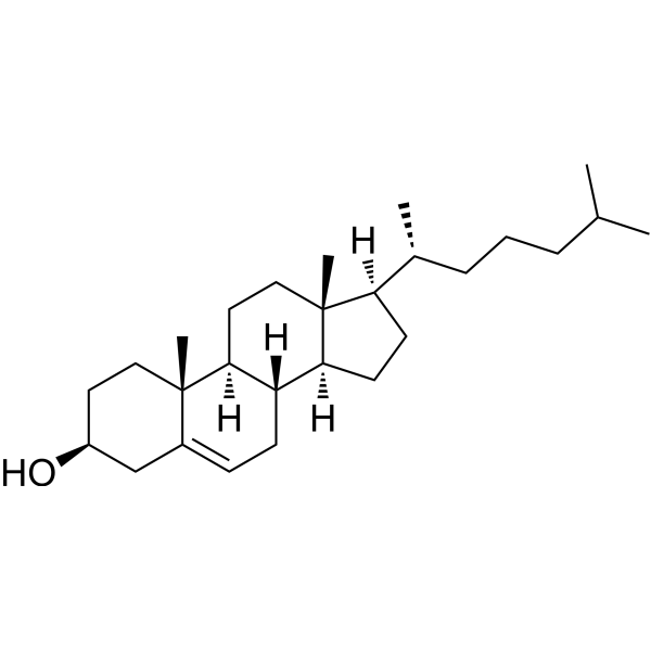
HY-N0322R
Cholesterol (Standard) -

HY-N0322E
Cholesterol (synthetic)ERRα Agonist -

HY-W010695R
Cholesteryl stearate (Standard)Biochemical Assay Reagent
All Endogenous Metabolite Isoforms
More

Biological Activity
Description
IC50 & Target
[1]|
Human Endogenous Metabolite |
In Vitro
Cholesteryl linoleate (5-40 μg/mL; 4 h) does not induce the adhesion of monocyte-like U937 cells, peripheral blood mononuclear cells, neutrophils, or neutrophil-like HL-60 cells to human umbilical vein endothelial cells[1].
Cholesteryl linoleate (4 h) does not upregulate the expression of E-selectin, ICAM-1, or VCAM-1 on the surface of human umbilical vein endothelial cells[1].
Cholesteryl linoleate in LDL is oxidized by J774A.1 cells expressing 12/15-LOX, primarily yielding 13S-hydroxycholesteryl linoleate, whereas mock-transfected cells lacking 12/15-LOX activity show no such oxidative effect[2].
Cholesteryl linoleate is oxidized in LDL by 12/15-LOX-expressing J774A.1 cells, which requires LRP but not the LDL receptor[2].
Cholesteryl linoleate in LDL is selectively taken up by mouse peritoneal macrophages and 12/15-LOX-expressing J774A.1 cells via LRP-mediated uptake[2].
Cholesteryl linoleate is a major cholesteryl ester present in normal human nasal fluid, with an average concentration in the low μg/mL range[3].
Cholesteryl linoleate (2-64 μg/mL; 16 h) liposomes inhibit the growth of Pseudomonas aeruginosa in a dose-dependent manner[3].
Cholesteryl linoleate (64 μg/mL; 3 h) liposomes exhibit bactericidal activity against Pseudomonas aeruginosa[3].
Cholesteryl linoleate (64 μg/mL; 16 h) liposomes inhibit the growth of Staphylococcus epidermidis, Enterococcus faecalis, and Enterobacter cloacae[3].
MedChemExpress (MCE) has not independently confirmed the accuracy of these methods. They are for reference only. Further protocols information, click here.
-
Cell Line:human umbilical vein endothelial cells (HUVECs)
-
Concentration:5, 10, 20 and 40 μg/mL
-
Incubation Time:4 h
-
Result:Did not induce the expression of E-selectin, ICAM-1, or VCAM-1 on the surface of HUVECs at concentrations effective for oxidized cholesteryl linoleate to induce monocyte-endothelial interactions.


Chemical Information
-
CAS No. 604-33-1
-
Appearance Solid
-
Molecular Weight 649.10
-
Formula C45H76O2
-
Color White to off-white
-
SMILES
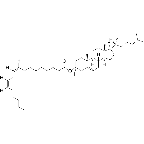
CC(C)CCC[C@@H](C)[C@H]1CC[C@@]2([H])[C@]3([H])CC=C4C[C@@H](OC(CCCCCCC/C=C\C/C=C\CCCCC)=O)CC[C@]4(C)[C@@]3([H])CC[C@]12C
-
Structure Classification
-
Initial Source
-
Shipping
Room temperature in continental US; may vary elsewhere.
-
Storage
Powder -20°C 3 years In solvent -80°C 6 months -20°C 1 month


Solvent & Solubility
In Vitro:
Ethanol : 2.5 mg/mL (3.85 mM; ultrasonic and warming and heat to 60°C)
Please refer to the solubility information to select the appropriate solvent. Once prepared, please aliquot and store the solution to prevent product inactivation from repeated freeze-thaw cycles.
Storage method and period of stock solution: -80°C, 6 months; -20°C, 1 month. When stored at -80°C, please use it within 6 months. When stored at -20°C, please use it within 1 month.
Please refer to the solubility information to select the appropriate solvent. Once prepared, please aliquot and store the solution to prevent product inactivation from repeated freeze-thaw cycles.
Storage method and period of stock solution: -80°C, 6 months; -20°C, 1 month. When stored at -80°C, please use it within 6 months. When stored at -20°C, please use it within 1 month.
Concentration (start) × Volume (start) = Concentration (final) × Volume (final)
In Vivo:
Select the appropriate dissolution method based on your experimental animal and administration route.
- For the following dissolution methods, please ensure to first prepare a clear stock solution using an In Vitro approach and then sequentially add co-solvents:
- To ensure reliable experimental results, the clarified stock solution can be appropriately stored based on storage conditions. As for the working solution for In Vivo experiments, it is recommended to prepare freshly and use it on the same day.
- The percentages shown for the solvents indicate their volumetric ratio in the final prepared solution. If precipitation or phase separation occurs during preparation, heat and/or sonication can be used to aid dissolution.
Add each solvent one by one: 10% DMSO 40% PEG300 5% Tween-80 45% Saline
Solubility: ≥ 2.5 mg/mL (3.85 mM); Clear solution
This protocol yields a clear solution of ≥ 2.5 mg/mL (saturation unknown).
Taking 1 mL working solution as an example, add 100 μL DMSO stock solution (25.0 mg/mL) to 400 μL PEG300, and mix evenly; then add 50 μL Tween-80 and mix evenly; then add 450 μL Saline to adjust the volume to 1 mL.
Preparation of Saline: Dissolve 0.9 g sodium chloride in ddH₂O and dilute to 100 mL to obtain a clear Saline solution.
Add each solvent one by one: 10% DMSO 90% (20% SBE-β-CD in Saline)
Solubility: ≥ 2.5 mg/mL (3.85 mM); Clear solution
This protocol yields a clear solution of ≥ 2.5 mg/mL (saturation unknown).
Taking 1 mL working solution as an example, add 100 μL DMSO stock solution (25.0 mg/mL) to 900 μL 20% SBE-β-CD in Saline, and mix evenly.
Preparation of 20% SBE-β-CD in Saline (4°C, storage for one week): 2 g SBE-β-CD powder is dissolved in 10 mL Saline, completely dissolve until clear.
In Vivo Dissolution Calculator
Please enter the basic information of animal experiments:
-
-
-
-
Recommended: Prepare an additional quantity of animals to account for potential losses during experiments.
Working solution concentration: 0.22 mg/mL

Protocols
-
Research Protocol for Infectious Diseases
Infectious-disease experiments test how pathogens interact with host barriers, innate immune receptors, inflammatory signaling, pathogen replication, and tissue injury; pattern-recognition receptors such as TLRs, RIG-I-like receptors, NOD-like receptors, and inflammasomes detect microbial molecules and activate NF-κB, interferon, and cytokine responses. The central hypothesis is that infection severity reflects the balance between pathogen burden and host response: protective inflammation restricts pathogen growth, whereas excessive or mislocalized inflammation contributes to tissue damage and disease phenotype. Unresolved questions include which host pathways are protective versus pathogenic, why some infection models fail to translate to human disease, and which combined readouts best predict clinically relevant infection outcomes.
-
RNA extraction experimental
By lysing cells, releasing RNA, and removing impurities such as proteins and DNA, high-purity RNA products are finally obtained. The commonly used traditional method is the guanidine isothiocyanate/phenol/chloroform method (Trizol), which is suitable for a variety of animal materials including animal tissues, microorganisms, cultured cells, etc., and most plant materials.
-
Endothelial Tube Formation Assay
Endothelial tube formation assay evaluates the ability of endothelial cells to attach, migrate, align, and organize into capillary-like networks when cultured on gelled basement membrane extract or Matrigel; the readout is the morphology and quantity of tube-like networks, which reflects an in vitro endothelial morphogenesis step related to angiogenesis. Basement membrane extract/Matrigel provides laminin-rich extracellular matrix cues that support endothelial differentiation into capillary-like structures, but it can contain biologically active growth factors, so growth-factor-reduced matrix is preferred when testing defined angiogenic stimulators or inhibitors.
-
Research Protocol for Cardiovascular Diseases
Cardiovascular disease can be modeled as maladaptive cardiac remodeling, where ischemic injury or pressure overload activates inflammatory signaling, fibroblast activation, extracellular-matrix deposition, cardiomyocyte hypertrophy, vascular remodeling, and progressive ventricular dysfunction. The TGF-β/SMAD axis is a central profibrotic pathway after myocardial injury and pressure overload, while innate immune and cytokine pathways regulate leukocyte recruitment, scar formation, and adverse remodeling. Key unresolved questions include which inflammatory signals are reparative versus harmful, when fibrosis is protective versus maladaptive, and whether pathway inhibition improves function without weakening necessary infarct healing or compensatory remodeling.
-
Bacterial live/dead nucleic-acid viability staining
The LIVE/DEAD bacterial viability staining method is based on differential permeability of nucleic-acid-binding fluorescent dyes, most commonly SYTO 9 and propidium iodide (PI), which enables discrimination of bacterial populations with intact versus compromised cytoplasmic membranes. SYTO 9 penetrates both intact and damaged bacterial membranes and binds nucleic acids to produce green fluorescence, whereas propidium iodide penetrates only cells with compromised membranes and fluoresces red while also reducing SYTO 9 signal through competitive binding and fluorescence interactions. The resulting fluorescence pattern is interpreted as a proxy for membrane integrity, which is widely used as an indicator of bacterial viability in microscopy, flow cytometry, and spectroscopic platforms. However, mechanistic studies show that SYTO 9 and PI interactions involve displacement and fluorescence resonance energy transfer effects, which can influence signal interpretation depending on dye ratios a


Purity & Documentation
-
Data Sheet (279 KB)
-
SDS (252 KB)
- English - EN (252 KB)
- Français - FR (252 KB)
- Deutsch - DE (252 KB)
- Norwegian - NO (252 KB)
- Español - ES (252 KB)
- Swedish - SV (252 KB)
- Italian - IT (252 KB)
- Korean - KR (252 KB)
- Portuguese - PT (252 KB)
-
Handling Instructions (2659 KB)
References
[1]. Huber J, et al. Oxidized cholesteryl linoleates stimulate endothelial cells to bind monocytes via the extracellular signal-regulated kinase 1/2 pathway. Arteriosclerosis, thrombosis, and vascular biology. 2002 Apr 01;22(4):581-6. [Content Brief]
[3]. Do TQ, et al. Lipids including cholesteryl linoleate and cholesteryl arachidonate contribute to the inherent antibacterial activity of human nasal fluid. J Immunol. 2008 Sep 15;181(6):4177-87. [Content Brief]


Complete Stock Solution Preparation Table
Please refer to the solubility information to select the appropriate solvent. Once prepared, please aliquot and store the solution to prevent product inactivation from repeated freeze-thaw cycles.
Storage method and period of stock solution: -80°C, 6 months; -20°C, 1 month. When stored at -80°C, please use it within 6 months. When stored at -20°C, please use it within 1 month.
| Optional Solvent | Concentration Solvent Mass | 1 mg | 5 mg | 10 mg | 25 mg |
|---|---|---|---|---|---|
| Ethanol | 1 mM | 1.5406 mL | 7.7030 mL | 15.4059 mL | 38.5149 mL |


Cholesteryl linoleate Related Classifications
HY-W010697 Related Classifications
 Powered by Bioz
Keywords
- Cholesteryl linoleate
- 604-33-1
- Endogenous Metabolite
- Bacterial
- human umbilical vein endothelial cells
- 12/15-lipoxygenase
- U937 cells
- macrophages
- respiratory epithelial secretions
- Pseudomonas aeruginosa
- atherosclerosis
- low-density lipoprotein particles
- LDL receptor-related protein
- human nasal fluid
- Inhibitor
- inhibitor
- inhibit