



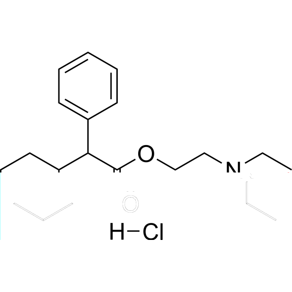
Drofenine hydrochloride
Based on 1 publication(s) in Google Scholar
Drofenine (Cycloadiphene; Hexahydroadiphenine) hydrochloride is an brain-penetrant antispasmodic agent. Drofenine hydrochloride is a Kv2.1 channel inhibitor with human IC50 of 9.53 μM. Drofenine hydrochloride is a butyrylcholinesterase (BChE) inhibitor with Ki of 0.003 mM, and is a TRPV3 activator. Drofenine hydrochloride blocks Kv2.1-dependent potassium efflux, inhibits Kv2.1/JNK/NF-κB and IkBa/NF-kB signaling, suppresses Kv2.1 mRNA/protein expression. Drofenine suppresses oligomeric Aβ-induced microglial NLRP3 inflammasome activation and neuronal Tau hyperphosphorylation, improves cognitive impairment, promotes neurite outgrowth. Drofenine hydrochloride induces calcium influx in keratinocytes and exert cytotoxicity against keratinocytes. Drofenine hydrochloride ameliorates diabetic peripheral neuropathy -like pathology. Drofenine hydrochloride can be used for the researches of Alzheimer's disease, diabetic peripheral neuropathy and smooth muscle spasm.
For research use only. We do not sell to patients.
- Purity : 98.0%
- CAS No.: 548-66-3
- Formula: C20H32ClNO2
- Molecular Weight:353.93
-
Storage:
4°C, sealed storage, away from moisture
* In solvent : -80°C, 6 months; -20°C, 1 month (sealed storage, away from moisture)

To place orders, for customer services and technical support, please contact: MedChemExpress USA
Tel: 609-228-6898 E-mail: [email protected] [email protected]


-
 Biological Activity
-
Chemical Information
-
Solvent & Solubility
- Protocols
- Purity & Documentation
- References
-
 Help & FAQs


-
HY-14566
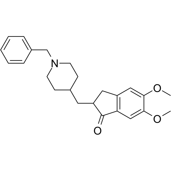
DonepezilSpecific AChE Inhibitor -
HY-B0034
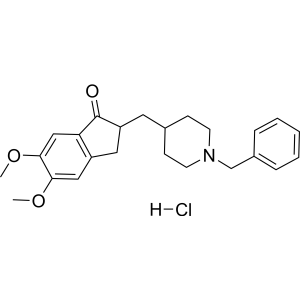
Donepezil HydrochlorideAChE Inhibitor -
HY-76299
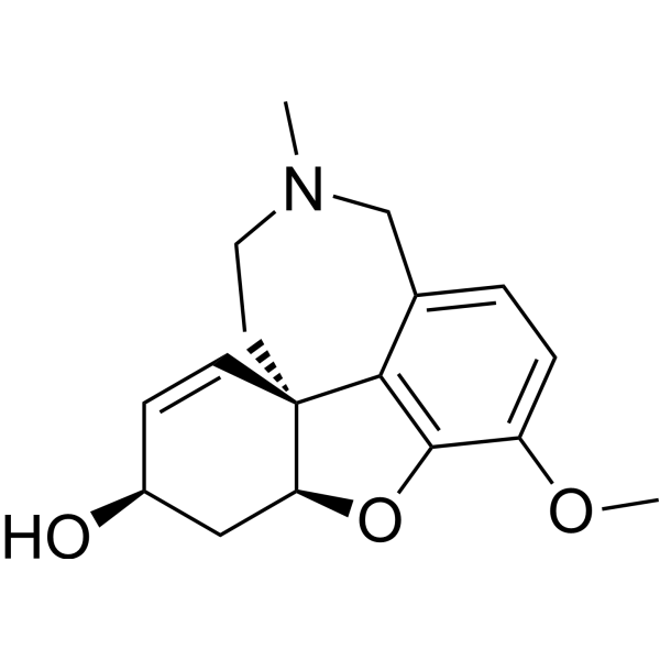
GalanthamineCholinesterase (ChE) Inhibitor -
HY-17368
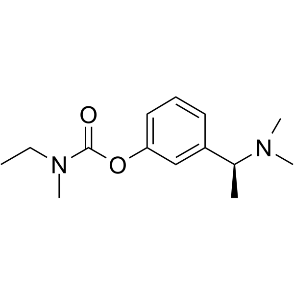
RivastigmineCholinesterase Inhibitor -
HY-11017
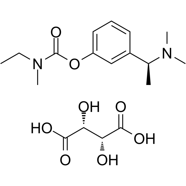
Rivastigmine tartrateCholinesterase Inhibitor -

HY-76299R
Galanthamine (Standard)Cholinesterase (ChE) Inhibitor -
HY-B2244
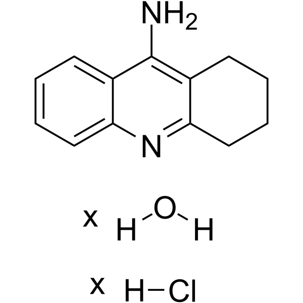
Tacrine hydrochloride hydrateAChE/BChE Inhibitor -

HY-17368R
Rivastigmine (Standard)Cholinesterase Inhibitor -
HY-B0882
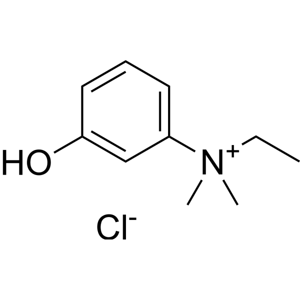
Edrophonium chlorideAChE Inhibitor -
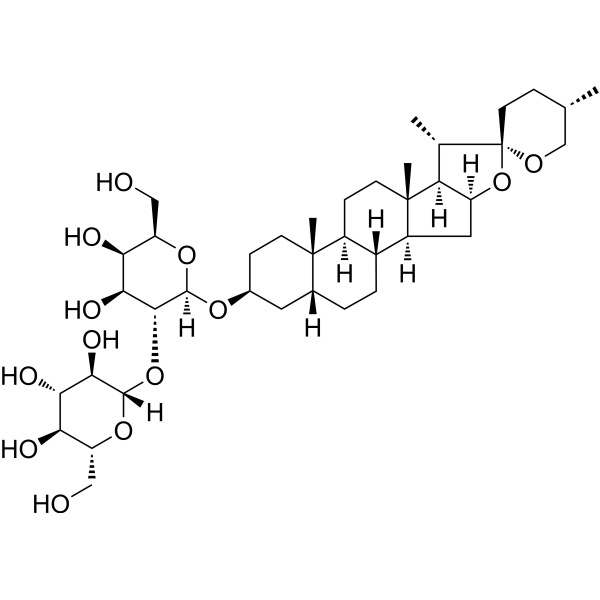
HY-N0810
Timosaponin AIIIAChE Inhibitor -
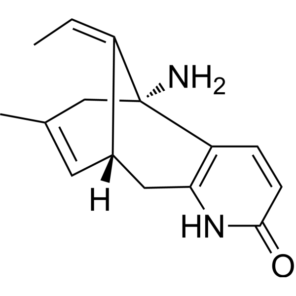
HY-17387
(-)-Huperzine AAChE/NMDA Receptor Inhibitor
Publications Citing Use of MedChemExpress (MCE) Drofenine hydrochloride
More

Biological Activity
Description
IC50 & Target
[1]|
BChE 3 μM () |
KV2.1 9.53 μM () |
TRPV3 |
NLRP3 |
In Vitro
Drofenine (10 μM; 0.5 h) hydrochloride suppresses oligomeric Aβ (o-Aβ)-enhanced Kv2.1-dependent outward potassium current and density in CHO-Kv2.1 cells by blocking o-Aβ-induced delays in Kv2.1 channel inactivation[1].
Drofenine (5-20 μM; 30 min) hydrochloride inhibits membrane potential in CHO-Kv2.1 cells without affecting normal CHO cells[2].
Drofenine (5-20 μM) hydrochloride does not reduce the viability of CHO-Kv2.1 cells[2].
Drofenine (1-10 μM) hydrochloride potently inhibits Kv2.1 channel currents in CHO-Kv2.1 cells with an IC50 of 9.53 μM[2].
Drofenine (1-10 μM) hydrochloride shows minimal inhibition of Kv2.2 channel currents in CHO-Kv2.2 cells at concentrations that effectively inhibit Kv2.1, indicating selectivity for Kv2.1[2].
Drofenine (0.01-0.1 mM) hydrochloride acts as a pure competitive inhibitor of purified human serum butyrylcholinesterase, with a Ki value of 0.003 mM[3].
Drofenine (30-1000 μM) hydrochloride acts as a selective agonist of human TRPV3 in TRPV3-overexpressing HEK-293 cells, activating calcium flux with an EC50 of 207 μM without activating TRPA1, TRPM8, TRPV1, TRPV2, or TRPV4 at concentrations up to 1000 μM[4].
Drofenine (30-1000 μM) hydrochloride acts as a potent agonist of endogenous TRPV3 in HaCaT human keratinocytes, inducing calcium flux with an EC50 of 605 μM[4].
Drofenine (10-1000 μM; 24 h) hydrochloride is cytotoxic to HaCaT human keratinocytes after 24 h of incubation, with an LC50 of 230 μM[4].
MedChemExpress (MCE) has not independently confirmed the accuracy of these methods. They are for reference only. Further protocols information, click here.
-
Cell Line:human immortalized keratinocyte (HaCaT) cells
-
Concentration:10-1000 μM
-
Incubation Time:24 h
-
Result:Was more cytotoxic to HaCaT cells than 2-APB and carvacrol, with an LC50 of 230 μM, compared to LC50 values of >300 μmol/L for both 2-APB (HY-W009724) and carvacrol (HY-N0711).
In Vivo
Drofenine (10-20 mg/kg; 4 total doses; Weeks 7, 8, 9, 10 post-STZ injection) hydrochloride suppresses Kv2.1 expression and function in DRG tissue, promotes DRG neuron neurite outgrowth, and ameliorates motor and sensory deficits in STZ (HY-13753)-induced type 1 DPN mice via Kv2.1 inhibition[2].
Drofenine (10-20 mg/kg; 4 total doses; Weeks 1, 2, 3, 4 of study period) hydrochloride suppresses Kv2.1 expression and function in DRG tissue, promotes DRG neuron neurite outgrowth, and ameliorates motor and sensory deficits in spontaneous type 2 db/db DPN mice[2].
MedChemExpress (MCE) has not independently confirmed the accuracy of these methods. They are for reference only.
-
Animal Model:5xFAD transgenic (APPSwFlLon, PSEN1-M146L-L286V) (female, 7 months old at treatment start, Alzheimer's disease model); wild-type (WT) (female)[1]
-
Dosage:10 mg/kg/day
-
Administration:i.p.; daily; 8 weeks
-
Result:Improved short-term working memory via increased discrimination index in novel object recognition test.
Improved spatial working memory via increased number of novel arm crossings in Y-maze test.
Improved spatial learning and long-term memory via reduced escape latency in Morris water maze training trials and increased platform crossing frequency in Morris water maze probe trials in 5xFAD mice; no cognitive improvement observed in Kv2.1-knockdown 5xFAD mice.
Reduced Iba1-positive microglial and GFAP-positive astrocytic gliosis in 5xFAD mouse brains.
Reduced NLRP3-ASC positive puncta, and reduced protein levels of NLRP3, p-NLRP3, ASC, Cas1 (p20), IL-1β, p-JNK, and p-NF-κB in 5xFAD mice.
Reduced microglial NF-κB nuclear translocation in 5xFAD mice; no anti-inflammatory effects observed in Kv2.1-knockdown 5xFAD mice.
Reduced AT8-positive hyperphosphorylated Tau (Ser202/Thr205) areas in the hippocampus of 5xFAD mice.
Reduced protein levels of p396-Tau, p231-Tau, p199-Tau, p-GSK3β, and p-CaMKII-α in 5xFAD mice; no effects on Tau phosphorylation or related kinases observed in Kv2.1-knockdown 5xFAD mice.
Improved long-term potentiation induction and maintenance in the hippocampal DG region of 5xFAD mice.
Restored protein levels of synaptic markers PSD95, synaptophysin (SYN), and VAMP2 in 5xFAD mice; no synaptic improvement observed in Kv2.1-knockdown 5xFAD mice.
Reduced TUNEL-positive apoptotic neurons in 5xFAD mice.
Reversed loosely arranged neuronal nuclei (Nissl staining) in the hippocampus of 5xFAD mice; no effects on neuronal survival observed in Kv2.1-knockdown 5xFAD mice.
Showed no significant hepatotoxicity or nephrotoxicity via no changes in serum ALT, AST, or urea levels in treated WT or 5xFAD mice.
-
Animal Model:C57BL/6 (male, 7 weeks old at study initiation, streptozotocin-induced type 1 diabetes)[2]
-
Dosage:10 mg/kg; 20 mg/kg
-
Administration:4 total doses; Weeks 7, 8, 9, 10 post-STZ injection
-
Result:Lowered Kv2.1/T-ERK ratios relative to vehicle-treated STZ mice.
Decreased relative Kv2.1 area in DRG tissue compared to vehicle-treated STZ mice.
Lowered relative Kv2.1 mRNA levels compared to vehicle-treated STZ mice.
Reduced elevated potassium currents in STZ mouse DRG neurons.
Increased total neurite length relative to vehicle-treated STZ mice.
Improved motor nerve conduction velocity relative to vehicle-treated STZ mice.
Reduced sensitivity to mechanical stimuli relative to vehicle-treated STZ mice.
Normalized thermal response relative to vehicle-treated STZ mice.
-
Animal Model:BKS Cg-m+/+Leprdb/J (db/db) (male, 17 weeks old at study initiation, spontaneous type 2 diabetes)[2]
-
Dosage:10 mg/kg; 20 mg/kg
-
Administration:4 total doses; Weeks 1, 2, 3, 4 of study period
-
Result:Lowered Kv2.1/T-ERK ratios relative to vehicle-treated db/db mice.
Decreased relative Kv2.1 area in DRG tissue compared to vehicle-treated db/db mice.
Lowered relative Kv2.1 mRNA levels compared to vehicle-treated db/db mice.
Reduced elevated potassium currents at +50 mV in db/db mouse DRG neurons.
Increased total neurite length relative to vehicle-treated db/db mice.
Improved motor nerve conduction velocity relative to vehicle-treated db/db mice.
Reduced sensitivity to mechanical stimuli relative to vehicle-treated db/db mice.
Normalized thermal response relative to vehicle-treated db/db mice.


Chemical Information
-
CAS No. 548-66-3
-
Appearance Solid
-
Molecular Weight 353.93
-
Formula C20H32ClNO2
-
Color White to off-white
-
SMILES
O=C(OCCN(CC)CC)C(C1CCCCC1)C2=CC=CC=C2.[H]Cl
-
Synonyms
Hexahydroadiphenine hydrochloride
-
Shipping
Room temperature in continental US; may vary elsewhere.
-
Storage
4°C, sealed storage, away from moisture
* In solvent : -80°C, 6 months; -20°C, 1 month (sealed storage, away from moisture)


Publications (1)
-
Journal Impact Factor
-
Most Recent
-
Biochem Biophys Res Commun
Inhibition of the transient receptor potential vanilloid 3 channel attenuates carbon tetrachloride-induced hepatic fibrosis. [Abstract]2021 Jun 18:558:86-93. PMID: 33906111


Solvent & Solubility
In Vitro:
DMSO : ≥ 75 mg/mL (211.91 mM; Hygroscopic DMSO has a significant impact on the solubility of product, please use newly opened DMSO)
H2O : ≥ 33.3 mg/mL (94.09 mM)
* "≥" means soluble, but saturation unknown.
Please refer to the solubility information to select the appropriate solvent. Once prepared, please aliquot and store the solution to prevent product inactivation from repeated freeze-thaw cycles.
Storage method and period of stock solution: -80°C, 6 months; -20°C, 1 month (sealed storage, away from moisture). When stored at -80°C, please use it within 6 months. When stored at -20°C, please use it within 1 month.
* Note: If you choose water as the stock solution, please dilute it to the working solution, then filter and sterilize it with a 0.22 μm filter before use.
Please refer to the solubility information to select the appropriate solvent. Once prepared, please aliquot and store the solution to prevent product inactivation from repeated freeze-thaw cycles.
Storage method and period of stock solution: -80°C, 6 months; -20°C, 1 month (sealed storage, away from moisture). When stored at -80°C, please use it within 6 months. When stored at -20°C, please use it within 1 month.
* Note: If you choose water as the stock solution, please dilute it to the working solution, then filter and sterilize it with a 0.22 μm filter before use.
Concentration (start) × Volume (start) = Concentration (final) × Volume (final)
In Vivo:
Select the appropriate dissolution method based on your experimental animal and administration route.
- For the following dissolution methods, please ensure to first prepare a clear stock solution using an In Vitro approach and then sequentially add co-solvents:
- To ensure reliable experimental results, the clarified stock solution can be appropriately stored based on storage conditions. As for the working solution for In Vivo experiments, it is recommended to prepare freshly and use it on the same day.
- The percentages shown for the solvents indicate their volumetric ratio in the final prepared solution. If precipitation or phase separation occurs during preparation, heat and/or sonication can be used to aid dissolution.
Add each solvent one by one: 10% DMSO 40% PEG300 5% Tween-80 45% Saline
Solubility: ≥ 2.5 mg/mL (7.06 mM); Clear solution
This protocol yields a clear solution of ≥ 2.5 mg/mL (saturation unknown).
Taking 1 mL working solution as an example, add 100 μL DMSO stock solution (25.0 mg/mL) to 400 μL PEG300, and mix evenly; then add 50 μL Tween-80 and mix evenly; then add 450 μL Saline to adjust the volume to 1 mL.
Preparation of Saline: Dissolve 0.9 g sodium chloride in ddH₂O and dilute to 100 mL to obtain a clear Saline solution.
Add each solvent one by one: 10% DMSO 90% (20% SBE-β-CD in Saline)
Solubility: ≥ 2.5 mg/mL (7.06 mM); Clear solution
This protocol yields a clear solution of ≥ 2.5 mg/mL (saturation unknown).
Taking 1 mL working solution as an example, add 100 μL DMSO stock solution (25.0 mg/mL) to 900 μL 20% SBE-β-CD in Saline, and mix evenly.
Preparation of 20% SBE-β-CD in Saline (4°C, storage for one week): 2 g SBE-β-CD powder is dissolved in 10 mL Saline, completely dissolve until clear.
For the following dissolution methods, please prepare the working solution directly:
It is recommended to prepare fresh solutions and use them promptly within a short period of time.
The percentages shown for the solvents indicate their volumetric ratio in the final prepared solution. If precipitation or phase separation occurs during preparation, heat and/or sonication can be used to aid dissolution.
Add each solvent one by one: PBS
Solubility: 16.67 mg/mL (47.10 mM); Clear solution; Need ultrasonic
In Vivo Dissolution Calculator
Please enter the basic information of animal experiments:
-
-
-
-
Recommended: Prepare an additional quantity of animals to account for potential losses during experiments.
Working solution concentration: 0.22 mg/mL
This product has good water solubility, please refer to the measured solubility data in water/PBS/Saline for details.

Protocols
-
RNA extraction experimental
By lysing cells, releasing RNA, and removing impurities such as proteins and DNA, high-purity RNA products are finally obtained. The commonly used traditional method is the guanidine isothiocyanate/phenol/chloroform method (Trizol), which is suitable for a variety of animal materials including animal tissues, microorganisms, cultured cells, etc., and most plant materials.
-
Western Blot
Western blotting (WB) is a commonly used experimental method in molecular biology, biochemistry, and immunogenetics for identifying and quantifying target proteins. It combines gel electrophoresis with immunoassay, enabling researchers to analyze protein expression, post-translational modifications, and molecular weight.
-
Mammalian live/dead viability and cytotoxicity staining
Live/dead viability and cytotoxicity staining assays are based on the simultaneous detection of intracellular esterase activity in metabolically active (viable) cells and membrane integrity loss in non-viable cells. In commonly used dual-staining approaches, membrane-permeant fluorogenic substrates are converted by intracellular esterases into fluorescent products in live cells, while impermeant DNA-binding dyes selectively enter cells with compromised plasma membranes and label nucleic acids in dead or dying cells, enabling discrimination between viable and non-viable populations by fluorescence microscopy or flow cytometry.
-
Cell differentiation
Cell differentiation refers to the process in which cells of the same origin gradually produce cell groups with different morphological structure and functional characteristics.
-
Research Protocol for Inflammation-related Diseases
The NLRP3 inflammasome is a cytosolic innate immune signaling platform that integrates priming signals and danger-signal activation to promote caspase-1 activation, maturation of IL-1β and IL-18, and gasdermin D-mediated pyroptotic cell death. The core experimental logic is to determine whether inflammatory disease phenotypes are driven by increased NLRP3 expression, ASC-containing inflammasome assembly, caspase-1 cleavage, GSDMD cleavage, and extracellular release of IL-1β/IL-18 rather than by nonspecific cell injury alone. The pathway is strongly linked to inflammation-related disease phenotypes because monosodium urate crystals activate NALP3/NLRP3 inflammasome signaling in gout-like crystal inflammation, cholesterol crystals activate NLRP3 inflammasomes in atherogenesis models, and DSS-induced intestinal inflammation has been reported to involve NLRP3 inflammasome activity. However, experimental colitis studies also show context-dependent protective effects of NLRP3 inflammasome co
-
PC12 NGF-induced neuronal-like differentiation
PC12 cells are a rat adrenal pheochromocytoma-derived clonal cell line that responds to nerve growth factor by stopping proliferation and extending neurites, producing a sympathetic neuron-like phenotype used to study neuronal differentiation and neurite outgrowth. NGF acts through TrkA-dependent signaling, and neurite outgrowth is associated with ERK/Akt signaling, microtubule organization, neuronal-marker expression, and increased electrophysiological neuronal features such as sodium-channel density. The main assay readout is morphological differentiation, usually measured as the percentage of neurite-bearing cells, neurite length, neurite number, or total neurite length per cell. Additional readouts include GAP-43, tyrosine hydroxylase, βIII-tubulin, neurofilament, synapsin I, synaptophysin, ERK phosphorylation, Akt phosphorylation, and sodium-channel current density.
-
Ca2+ Staining Technique
Ca2+ staining is an experimental technique that utilizes specific fluorescent probes (such as Fluo-4 AM, Fura-2, etc.) to qualitatively or quantitatively detect dynamic changes in intracellular Ca2+ concentrations; this is achieved by monitoring the changes in fluorescent signals generated when these probes bind to free intracellular calcium ions. The underlying principle relies primarily on the presence of chelating groups within the probe's molecular structure that possess high affinity for calcium ions.
-
Protocol for Hematoxylin-Eosin (H&E) Staining
Hematoxylin-eosin staining is a routine histological method that stains nuclei mainly blue-purple with hemalum and stains cytoplasm, extracellular matrix, and many stromal components pink with eosin, allowing tissue architecture, cell morphology, necrosis, inflammation, fibrosis, tumor growth pattern, and treatment-associated injury to be evaluated by light microscopy. In cancer cells, primary neurons, mouse tumor models, intestinal organoids, inflammatory macrophage preparations, and drug-screening tissues, H&E is a morphology assay rather than a molecular assay; it should be interpreted with complementary molecular or immunostaining assays when the biological question concerns specific proteins, RNA levels, ferroptosis, mitophagy, or immune phenotypes.
-
Protocol For Protein Expression And Purification
Recombinant protein expression in Escherichia coli followed by purification of a His-tagged soluble protein by immobilized metal affinity chromatography (IMAC), with optional MBP fusion and TEV tag removal when the construct includes these elements. The biological readout is production of the encoded target protein, detected as an inducible band at the expected molecular mass by SDS-PAGE and quantified by total protein assay or chromatographic absorbance; the purification readout is enrichment of the target protein in elution fractions after selective binding of polyhistidine residues to immobilized Ni2+/metal-chelate resin and elution by imidazole-containing buffer. Expression is driven by an inducible bacterial expression system, commonly T7/lac-based, in which IPTG or lactose/auto-induction activates transcription and translation of the cloned gene; lower induction temperature, lower inducer concentration, induction timing, and solubility-enhancing fusion tags can influence the frac
-
Alzheimer’s Disease Modeling
Alzheimer’s Disease (AD) is a neurodegenerative disorder characterized by a progressive decline in cognitive functions and loss of specific types of neurons and synapses. Alzheimer's symptoms can be simulated in mice by injecting drugs (such as Aβ) or genetically modified.
-
Cell Viability Determination by MTT Colorimetric Assay
The following protocol uses the MTT colorimetric assay as a classic literature-established method for assessing cell viability/metabolic activity in cultured mammalian cells. MTT[3-(4,5-dimethylthiazol-2-yl)-2,5-diphenyltetrazolium bromide] is reduced by metabolically active cells to a colored formazan product; the amount of formazan is quantified spectrophotometrically and provides an indirect measure of metabolically active viable cells. Importantly, MTT reduction reflects cellular oxidoreductase/metabolic activity rather than an absolute direct count of living cells, so changes in cellular metabolism can alter the signal independently of cell number.
-
PC12 NGF-Induced Neuronal Differentiation Culture
PC12 cells are a rat adrenal pheochromocytoma clonal line that responds to NGF by stopping proliferation and extending branching neurite-like processes; after longer NGF exposure, cells develop long processes and neuronal-like ultrastructural and functional features. NGF-induced differentiation is read out mainly by neurite outgrowth, reduced proliferation, microtubule assembly, and neuronal differentiation-associated proteins such as MAPs, tau, GAP-43, and synapsin-1.
-
Cell Cytotoxicity Assay
Cytotoxicity assays are usually based on the assessment of cell membrane damage, which can also be indirectly detected by measuring cell viability. Detection methods include MTT assay, CKK-8 assay, LDH assay and ATP assay, etc.
-
SH-SY5Y Neuronal Differentiation Culture
SH-SY5Y neuronal differentiation culture uses sequential exposure to retinoic acid and neurotrophic factors to reduce proliferative neuroblastoma-like behavior and induce neuron-like morphology, including neurite extension, neuronal marker expression, and, in RA/BDNF protocols, greater synaptic-marker expression than undifferentiated culture. Retinoic acid is commonly used as the initiating differentiation cue, while BDNF in serum-reduced or serum-free medium supports later maturation and neurotrophic-factor-dependent neuron-like survival.
-
SH-SY5Y neuronal-like differentiation
SH-SY5Y neuronal-like differentiation uses defined culture conditions to shift proliferative human neuroblastoma cells toward a neuron-like state, mainly assessed by reduced proliferation, neurite extension, neuronal-marker expression, and, in some protocols, increased dependence on neurotrophic support. Retinoic acid (RA) is commonly used for the first differentiation phase, and sequential RA followed by brain-derived neurotrophic factor (BDNF) in serum-free medium is a well-characterized approach for generating neuron-like SH-SY5Y cultures with extensive neurite outgrowth. The primary readouts are morphology-based neurite outgrowth and marker-based confirmation using proteins such as βIII-tubulin, MAP2, GAP43, synaptophysin, NeuN, NSE, TH, or related neuronal/synaptic markers, depending on the study endpoint.
-
Pyroptosis Solutions
Pyroptosis is a lytic inflammatory cell-death pathway executed by gasdermin pores, most classically through inflammasome-mediated activation of caspase-1, cleavage of gasdermin D, membrane pore formation, LDH release, and secretion of IL-1β and IL-18. The canonical pathway is commonly modeled by priming cells with an inflammatory signal such as LPS to induce pro-IL-1β and inflammasome components, followed by an activation signal such as ATP or nigericin to activate NLRP3, ASC speck formation, caspase-1 cleavage, GSDMD cleavage, cytokine release, and pyroptotic membrane rupture. The non-canonical pathway is triggered when cytosolic LPS activates mouse caspase-11 or human caspase-4/5, leading to GSDMD cleavage and pyroptosis, and this can secondarily activate NLRP3-dependent IL-1β release. Pyroptosis is linked to inflammatory injury, infection, cancer, liver disease, ocular disease, placental inflammation, and other disease phenotypes, but unresolved questions include which gasdermin fam


Purity & Documentation
-
Data Sheet (286 KB)
-
SDS (761 KB)
- English - EN (761 KB)
- Français - FR (761 KB)
- Deutsch - DE (761 KB)
- Norwegian - NO (761 KB)
- Español - ES (761 KB)
- Swedish - SV (761 KB)
- Italian - IT (761 KB)
- Korean - KR (761 KB)
- Portuguese - PT (761 KB)
-
Handling Instructions (2659 KB)
References
[1]. Lu J, et al. Drofenine as a Kv2.1 inhibitor alleviated AD-like pathology in mice through Aβ/Kv2.1/microglial NLRP3/neuronal Tau axis. Acta Pharm Sin B. 2025;15(1):371-391. [Content Brief]
[2]. Xu X, et al. Antispasmodic Drug Drofenine as an Inhibitor of Kv2.1 Channel Ameliorates Peripheral Neuropathy in Diabetic Mice. iScience. 2020;23(10):101617. Published 2020 Sep 28. [Content Brief]
[3]. Bodur E, et al. Inhibition effects of benactyzine and drofenine on human serum butyrylcholinesterase. Arch Biochem Biophys. 2001 Feb 1;386(1):25-29. [Content Brief]
[4]. Deering‐Rice C E, et al. Drofenine: a 2‐APB analog with improved selectivity for human TRPV3[J]. Pharmacology research & perspectives, 2014, 2(5): e00062. [Content Brief]


Complete Stock Solution Preparation Table
Please refer to the solubility information to select the appropriate solvent. Once prepared, please aliquot and store the solution to prevent product inactivation from repeated freeze-thaw cycles.
Storage method and period of stock solution: -80°C, 6 months; -20°C, 1 month (sealed storage, away from moisture). When stored at -80°C, please use it within 6 months. When stored at -20°C, please use it within 1 month.
| Optional Solvent | Concentration Solvent Mass | 1 mg | 5 mg | 10 mg | 25 mg |
|---|---|---|---|---|---|
| H2O / DMSO | 1 mM | 2.8254 mL | 14.1271 mL | 28.2542 mL | 70.6354 mL |
| 5 mM | 0.5651 mL | 2.8254 mL | 5.6508 mL | 14.1271 mL | |
| 10 mM | 0.2825 mL | 1.4127 mL | 2.8254 mL | 7.0635 mL | |
| 15 mM | 0.1884 mL | 0.9418 mL | 1.8836 mL | 4.7090 mL | |
| 20 mM | 0.1413 mL | 0.7064 mL | 1.4127 mL | 3.5318 mL | |
| 25 mM | 0.1130 mL | 0.5651 mL | 1.1302 mL | 2.8254 mL | |
| 30 mM | 0.0942 mL | 0.4709 mL | 0.9418 mL | 2.3545 mL | |
| 40 mM | 0.0706 mL | 0.3532 mL | 0.7064 mL | 1.7659 mL | |
| 50 mM | 0.0565 mL | 0.2825 mL | 0.5651 mL | 1.4127 mL | |
| 60 mM | 0.0471 mL | 0.2355 mL | 0.4709 mL | 1.1773 mL | |
| 80 mM | 0.0353 mL | 0.1766 mL | 0.3532 mL | 0.8829 mL | |
| DMSO | 100 mM | 0.0283 mL | 0.1413 mL | 0.2825 mL | 0.7064 mL |
* Note: If you choose water as the stock solution, please dilute it to the working solution, then filter and sterilize it with a 0.22 μm filter before use.


Drofenine hydrochloride Related Classifications
HY-B1239 Related Classifications
 Powered by Bioz