



KHK-IN-1
Based on 1 publication(s) in Google Scholar
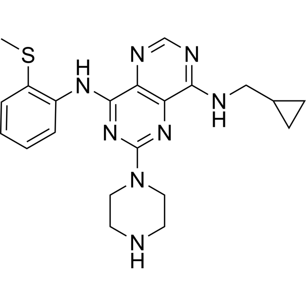
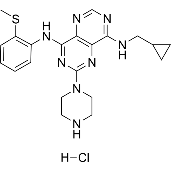
KHK-IN-1 (compound 8) is a selective and cell membrane permeable ketohexokinase (KHK) inhibitor (IC50=12 nM; F=34%). KHK-IN-1 inhibits the production of F1P in HepG2 cell lysates (IC50=400 nM). KHK-IN-1 has potential for the study of diabetes and obesity.
Para uso exclusivo en investigación. No vendemos a pacientes.
- Pureza : 99.00%
- No. CAS: 1303469-70-6
- Fòrmula: C21H26N8S
- Peso molecular:422.55
-
Almacenamiento:Powder -20°C, 3 years , 4°C, 2 years ; In solvent -80°C, 6 months , -20°C, 1 month

To place orders, for customer services and technical support, please contact: MedChemExpress USA
Tel: 609-228-6898 E-mail: [email protected] [email protected]


-
 Actividad biológica
-
Chemical Information
- Protocolo
- Pureza y Documentación
- Referencias
-
 Help & FAQs



Publications Citing Use of MedChemExpress (MCE) KHK-IN-1
More

Actividad biológica
Descripciòn
IC50 & Target
IC50=12 nM (KHK)[1].
Cellular Effect
|
Cell Line

|
Type | Value | Description | References |
|---|---|---|---|---|
| HepG2 | IC50 |
<500 nM
Compound: 1
|
Inhibition of KHKC in human HepG2 cells assessed as level of fructose-1-phosphate
Inhibition of KHKC in human HepG2 cells assessed as level of fructose-1-phosphate
|
[PMID: 22795331] |
| HepG2 | IC50 |
400 nM
Compound: 8
|
Inhibition of ketokinase isoform C in human HepG2 cells assessed as levels of fructose-1-phosphate preincubated for 30 mins followed by substrate addition measured after 3 hrs by LC-MS analysis
Inhibition of ketokinase isoform C in human HepG2 cells assessed as levels of fructose-1-phosphate preincubated for 30 mins followed by substrate addition measured after 3 hrs by LC-MS analysis
|
[PMID: 24900346] |
In Vitro
KHK-IN-1 stable in human and rat liver microsome preparations (88 and 72% remaining at 10 min) and do not significantly inhibit cytochrome P450s from human liver microsomes (1A2, 2C19, 2D6, 2C9, and 3A4)[1].
KHK-IN-1 (0-10 μM; incubate 30 min, then add to 15 mM fructose and incubate for another 3 h) inhibits production of F1P in HepG2 cell lysates with an IC50 value of 400 nM[1].
MedChemExpress (MCE) has not independently confirmed the accuracy of these methods. They are for reference only. Further protocols information, click here.
-
Cell Line:HepG2 cells
-
Concentration:0-10 µM
-
Incubation Time:Incubate 30 min, then add to 15 mM fructose and incubate for another 3 h
-
Result:Exhibited inhibition of F1P production in HepG2 cell lysates (IC50=400 nM).
In Vivo
MedChemExpress (MCE) has not independently confirmed the accuracy of these methods. They are for reference only.
-
Animal Model:Male Sprague-Dawley rats (~250 g)[1].
-
Dosage:10 mg/kg
-
Administration:Oral gavage; single
-
Result:Exhibited reasonable oral bioavailability in rats (F=34%; oral t1/2=4 h), but had a high volume of distribution (Vdss= 32 L/kg) and a high rate of clearance (CL=160 mL/min/kg).


Chemical Information
-
No. CAS 1303469-70-6
-
Appearance Solid
-
Peso molecular 422.55
-
Fòrmula C21H26N8S
-
Color Light yellow to yellow
-
SMILES
CSC(C=CC=C1)=C1NC2=NC(N3CCNCC3)=NC4=C2N=CN=C4NCC5CC5
-
Envío
Room temperature in continental US; may vary elsewhere.
-
Almacenamiento
Powder -20°C 3 years 4°C 2 years In solvent -80°C 6 months -20°C 1 month


Publications (1)
-
Journal Impact Factor
-
Most Recent
-
Exp Cell Res
Ketohexokinase-A deficiency attenuates the proliferation via reducing β-catenin in gastric cancer cells. [Abstract]2024 Apr 16;438(1):114038. PMID: 38614422

Protocolo
-
Research Protocol for Metabolic Diseases
AMP-activated protein kinase, AMPK, is a conserved cellular energy sensor that responds to reduced cellular energy status and coordinates metabolism by increasing ATP-generating catabolic pathways while suppressing ATP-consuming anabolic processes. In metabolic disease research, the AMPK pathway is experimentally relevant because it regulates hepatic lipid synthesis, fatty acid oxidation, glucose production, skeletal-muscle glucose disposal, mTORC1-linked biosynthesis, autophagy, mitochondrial homeostasis, and whole-body energy balance. The central pathway logic is that energy stress, metformin, exercise-like stimulation, or direct AMPK activators increase AMPKα Thr172 phosphorylation and downstream substrate phosphorylation, including ACC and RAPTOR. Phosphorylation of ACC suppresses lipogenesis and supports fatty acid oxidation, whereas phosphorylation of RAPTOR suppresses mTORC1 signaling and links cellular energy status to growth and protein synthesis control. The pathway is linked


Pureza y Documentación
-
Ficha de datos (275 KB)
-
SDS (393 KB)
- English - EN (393 KB)
- Français - FR (393 KB)
- Deutsch - DE (393 KB)
- Norwegian - NO (393 KB)
- Español - ES (393 KB)
- Swedish - SV (393 KB)
- Italian - IT (393 KB)
- Korean - KR (393 KB)
- Portuguese - PT (393 KB)
-
Instrucciones de manejo (2659 KB)
Referencias

Calculators
Concentration (start) × Volume (start) = Concentration (final) × Volume (final)


KHK-IN-1 Related Classifications
HY-12841 Related Classifications
 Powered by Bioz