



NGI-1
Based on 22 publication(s) in Google Scholar
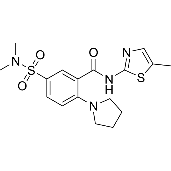
NGI-1 (ML414) is a potent oligosaccharyltransferase (OST) inhibitor, directly targeting and blocking the function of the OST catalytic subunits STT3A and STT3B. NGI-1 is a cell permeable inhibitor and can effectively reduce virus infectivity without affecting cell viability.
For research use only. We do not sell to patients.
- Purity : 99.97%
- CAS No.: 790702-57-7
- Formula: C17H22N4O3S2
- Molecular Weight:394.51
-
Storage:Powder -20°C, 3 years , 4°C, 2 years ; In solvent -80°C, 2 years , -20°C, 1 year

To place orders, for customer services and technical support, please contact: MedChemExpress USA
Tel: 609-228-6898 E-mail: [email protected] [email protected]


-
 Biological Activity
-
Chemical Information
-
Solvent & Solubility
- Protocols
- Purity & Documentation
- References
-
 Help & FAQs


-
HY-A0098
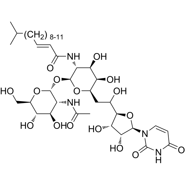
TunicamycinER Stress Inducer -
HY-106161
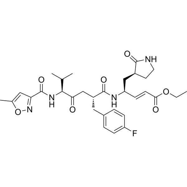
RupintrivirHRV 3C Protease Inhibitor -
HY-16704
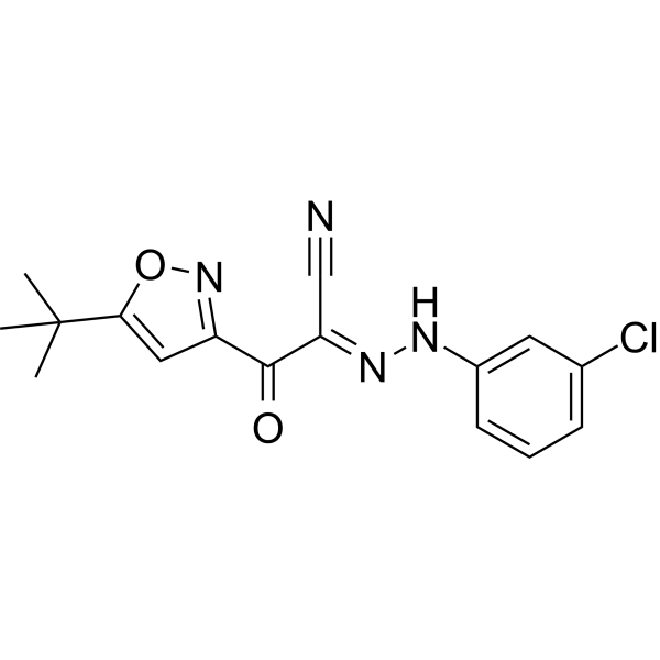
ESI-09Virus Protease Inhibitor -
HY-13966
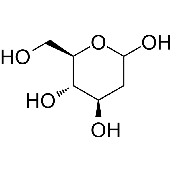
2-Deoxy-D-glucoseGlycolysis Inhibitor -
HY-Y0873
PEG300Neutral Polymer -
HY-129389
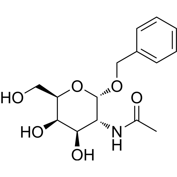
Benzyl-α-GalNAcO-Glycosylation Inhibitor -
HY-119738
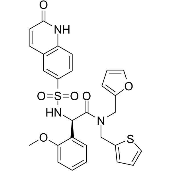
OSMI-1O-GlcNAc Transferase Inhibitor -
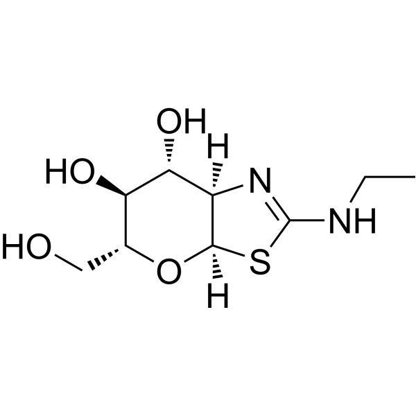
HY-12588
Thiamet GO-GlcNAcase Inhibitor -
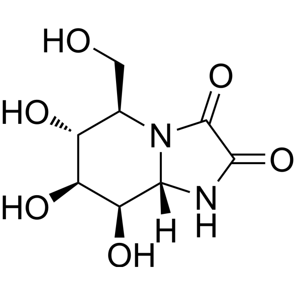
HY-19332
Kifunensineα-Mannosidases Inhibitor -
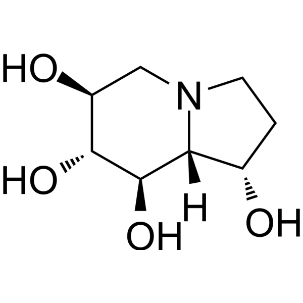
HY-N2022
Castanospermineα/β-Glucosidases Inhibitor -

HY-13259
MG-132Proteasome Inhibitor -

HY-15141
StaurosporinePKC/PKA Inhibitor
Publications Citing Use of MedChemExpress (MCE) NGI-1
More- Cancer Discov. 2020 Dec;10(12):1872-1893. [Abstract]
- Nat Metab. 2026 Jun 9;8(6):1410-1425.
- Nat Commun. 2025 Oct 1;16(1):8736. [Abstract]
- Nat Commun. 2025 Apr 10;16(1):3391. [Abstract]
- Cell Chem Biol. 2026 Jun 16:S2451-9456(26)00196-0. [Abstract]
- Cell Chem Biol. 2025 Oct 22:S2451-9456(25)00306-X. [Abstract]
- Int J Biol Macromol. 2025 Feb:289:138846. [Abstract]
- Mol Ther Oncol. 2025 Mar 8;33(2):200964. [Abstract]
- MAbs. 2025 Dec;17(1):2574406. [Abstract]
- Anal Chem. 2026 Mar 24;98(11):8143-8154. [Abstract]
- Sci Signal. 2026 Mar 3;19(927):eadz6443. [Abstract]
- Front Mol Biosci. 2022 Apr 27;9:899192. [Abstract]
- FEBS J. 2025 Jun 26. [Abstract]
- J Virol. 2025 Mar 18;99(3):e0001825. [Abstract]
- J Biol Chem. 2023 Oct;299(10):105211. [Abstract]
- J Virol. 2019 Nov 13;93(23):e01443-19. [Abstract]
- Biochem Biophys Res Commun. 2020 Nov 26;533(1):77-82. [Abstract]
- The State University of New Jersey. 2026 Jul.
- bioRxiv. 2026 Jan 26:2026.01.23.701155. [Abstract]
- bioRxiv. 2025 Aug 08.
- bioRxiv. 2025 Jul 26:2025.07.22.666143. [Abstract]
- SSRN. 2025 Apr 4.
 Customer Validation & Images

-
Flow Cytometry
-
WB
-
IP
-
Flow Cytometry
-
In Vivo Efficacy Study


Biological Activity
Description
IC50 & Target
OST[1]
Cellular Effect
|
Cell Line

|
Type | Value | Description | References |
|---|---|---|---|---|
| Hepatocyte | IC50 |
20 μM
Compound: 1
|
Cytotoxicity against human Hepatocyte cultured as 3D spheroids incubated for 14 days by CellTiter-Glo luminescence assay
Cytotoxicity against human Hepatocyte cultured as 3D spheroids incubated for 14 days by CellTiter-Glo luminescence assay
|
[PMID: 39136957] |
In Vitro
NGI-1 inhibits the glycosylation of LASV GP mediated by STT3A-OST (in STT3B- and MAGT1-TUSC3- cells) or STT3B-OST (in STT3A- cells) and impaires its proteolytic cleavage in a dose-dependent manner[1].
NGI-1 blocks EGFR N-linked glycosylation in lung adenocarcinoma cells as assessed. In controls EGFR is biotinylated, consistent with its plasma membrane expression, but in NGI-1 treated cells the EGFR is predominantly found in the non-biotinylated intracellular fraction suggesting a change in cellular localization[2].
MedChemExpress (MCE) has not independently confirmed the accuracy of these methods. They are for reference only.
-
Cell Line:STT3A-, STT3B- and MAGT1-TUSC3- cells
-
Concentration:1, 2, 5 μM
-
Incubation Time:36 hours
-
Result:Inhibited the glycosylation of LASV GP mediated by STT3A-OST (in STT3B- and MAGT1-TUSC3- cells) or STT3B-OST (in STT3A- cells) and impaired its proteolytic cleavage in a dose-dependent manner.


Chemical Information
-
CAS No. 790702-57-7
-
Appearance Solid
-
Molecular Weight 394.51
-
Formula C17H22N4O3S2
-
Color White to off-white
-
SMILES
O=C(NC1=NC=C(C)S1)C2=CC(S(=O)(N(C)C)=O)=CC=C2N3CCCC3
-
Synonyms
ML414
-
Shipping
Room temperature in continental US; may vary elsewhere.
-
Storage
Powder -20°C 3 years 4°C 2 years In solvent -80°C 2 years -20°C 1 year


Publications (22)
-
Journal Impact Factor
-
Most Recent
-
Cancer Discov
Pharmacologic Suppression of B7-H4 Glycosylation Restores Antitumor Immunity in Immune-Cold Breast Cancers. [Abstract]2020 Dec;10(12):1872-1893. PMID: 32938586
NGI-1 purchased from MedChemExpress. Usage Cited in: Cancer Discov. 2020 Dec;10(12):1872-1893. [Abstract]
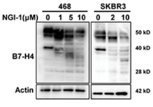
MDA-MB-468 and SKBR3 cells were treated with 10 μM OST inhibitor NGI-1 for 24 h. The expression of B7-H4 was examined by immunoblotting.
NGI-1 purchased from MedChemExpress. Usage Cited in: Cancer Discov. 2020 Dec;10(12):1872-1893. [Abstract]
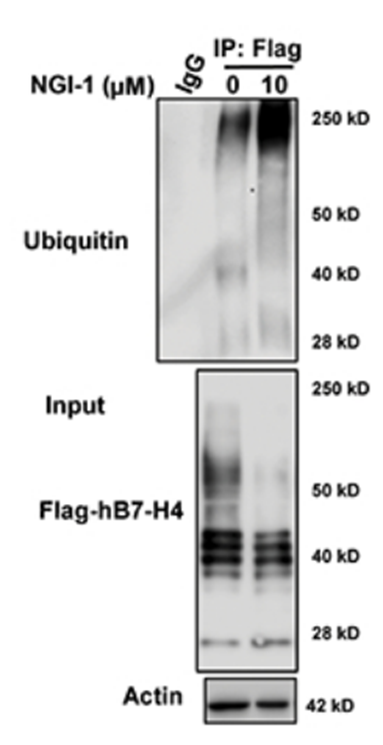
Blockade of B7-H4 glycosylation by NGI-1 enhances B7-H4 ubiquitination. 293T cells were transfected with Flag-hB7-H4 in the presence or absence of 10 μM NGI-1 for 24 h. Then Flag-hB7-H4 was immunoprecipitated followed by immunoblotting using antibody against ubiquitin.
NGI-1 purchased from MedChemExpress. Usage Cited in: Cancer Discov. 2020 Dec;10(12):1872-1893. [Abstract]
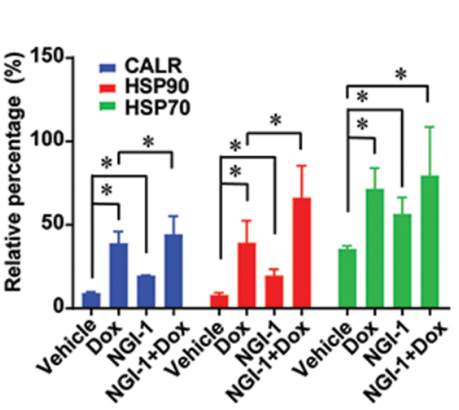
NGI-1 ( 10 μM, 24 h) significantly increased the DOX-induced expression of cell surface CALR and that of other DAMPs including HSP90 and HSP70 on the cell surface, as detectable by immunofluorescence and flow cytometry.
NGI-1 purchased from MedChemExpress. Usage Cited in: Cancer Discov. 2020 Dec;10(12):1872-1893. [Abstract]
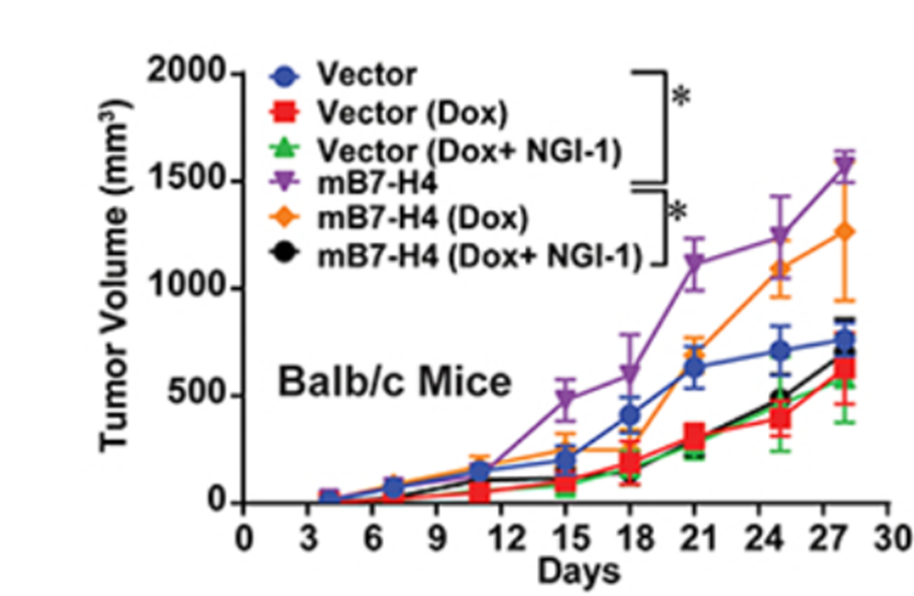
In vivo vaccination experiments showed that combined DOX/NGI-1 (10 μM) treatment yields a more potent vaccine than DOX treatment alone.
-
-
Nat Commun
Self-cleavage of the GAIN domain of adhesion G protein-coupled receptors requires multiple domain-extrinsic factors. [Abstract]2025 Oct 1;16(1):8736. PMID: 41034233 -
Nat Commun
FRET imaging of glycoRNA on small extracellular vesicles enabling sensitive cancer diagnostics. [Abstract]2025 Apr 10;16(1):3391. PMID: 40210865 -
Cell Chem Biol
2026 Jun 16:S2451-9456(26)00196-0. PMID: 42302779 -
Cell Chem Biol
STT3A is essential for Wnt signaling and represents a target for cancers driven by RNF43 deficiency. [Abstract]2025 Oct 22:S2451-9456(25)00306-X. PMID: 41130209
NGI-1 purchased from MedChemExpress. Usage Cited in: Cell Chem Biol. 2025 Oct 22:S2451-9456(25)00306-X. [Abstract]
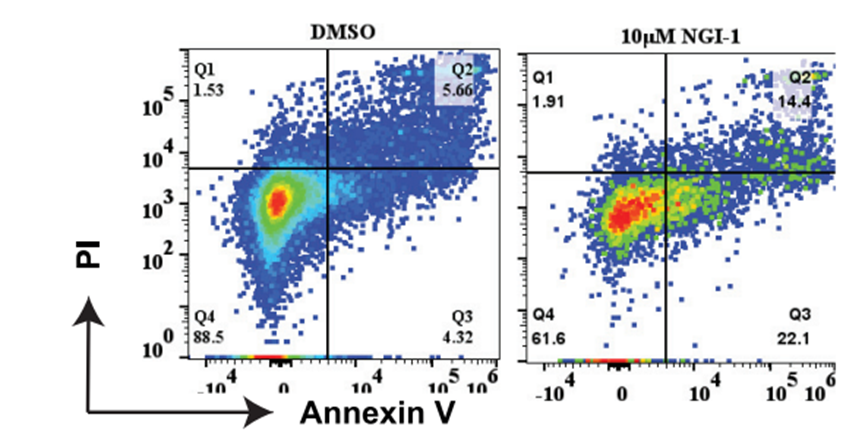
Flow cytometry describes the Annexin V staining of MM cell lines upon NGI-1 (10 μM) treatment resulted in a notable increase in cell death.
-
Int J Biol Macromol
Site-specific analysis and functional characterization of N-linked glycosylation for β-Klotho protein. [Abstract]2025 Feb:289:138846. PMID: 39701265 -
Mol Ther Oncol
The oligosaccharyltransferase complex is an essential component of multiple myeloma plasma cells. [Abstract]2025 Mar 8;33(2):200964. PMID: 40200920 -
MAbs
Computational analysis reveals non-consensus N-glycosylation sequons in antibody Fab region. [Abstract]2025 Dec;17(1):2574406. PMID: 41090251 -
Anal Chem
2026 Mar 24;98(11):8143-8154. PMID: 41810699 -
Sci Signal
Glucose metabolism sustains aberrant STAT3 signaling in colorectal cancer through glycosylated local signaling factors. [Abstract]2026 Mar 3;19(927):eadz6443. PMID: 41774818 -
Front Mol Biosci
Nascent Glycoproteome Reveals That N-Linked Glycosylation Inhibitor-1 Suppresses Expression of Glycosylated Lysosome-Associated Membrane Protein-2. [Abstract]2022 Apr 27;9:899192. PMID: 35573732 -
FEBS J
2025 Jun 26. PMID: 40568888 -
J Virol
STT3B promotes porcine epidemic diarrhea virus replication by regulating N-glycosylation of PEDV S protein. [Abstract]2025 Mar 18;99(3):e0001825. PMID: 39945486 -
J Biol Chem
Golgi α-Mannosidases Regulate Cell Surface N-Glycan Type and Ectodomain Shedding of the Transmembrane Protease Corin. [Abstract]2023 Oct;299(10):105211. PMID: 37660903 -
J Virol
Comprehensive Interactome Analysis Reveals that STT3B Is Required for N-Glycosylation of Lassa Virus Glycoprotein. [Abstract]2019 Nov 13;93(23):e01443-19. PMID: 31511384 -
Biochem Biophys Res Commun
N-glycosylation of Siglec-15 decreases its lysosome-dependent degradation and promotes its transportation to the cell membrane. [Abstract]2020 Nov 26;533(1):77-82. PMID: 32921411 -
-
bioRxiv
OSTM1 is a ubiquitin E3 ligase that suppresses B-cell malignancy by activating the cAMP/PKA/CREB pathway. [Abstract]2026 Jan 26:2026.01.23.701155. PMID: 41659680 -
-
bioRxiv
Glucose Metabolism Sustains Aberrant STAT3 Signaling in Colorectal Cancer via Glycosylated Paracrine Factors. [Abstract]2025 Jul 26:2025.07.22.666143. PMID: 40777356 -


Solvent & Solubility
In Vitro:
DMSO : 25 mg/mL (63.37 mM; ultrasonic and warming and heat to 60°C; Hygroscopic DMSO has a significant impact on the solubility of product, please use newly opened DMSO)
Please refer to the solubility information to select the appropriate solvent. Once prepared, please aliquot and store the solution to prevent product inactivation from repeated freeze-thaw cycles.
Storage method and period of stock solution: -80°C, 2 years; -20°C, 1 year. When stored at -80°C, please use it within 2 years. When stored at -20°C, please use it within 1 year.
Please refer to the solubility information to select the appropriate solvent. Once prepared, please aliquot and store the solution to prevent product inactivation from repeated freeze-thaw cycles.
Storage method and period of stock solution: -80°C, 2 years; -20°C, 1 year. When stored at -80°C, please use it within 2 years. When stored at -20°C, please use it within 1 year.
Concentration (start) × Volume (start) = Concentration (final) × Volume (final)


Purity & Documentation
-
Data Sheet (276 KB)
-
SDS (396 KB)
- English - EN (396 KB)
- Français - FR (396 KB)
- Deutsch - DE (396 KB)
- Norwegian - NO (396 KB)
- Español - ES (396 KB)
- Swedish - SV (396 KB)
- Italian - IT (396 KB)
- Korean - KR (396 KB)
- Portuguese - PT (396 KB)
-
Handling Instructions (2659 KB)
References
[1]. Zhu S, et al. Comprehensive Interactome Analysis Reveals that STT3B is Required for the N-Glycosylation of Lassa Virus Glycoprotein. J Virol. 2019 Sep 11. pii: JVI.01443-19. [Content Brief]
[2]. Lopez-Sambrooks C, et al. Oligosaccharyltransferase inhibition induces senescence in RTK-driven tumor cells. Nat Chem Biol. 2016 Dec;12(12):1023-1030. [Content Brief]


Complete Stock Solution Preparation Table
Please refer to the solubility information to select the appropriate solvent. Once prepared, please aliquot and store the solution to prevent product inactivation from repeated freeze-thaw cycles.
Storage method and period of stock solution: -80°C, 2 years; -20°C, 1 year. When stored at -80°C, please use it within 2 years. When stored at -20°C, please use it within 1 year.
| Optional Solvent | Concentration Solvent Mass | 1 mg | 5 mg | 10 mg | 25 mg |
|---|---|---|---|---|---|
| DMSO | 1 mM | 2.5348 mL | 12.6739 mL | 25.3479 mL | 63.3697 mL |
| 5 mM | 0.5070 mL | 2.5348 mL | 5.0696 mL | 12.6739 mL | |
| 10 mM | 0.2535 mL | 1.2674 mL | 2.5348 mL | 6.3370 mL | |
| 15 mM | 0.1690 mL | 0.8449 mL | 1.6899 mL | 4.2246 mL | |
| 20 mM | 0.1267 mL | 0.6337 mL | 1.2674 mL | 3.1685 mL | |
| 25 mM | 0.1014 mL | 0.5070 mL | 1.0139 mL | 2.5348 mL | |
| 30 mM | 0.0845 mL | 0.4225 mL | 0.8449 mL | 2.1123 mL | |
| 40 mM | 0.0634 mL | 0.3168 mL | 0.6337 mL | 1.5842 mL | |
| 50 mM | 0.0507 mL | 0.2535 mL | 0.5070 mL | 1.2674 mL | |
| 60 mM | 0.0422 mL | 0.2112 mL | 0.4225 mL | 1.0562 mL |


NGI-1 Related Classifications
HY-117383 Related Classifications
 Powered by Bioz