



Oxybenzone
Based on 7 publication(s) in Google Scholar
Oxybenzone (Benzophenone 3) is a commonly used UV filter in sun tans and skin protectants. Oxybenzone act as endocrine disrupting chemicals (EDCs) and can pass through the placental and blood-brain barriers. Benzophenone-3 impairs autophagy, alters epigenetic status, and disrupts retinoid X receptor signaling in apoptotic neuronal cells.
For research use only. We do not sell to patients.
- Purity : 99.93%
- CAS No.: 131-57-7
- Formula: C14H12O3
- Molecular Weight:228.25
-
Storage:Powder -20°C, 3 years , 4°C, 2 years ; In solvent -80°C, 2 years , -20°C, 1 year

To place orders, for customer services and technical support, please contact: MedChemExpress USA
Tel: 609-228-6898 E-mail: [email protected] [email protected]


-
 Biological Activity
-
Chemical Information
-
Solvent & Solubility
- Protocols
- Purity & Documentation
- References
-
 Help & FAQs


-
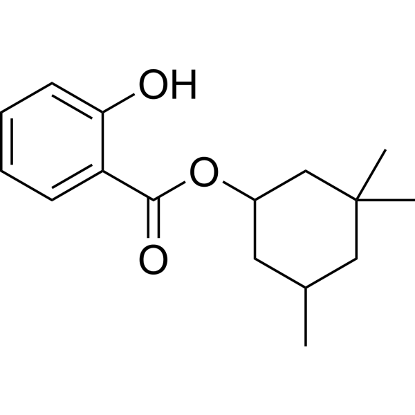
HY-B0928
HomosalateSun-Screening Agent -
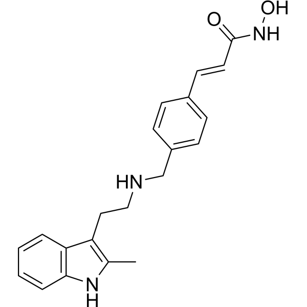
HY-10224
PanobinostatHDAC Inhibitor -
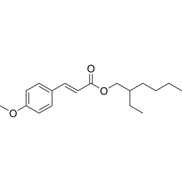
HY-B1234
OctinoxateIngredient of Sunscreens, Thyroid Hormone Receptor Agonist, ultraviolet filter -
HY-A0087
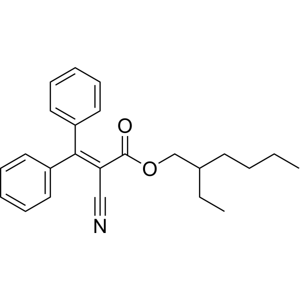
OctocryleneOrganic Ultraviolet (UV) Filter -
HY-B1119
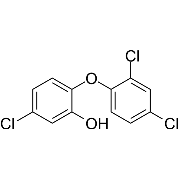
TriclosanAntibacterial Agent -
HY-B0167
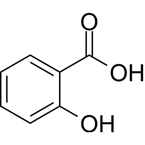
Salicylic acidCOX-2 Inhibitor -
HY-18260
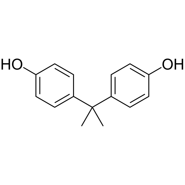
Bisphenol AEndocrine-Disrupting Compound -
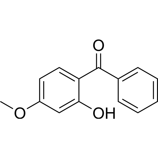
HY-A0067R
Oxybenzone (Standard)Ultraviolet Light Filter -
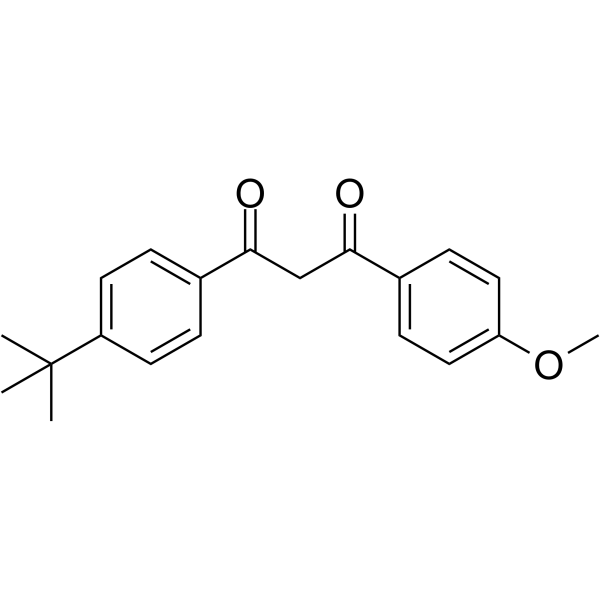
HY-B0316
AvobenzoneLong-Wave UVA Filter -
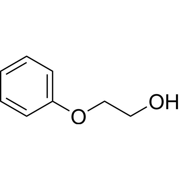
HY-B1729
PhenoxyethanolPreservative -
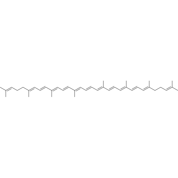
HY-N0287
LycopeneAntioxidant -

HY-21191
Perfluorobutanesulfonic acidBiochemical Assay Reagents
Publications Citing Use of MedChemExpress (MCE) Oxybenzone
More- Environ Pollut. 2025 May 1:372:126064. [Abstract]
- Ecotoxicol Environ Saf. 2026 Jul 10:322:120480.
- Ecotoxicol Environ Saf. 2026 Apr 1:314:120006. [Abstract]
- Ecotoxicol Environ Saf. 2025 Mar 1:292:117982. [Abstract]
- Chem Biol Interact. 2025 Nov 1:421:111757. [Abstract]
- University of Pennsylvania. 2025.
- Chemosphere. 2021 Oct:280:130670. [Abstract]


Biological Activity
Description
Cellular Effect
|
Cell Line

|
Type | Value | Description | References |
|---|---|---|---|---|
| Peritoneal macrophage cell | CC50 |
24.6 μg/mL
Compound: 1a, LFQM-115
|
Cytotoxicity against mouse peritoneal macrophages after 72 hrs by MTT assay
Cytotoxicity against mouse peritoneal macrophages after 72 hrs by MTT assay
|
[PMID: 23623672] |
In Vitro
Oxybenzone (Benzophenone 3) (25 μM; 24 hours) decreases the relative RXRβ and RXRγ protein levels by 61 and 56%, respectively and increases the relative RXRα protein level by 49%[3].
Oxybenzone (25-100 μM; 24 hours) induces an increase in caspase-3 levels in primary cultures of mouse neocortical cells at 7 DIV. Oxybenzone-induced apoptosis involves the activation of RXRα signaling and the impairment of RXRβ/RXRγ signaling. Oxybenzone (25 μM; 24 hours) inhibits global DNA methylation as well as reduced HDAC and HAT activities in mouse embryonic neuronal cells[3].
MedChemExpress (MCE) has not independently confirmed the accuracy of these methods. They are for reference only. Further protocols information, click here.
-
Cell Line:Mouse neocortical cells at 7 DIV
-
Concentration:25 μM
-
Incubation Time:24 hours
-
Result:Exposure to Oxybenzone (25 μM) for 24 h decreased the relative RXRβ and RXRγ protein levels by 61 and 56%, respectively. Treatment with Oxybenzone (25 μM) increased the relative RXRα protein level by 49%.


Chemical Information
-
CAS No. 131-57-7
-
Appearance Solid
-
Molecular Weight 228.25
-
Formula C14H12O3
-
Color Light yellow to yellow
-
SMILES
O=C(C1=CC=C(OC)C=C1O)C2=CC=CC=C2
-
Synonyms
Benzophenone 3
-
Shipping
Room temperature in continental US; may vary elsewhere.
-
Storage
Powder -20°C 3 years 4°C 2 years In solvent -80°C 2 years -20°C 1 year


Publications (7)
-
Journal Impact Factor
-
Most Recent
-
Environ Pollut
Mechanism of chondrocyte injury induced by Benzophenone-3 through modulation of the IL-6/JAK2/STAT3 pathway. [Abstract]2025 May 1:372:126064. PMID: 40090449 -
-
Ecotoxicol Environ Saf
Mechanism of benzophenone-3 in promoting proliferation and migration of prostate cancer cells via the acyl-CoA dehydrogenase 9 axis. [Abstract]2026 Apr 1:314:120006. PMID: 41861636 -
Ecotoxicol Environ Saf
Pubertal low dose exposure to benzophenone-3 (BP-3) alters murine mammary stem cell functions. [Abstract]2025 Mar 1:292:117982. PMID: 40020383 -
Chem Biol Interact
2025 Nov 1:421:111757. PMID: 41033242 -
-
Chemosphere
Benzophenone-3 induced abnormal development of enteric nervous system in zebrafish through MAPK/ERK signaling pathway. [Abstract]2021 Oct:280:130670. PMID: 33971419


Solvent & Solubility
In Vitro:
DMSO : 100 mg/mL (438.12 mM; Need ultrasonic; Hygroscopic DMSO has a significant impact on the solubility of product, please use newly opened DMSO)
H2O : < 0.1 mg/mL (insoluble)
Please refer to the solubility information to select the appropriate solvent. Once prepared, please aliquot and store the solution to prevent product inactivation from repeated freeze-thaw cycles.
Storage method and period of stock solution: -80°C, 2 years; -20°C, 1 year. When stored at -80°C, please use it within 2 years. When stored at -20°C, please use it within 1 year.
Please refer to the solubility information to select the appropriate solvent. Once prepared, please aliquot and store the solution to prevent product inactivation from repeated freeze-thaw cycles.
Storage method and period of stock solution: -80°C, 2 years; -20°C, 1 year. When stored at -80°C, please use it within 2 years. When stored at -20°C, please use it within 1 year.
Concentration (start) × Volume (start) = Concentration (final) × Volume (final)
In Vivo:
Select the appropriate dissolution method based on your experimental animal and administration route.
- For the following dissolution methods, please ensure to first prepare a clear stock solution using an In Vitro approach and then sequentially add co-solvents:
- To ensure reliable experimental results, the clarified stock solution can be appropriately stored based on storage conditions. As for the working solution for In Vivo experiments, it is recommended to prepare freshly and use it on the same day.
- The percentages shown for the solvents indicate their volumetric ratio in the final prepared solution. If precipitation or phase separation occurs during preparation, heat and/or sonication can be used to aid dissolution.
Add each solvent one by one: 10% DMSO 40% PEG300 5% Tween-80 45% Saline
Solubility: ≥ 2.5 mg/mL (10.95 mM); Clear solution
This protocol yields a clear solution of ≥ 2.5 mg/mL (saturation unknown).
Taking 1 mL working solution as an example, add 100 μL DMSO stock solution (25.0 mg/mL) to 400 μL PEG300, and mix evenly; then add 50 μL Tween-80 and mix evenly; then add 450 μL Saline to adjust the volume to 1 mL.
Preparation of Saline: Dissolve 0.9 g sodium chloride in ddH₂O and dilute to 100 mL to obtain a clear Saline solution.
Add each solvent one by one: 10% DMSO 90% (20% SBE-β-CD in Saline)
Solubility: ≥ 2.5 mg/mL (10.95 mM); Clear solution
This protocol yields a clear solution of ≥ 2.5 mg/mL (saturation unknown).
Taking 1 mL working solution as an example, add 100 μL DMSO stock solution (25.0 mg/mL) to 900 μL 20% SBE-β-CD in Saline, and mix evenly.
Preparation of 20% SBE-β-CD in Saline (4°C, storage for one week): 2 g SBE-β-CD powder is dissolved in 10 mL Saline, completely dissolve until clear.
For the following dissolution methods, please prepare the working solution directly:
It is recommended to prepare fresh solutions and use them promptly within a short period of time.
The percentages shown for the solvents indicate their volumetric ratio in the final prepared solution. If precipitation or phase separation occurs during preparation, heat and/or sonication can be used to aid dissolution.
Add each solvent one by one: PBS
Solubility: 5 mg/mL (21.91 mM); Clear solution; Need ultrasonic and warming and heat to 60°C
In Vivo Dissolution Calculator
Please enter the basic information of animal experiments:
-
-
-
-
Recommended: Prepare an additional quantity of animals to account for potential losses during experiments.
Please enter your animal formula composition:
-
%DMSO +
Recommended: Keep the proportion of DMSO in working solution below 2% if your animal is weak.
-
%+
-
+%Tween-80 + +
-
%Saline +
The co-solvents required include: DMSO, . All of co-solvents are available by MedChemExpress (MCE). , Tween 80. All of co-solvents are available by MedChemExpress (MCE).
Working solution concentration: 0.22 mg/mL
Method for preparing stock solution: mg drug dissolved in μL DMSO. Stock solution concentration: mg/mL.
1. Take μL DMSO stock solution;
2. Add μL .
μL , mix evenly;
3. Then add μL Tween 80, mix evenly;
4. Then add μL
Please ensure that the stock solution in the first step is dissolved to a clear state, and add co-solvents in sequence. You can use ultrasonic heating (ultrasonic cleaner, recommended frequency 20-40 kHz), vortexing, etc. to assist dissolution.

Protocols
-
Apoptosis
Apoptosis, also called programmed cell death, is generally characterized by distinct morphological characteristics.
-
Autophagy
Autophagy is a process in which eukaryotic cells use lysosomes to degrade their own cytoplasmic proteins and damaged organelles under the regulation of autophagy related gene (Atg). Microtubule-associated proteins light chain 3 (LC3) is recognized as autophagy marker, which transfers from cytoplasmic LC3 (LC3-I) to membrane type (LC3-II). LC3-II/I ratio could be detected by Western Blot and fluorescence microscopy.
-
Apoptosis Solutions
Apoptosis is a regulated, generally non-lytic cell-death pathway that removes unwanted, damaged, infected, or abnormal cells through coordinated morphological changes, caspase activation, DNA fragmentation, and membrane remodeling. The intrinsic apoptosis pathway is controlled mainly by mitochondrial outer membrane permeabilization, BCL-2 family proteins, cytochrome c release, apoptosome formation, caspase-9 activation, and downstream executioner caspase-3/7 activation. The extrinsic apoptosis pathway is initiated by death receptors such as Fas, TNFR, and TRAIL receptors, which recruit adaptor proteins and activate caspase-8 before engaging executioner caspases or mitochondrial amplification through BID cleavage. Apoptosis is linked to many phenotypes, including cancer cell killing, tissue homeostasis, immune regulation, neurodegeneration, infection response, and treatment-induced cytotoxicity; unresolved questions include how apoptosis interacts with necroptosis, pyroptosis, ferroptos
-
Lysosome and acidic-vesicle live-cell staining
Lysosome and acidic-vesicle live-cell staining detects acidic intracellular compartments by using membrane-permeant acidotropic probes that accumulate in low-pH vesicles, including lysosomes, late endosomes, autolysosomes, and acidic phagosomes. LysoTracker staining is commonly used as an intensity-based readout of acidic lysosomal compartment abundance or enlargement, while acridine orange produces green fluorescence in less concentrated compartments and red fluorescence after concentration-dependent accumulation in acidic vesicular organelles. Loss or reduction of acridine-orange red signal can be used as a readout of lysosomal membrane permeabilization or reduced acidic-vesicle integrity. This protocol is designed for live cultured cells and can be adapted for fluorescence microscopy, high-content imaging, plate-reader readout, or flow cytometry when the selected literature supports the readout. Because these dyes report acidotropic accumulation rather than lysosome identity alone,
-
Macroautophagy Solutions
Macroautophagy is a conserved lysosome-dependent degradation pathway in which cytoplasmic material is sequestered into double-membrane autophagosomes and delivered to lysosomes for degradation and recycling. The pathway supports cellular homeostasis during nutrient limitation, organelle stress, protein-aggregate accumulation, infection, differentiation, and tissue remodeling by coupling cargo sequestration, autophagosome maturation, lysosomal fusion, and degradation of cargo-derived macromolecules. The core molecular sequence includes initiation by nutrient- and stress-regulated autophagy machinery, autophagosome nucleation, LC3/ATG8-family conjugation to autophagosomal membranes, cargo selection through receptors such as SQSTM1/p62, autophagosome-lysosome fusion, and lysosomal degradation. LC3 was identified as a mammalian homolog of yeast Atg8 that localizes to autophagosomal membranes after processing, and p62/SQSTM1 was shown to connect ubiquitinated cargo with autophagic degradati


Purity & Documentation
-
Data Sheet (276 KB)
-
SDS (458 KB)
- English - EN (458 KB)
- Français - FR (458 KB)
- Deutsch - DE (458 KB)
- Norwegian - NO (458 KB)
- Español - ES (458 KB)
- Swedish - SV (458 KB)
- Italian - IT (458 KB)
- Korean - KR (458 KB)
- Portuguese - PT (458 KB)
-
Handling Instructions (2659 KB)
References
[1]. Wnuk A, et al. Prenatal exposure to benzophenone-3 (BP-3) induces apoptosis, disrupts estrogen receptor expression and alters the epigenetic status of mouse neurons. J Steroid Biochem Mol Biol. 2018;182:106-118. [Content Brief]
[2]. DiNardo JC, et al. Can oxybenzone cause Hirschsprung's disease?. Reprod Toxicol. 2019;86:98-100. [Content Brief]
[3]. Wnuk A, et al. Benzophenone-3 Impairs Autophagy, Alters Epigenetic Status, and Disrupts Retinoid X Receptor Signaling in Apoptotic Neuronal Cells. Mol Neurobiol. 2018;55(6):5059-5074. [Content Brief]


Complete Stock Solution Preparation Table
Please refer to the solubility information to select the appropriate solvent. Once prepared, please aliquot and store the solution to prevent product inactivation from repeated freeze-thaw cycles.
Storage method and period of stock solution: -80°C, 2 years; -20°C, 1 year. When stored at -80°C, please use it within 2 years. When stored at -20°C, please use it within 1 year.
| Optional Solvent | Concentration Solvent Mass | 1 mg | 5 mg | 10 mg | 25 mg |
|---|---|---|---|---|---|
| DMSO | 1 mM | 4.3812 mL | 21.9058 mL | 43.8116 mL | 109.5290 mL |
| 5 mM | 0.8762 mL | 4.3812 mL | 8.7623 mL | 21.9058 mL | |
| 10 mM | 0.4381 mL | 2.1906 mL | 4.3812 mL | 10.9529 mL | |
| 15 mM | 0.2921 mL | 1.4604 mL | 2.9208 mL | 7.3019 mL | |
| 20 mM | 0.2191 mL | 1.0953 mL | 2.1906 mL | 5.4765 mL | |
| 25 mM | 0.1752 mL | 0.8762 mL | 1.7525 mL | 4.3812 mL | |
| 30 mM | 0.1460 mL | 0.7302 mL | 1.4604 mL | 3.6510 mL | |
| 40 mM | 0.1095 mL | 0.5476 mL | 1.0953 mL | 2.7382 mL | |
| 50 mM | 0.0876 mL | 0.4381 mL | 0.8762 mL | 2.1906 mL | |
| 60 mM | 0.0730 mL | 0.3651 mL | 0.7302 mL | 1.8255 mL | |
| 80 mM | 0.0548 mL | 0.2738 mL | 0.5476 mL | 1.3691 mL | |
| 100 mM | 0.0438 mL | 0.2191 mL | 0.4381 mL | 1.0953 mL |


Oxybenzone Related Classifications
HY-A0067 Related Classifications
 Powered by Bioz