


Salinomycin
Based on 52 publication(s) in Google Scholar
Salinomycin (Procoxacin), a polyether potassium ionophore antibiotic, selectively inhibits the growth of gram-positive bacteria. Salinomycin is a potent inhibitor of Wnt/β-catenin signaling, blocks Wnt-induced LRP6 phosphorylation. Salinomycin shows selective activity against human cancer stem cells.
For research use only. We do not sell to patients.
- Purity: 99.74%
- CAS No.: 53003-10-4
- Formula: C42H70O11
- Molecular Weight:751.00
-
Storage:Powder -20°C, 3 years , 4°C, 2 years ; In solvent -80°C, 6 months , -20°C, 1 month

To place orders, for customer services and technical support, please contact: MedChemExpress USA
Tel: 609-228-6898 E-mail: [email protected] [email protected]


-
 Biological Activity
-
Chemical Information
-
Solvent & Solubility
- Protocol
- Purity & Documentation
- References
-
 Help & FAQs
-


Anti-Infection Compound Library
HY-L002
-
Apoptosis Compound Library
HY-L003
-
Stem Cell Signaling Compound Library
HY-L017
-
Wnt/Hedgehog/Notch Compound Library
HY-L020
-
Anti-Cancer Compound Library
HY-L025
-
Autophagy Compound Library
HY-L029
-
Anti-Aging Compound Library
HY-L034
-
Differentiation Inducing Compound Library
HY-L038
-
Antibacterial Compound Library
HY-L049
-
Cytoskeleton Compound Library
HY-L060
-
Antibiotics Library
HY-L067
-
Neuroprotective Compound Library
HY-L070
-
Anti-Breast Cancer Compound Library
HY-L074
-
Antiparasitic Compound library
HY-L082
-
Mitochondria-Targeted Compound Library
HY-L089
-
Transcription Factor-Targeted Library
HY-L090
-
Anti-Liver Cancer Compound Library
HY-L101
-
Rare Diseases Drug Library
HY-L102
-
Anti-Colorectal Cancer Compound Library
HY-L103
-
Human Metabolite Library
HY-L123
-
Anti-Prostate Cancer Compound Library
HY-L124
-
Anti-Pulmonary Fibrosis Compound Library
HY-L125
-
Cancer Stem Cells Compound Library
HY-L135
-
Heterocyclic Compound Library
HY-L138
-
Antihypertensive Compound Library
HY-L145
-
Highly Selective Inhibitors Library
HY-L158
-
Highly Selective Activators Library
HY-L159
-
Cell Death Library
HY-L162
-
Extracellular Vesicles (EVs) Compound Library
HY-L168
-
Anti-Ovarian Cancer Compound Library
HY-L173
-
Multi-Target Compound Library
HY-L176
-
Radiosensitizer Library
HY-L179
-
Mitophagy Compound Library
HY-L180
-
Bioactive Compound Library Max
HY-L181
-
Anti-Aging Compound Library Mini
HY-L034M
-
MCE Bioactive Compound Library
HY-L001V
-
Bioactive Compound Library
HY-L001
-
Anti-Gastric Cancer Compound Library
HY-L184
-
Anti-Fibrosis Compound Library
HY-L185
-
Non-Alcoholic Fatty Liver Disease (NAFLD) Compound Library
HY-L199
-
Cell Proliferation Compound Library
HY-L201
-
High-Throughput Bioactive Compound Library
HY-L205
-
Anti-Rheumatic Arthritis Compound Library
HY-L210
-
Mass Spectrometry Human Metabolite Library
HY-L215
-
RNA Binding Bioactive Compound Library
HY-L248


Publications Citing Use of MedChemExpress (MCE) Salinomycin
More- Nat Commun. 2025 Jul 18;16(1):6621. [Abstract]
- Adv Sci (Weinh). 2025 Sep 9:e13476. [Abstract]
- Adv Sci (Weinh). 2025 Feb 8:e2415229. [Abstract]
- J Control Release. 2020 Oct 10;326:387-395. [Abstract]
- Cell Death Dis. 2023 Mar 11;14(3):193. [Abstract]
- Pharmacol Res. 2020 May;155:104751. [Abstract]
- Biosens Bioelectron. 2024 Jul 1:255:116245. [Abstract]
- Cell Commun Signal. 2018 Nov 23;16(1):89. [Abstract]
- Acta Biomater. 2022 Oct 15:152:380-392. [Abstract]
- Int J Nanomedicine. 2020 Sep 15;15:6839-6854. [Abstract]
- Mol Med. 2025 Jan 22;31(1):19. [Abstract]
- EMBO Mol Med. 2019 Oct;11(10):e9930. [Abstract]
- Cell Rep. 2025 Jul 22;44(7):115922. [Abstract]
- Anal Chem. 2023 Apr 11;95(14):6191. [Abstract]
- Talanta. 2025 Apr 10:293:128128. [Abstract]
- Eur J Med Chem. 2024 Oct 5:276:116701. [Abstract]
- Cell Biosci. 2021 Aug 4;11(1):156. [Abstract]
- Biomed J. 2020 Aug;43(4):368-374. [Abstract]
- RSC Adv. 2021 Nov 2;11(56):35331-35341. [Abstract]
- J Mol Cell Biol. 2025 Dec 2:mjaf048. [Abstract]
- Hepatol Commun. 2025 May 16;9(6):e0706. [Abstract]
- Int J Pharm. 2023 Mar 25:635:122779. [Abstract]
- Eur J Pharmacol. 2025 Jul 5:998:177533. [Abstract]
- Cell Oncol (Dordr). 2024 Dec;47(6):2201-2215. [Abstract]
- Int J Mol Sci. 2023 Mar 21;24(6):5939. [Abstract]
- Int J Mol Med. 2019 Oct;44(4):1289-1298. [Abstract]
- Int J Mol Sci. 2019 Jun 12;20(12):2861. [Abstract]
- Nanoscale. 2024 Nov 7;16(43):20131-20146. [Abstract]
- AAPS PharmSciTech. 2024 Sep 7;25(7):211. [Abstract]
- Sci Rep. 2026 Feb 9;16(1):7909. [Abstract]
- Cancers. 2020 Aug 5;12(8):2176. [Abstract]
- Oncol Rep. 2018 Aug;40(2):877-886. [Abstract]
- ACS Infect Dis. 2021 Aug 13;7(8):2390-2401. [Abstract]
- Antiviral Res. 2026 May:249:106403. [Abstract]
- FASEB J. 2025 Dec 31;39(24):e71350. [Abstract]
- Biochim Biophys Acta Mol Cell Res. 2025 May 11:119989. [Abstract]
- Viruses. 2022 Aug 6;14(8):1734. [Abstract]
- J Bioenerg Biomembr. 2021 Oct;53(5):513-523. [Abstract]
- Int J Hyperthermia. 2026 Dec;43(1):2643510. [Abstract]
- Exp Ther Med. 2020 Mar;19(3):1835-1843. [Abstract]
- Leuk Lymphoma. 2023 Jun;64(6):1151-1160. [Abstract]
- Res Sq. 2026 Jan 9.
- SSRN. 2025 Dec 2.
- SSRN. 2024 Nov 12.
- bioRxiv. 2024 September 18.
- bioRxiv. 2024 September 07.
- Chemosphere. 2024 Jul 3:142758. [Abstract]
- bioRxiv. 2023 Jun 25.
- bioRxiv. 2023 Jan 17.
- Research Square Print. 2023 Jan 6th.
- Norwegian University of Science and Technology. 2021 Oct.
- bioRxiv. 2020 Jun.
 Customer Validation & Images

-
Others
-
WB
-
WB
-
WB
All Antibiotic Isoforms
MoreAll Parasite Isoforms
More

Biological Activity
|
Coccidia |
|
Cell Line
|
Type | Value | Description | References |
|---|---|---|---|---|
| 2008 | IC50 |
3.2 μM
Compound: Salinomycin
|
Cytotoxicity against human OV2008 cells assessed as reduction in cell viability incubated for 72 hrs by MTT assay
Cytotoxicity against human OV2008 cells assessed as reduction in cell viability incubated for 72 hrs by MTT assay
|
[PMID: 28651817] |
| 2008 | IC50 |
4.78 μM
Compound: Salinomycin
|
Cytotoxicity against human OV2008 cells assessed as reduction in cell viability incubated for 48 hrs by MTT assay
Cytotoxicity against human OV2008 cells assessed as reduction in cell viability incubated for 48 hrs by MTT assay
|
[PMID: 28651817] |
| 2008 | IC50 |
7.44 μM
Compound: Salinomycin
|
Cytotoxicity against human OV2008 cells assessed as reduction in cell viability incubated for 24 hrs by MTT assay
Cytotoxicity against human OV2008 cells assessed as reduction in cell viability incubated for 24 hrs by MTT assay
|
[PMID: 28651817] |
| 4T1 | IC50 |
3.78 μM
Compound: 1
|
Cytotoxicity against mouse 4T1 cells assessed as cell growth inhibition after 48 hrs by MTS assay
Cytotoxicity against mouse 4T1 cells assessed as cell growth inhibition after 48 hrs by MTS assay
|
[PMID: 29466777] |
| 4T1 | IC50 |
3.78 μM
Compound: 1; SAL
|
Antiproliferative activity against mouse 4T1 cells by MTT assay
Antiproliferative activity against mouse 4T1 cells by MTT assay
|
[PMID: 30684870] |
| A2780 | IC50 |
0.16 μM
Compound: Sal
|
Antitumor activity against CD44 negative human A2780 cells assessed as reduction in cell viability incubated for 72 hrs by CCK8 assay
Antitumor activity against CD44 negative human A2780 cells assessed as reduction in cell viability incubated for 72 hrs by CCK8 assay
|
[PMID: 39067438] |
| A-431 | IC50 |
5.27 μM
Compound: Sal
|
Cytotoxicity against human A-431 cells assessed as inhibition of cell growth incubated for 72 hrs by CCK8 assay
Cytotoxicity against human A-431 cells assessed as inhibition of cell growth incubated for 72 hrs by CCK8 assay
|
[PMID: 39067438] |
| A549 | IC50 |
3.98 μM
Compound: 1
|
Cytotoxicity against human A549 cells assessed as cell growth inhibition after 48 hrs by MTS assay
Cytotoxicity against human A549 cells assessed as cell growth inhibition after 48 hrs by MTS assay
|
[PMID: 29466777] |
| A549 | IC50 |
3.98 μM
Compound: 1; SAL
|
Antiproliferative activity against human A549 cells by MTT assay
Antiproliferative activity against human A549 cells by MTT assay
|
[PMID: 30684870] |
| A549 | IC50 |
25.08 μM
Compound: Sal
|
Cytotoxicity against human A549 cells assessed as inhibition of cell growth incubated for 72 hrs by CCK8 assay
Cytotoxicity against human A549 cells assessed as inhibition of cell growth incubated for 72 hrs by CCK8 assay
|
[PMID: 39067438] |
| ACHN | IC50 |
26.03 μM
Compound: Sal
|
Cytotoxicity against human ACHN cells assessed as inhibition of cell growth incubated for 72 hrs by CCK8 assay
Cytotoxicity against human ACHN cells assessed as inhibition of cell growth incubated for 72 hrs by CCK8 assay
|
[PMID: 39067438] |
| BALB/3T3 | IC50 |
28.08 μM
Compound: 1
|
Cytotoxicity against mouse BALB/3T3 cells incubated for 72 hrs by MTT assay
Cytotoxicity against mouse BALB/3T3 cells incubated for 72 hrs by MTT assay
|
[PMID: 23079523] |
| BALB/3T3 | IC50 |
25.8 μM
Compound: SAL
|
Antiproliferative activity mouse BALB/3T3 cells
Antiproliferative activity mouse BALB/3T3 cells
|
[PMID: 24631190] |
| BALB/3T3 | IC50 |
9.33 μM
Compound: SAL
|
Antiproliferative activity against mouse BALB/3T3 cells assessed as growth inhibition after 72 hrs by SRB method
Antiproliferative activity against mouse BALB/3T3 cells assessed as growth inhibition after 72 hrs by SRB method
|
[PMID: 25644674] |
| BALB/3T3 | IC50 |
27.76 μM
Compound: SAL
|
Antiproliferative activity against mouse BALB/3T3 cells after 72 hrs
Antiproliferative activity against mouse BALB/3T3 cells after 72 hrs
|
[PMID: 26163197] |
| BEAS-2B | IC50 |
13.07 μM
Compound: 1
|
Cytotoxicity against human BEAS2B cells assessed as cell growth inhibition after 48 hrs by MTS assay
Cytotoxicity against human BEAS2B cells assessed as cell growth inhibition after 48 hrs by MTS assay
|
[PMID: 29466777] |
| BT-474 | CC50 |
14 μM
Compound: Salinomycin
|
Cytotoxicity against human BT474 cells after 48 hrs by MTS assay
Cytotoxicity against human BT474 cells after 48 hrs by MTS assay
|
10.1039/C6MD00328A |
| Caco-2 | IC50 |
12.99 μM
Compound: 1; SAL
|
Antiproliferative activity against human Caco2 cells by MTT assay
Antiproliferative activity against human Caco2 cells by MTT assay
|
[PMID: 30684870] |
| Caco-2 | IC50 |
15.67 μM
Compound: 4; SAL
|
Antiproliferative activity against human Caco-2 cells measured after 24 hrs by MTT assay
Antiproliferative activity against human Caco-2 cells measured after 24 hrs by MTT assay
|
[PMID: 38142509] |
| CAL-27 | IC50 |
30.15 μM
Compound: Sal
|
Cytotoxicity against human CAL-27 cells assessed as inhibition of cell growth incubated for 72 hrs by CCK8 assay
Cytotoxicity against human CAL-27 cells assessed as inhibition of cell growth incubated for 72 hrs by CCK8 assay
|
[PMID: 39067438] |
| DU-145 | IC50 |
47.06 μM
Compound: Sal
|
Cytotoxicity against human DU-145 cells assessed as inhibition of cell growth incubated for 72 hrs by CCK8 assay
Cytotoxicity against human DU-145 cells assessed as inhibition of cell growth incubated for 72 hrs by CCK8 assay
|
[PMID: 39067438] |
| DU-145 | CC50 |
>20 μM
Compound: Salinomycin
|
Cytotoxicity against human DU145 cells after 48 hrs by MTS assay
Cytotoxicity against human DU145 cells after 48 hrs by MTS assay
|
10.1039/C6MD00328A |
| HCCLM3 | IC50 |
7.99 μM
Compound: Sal
|
Cytotoxicity against human HCCLM3 cells assessed as inhibition of cell growth incubated for 72 hrs by CCK8 assay
Cytotoxicity against human HCCLM3 cells assessed as inhibition of cell growth incubated for 72 hrs by CCK8 assay
|
[PMID: 39067438] |
| HEK293 | IC50 |
195 nM
Compound: 1
|
Cytotoxicity against HEK293 cells assessed as cell growth inhibition after 72 hrs by MTT assay
Cytotoxicity against HEK293 cells assessed as cell growth inhibition after 72 hrs by MTT assay
|
[PMID: 29466777] |
| HEK293 | IC50 |
329.5 nM
Compound: 1
|
Cytotoxicity against doxycycline inducible T-Rex/K-Ras G12V HEK293 cells assessed as cell growth inhibition after 72 hrs by MTT assay
Cytotoxicity against doxycycline inducible T-Rex/K-Ras G12V HEK293 cells assessed as cell growth inhibition after 72 hrs by MTT assay
|
[PMID: 29466777] |
| HeLa | IC50 |
4.49 μM
Compound: 1
|
Cytotoxicity against human HeLa cells assessed as cell growth inhibition after 48 hrs by MTS assay
Cytotoxicity against human HeLa cells assessed as cell growth inhibition after 48 hrs by MTS assay
|
[PMID: 29466777] |
| HeLa | IC50 |
0.32 μM
Compound: 1; SAL
|
Antiproliferative activity against human HeLa cells by MTT assay
Antiproliferative activity against human HeLa cells by MTT assay
|
[PMID: 30684870] |
| HeLa | IC50 |
25.01 μM
Compound: Sal
|
Cytotoxicity against human HeLa cells assessed as inhibition of cell growth incubated for 72 hrs by CCK8 assay
Cytotoxicity against human HeLa cells assessed as inhibition of cell growth incubated for 72 hrs by CCK8 assay
|
[PMID: 39067438] |
| Hepatocyte | IC50 |
0.016 ppm
Compound: Salinomycin
|
Inhibitory activity against second generation merozoids of Eimeria tenella in chick hepatocyte assay in the presence of iproniazid; Value given as 0.08-0.016
Inhibitory activity against second generation merozoids of Eimeria tenella in chick hepatocyte assay in the presence of iproniazid; Value given as 0.08-0.016
|
[PMID: 9873375] |
| Hepatocyte | IC50 |
0.016 ppm
Compound: Salinomycin
|
Inhibitory activity against second generation merozoids of Eimeria tenella in chick hepatocyte assay in the presence of Tranylcypromine; Value given as 0.08-0.016
Inhibitory activity against second generation merozoids of Eimeria tenella in chick hepatocyte assay in the presence of Tranylcypromine; Value given as 0.08-0.016
|
[PMID: 9873375] |
| Hepatocyte | IC50 |
0.016 ppm
Compound: Salinomycin
|
Inhibitory activity against second generation merozoids of Eimeria tenella in chick hepatocyte assay in the absence of inhibitor; Value given as 0.08-0.016
Inhibitory activity against second generation merozoids of Eimeria tenella in chick hepatocyte assay in the absence of inhibitor; Value given as 0.08-0.016
|
[PMID: 9873375] |
| Hepatocyte | IC50 |
0.016p.p.. M
Compound: Salinomycin
|
Inhibitory activity against second generation merozoids of Eimeria tenella in chick hepatocyte assay in the absence of inhibitor; Value given as 0.08-0.016
Inhibitory activity against second generation merozoids of Eimeria tenella in chick hepatocyte assay in the absence of inhibitor; Value given as 0.08-0.016
|
[PMID: 9873375] |
| HepG2 | IC50 |
24.5 μM
Compound: Salinomycin
|
Cytotoxicity against human HepG2 cells assessed as reduction in cell viability incubated for 72 hrs by MTT assay
Cytotoxicity against human HepG2 cells assessed as reduction in cell viability incubated for 72 hrs by MTT assay
|
[PMID: 30871771] |
| HGC-27 | IC50 |
2.57 μM
Compound: Sal
|
Cytotoxicity against human HGC27 cells assessed as decrease in cell proliferation after 48 hrs by MTT assay
Cytotoxicity against human HGC27 cells assessed as decrease in cell proliferation after 48 hrs by MTT assay
|
[PMID: 28262526] |
| HGC-27 | IC50 |
2.57 μM
Compound: 1; SAL
|
Antiproliferative activity against human HGC27 cells by MTT assay
Antiproliferative activity against human HGC27 cells by MTT assay
|
[PMID: 30684870] |
| HGC-27 | IC50 |
8.18 μM
Compound: 1; SAL
|
Antiproliferative activity against human HGC27 cells after 48 hrs by MTT assay
Antiproliferative activity against human HGC27 cells after 48 hrs by MTT assay
|
[PMID: 30684870] |
| HL-60 | IC50 |
0.44 μM
Compound: 1
|
Cytotoxicity against human HL60 cells incubated for 72 hrs by MTT assay
Cytotoxicity against human HL60 cells incubated for 72 hrs by MTT assay
|
[PMID: 23079523] |
| HL-60 | IC50 |
1.2 μM
Compound: SAL
|
Antiproliferative activity against human HL60 cells
Antiproliferative activity against human HL60 cells
|
[PMID: 24631190] |
| HL-60 | IC50 |
0.29 μM
Compound: SAL
|
Antiproliferative activity against human HL60 cells assessed as growth inhibition after 72 hrs by MTT assay
Antiproliferative activity against human HL60 cells assessed as growth inhibition after 72 hrs by MTT assay
|
[PMID: 25644674] |
| HL-60 | IC50 |
0.58 μM
Compound: 1
|
Cytotoxicity against human HL60 cells assessed as cell growth inhibition after 48 hrs by MTS assay
Cytotoxicity against human HL60 cells assessed as cell growth inhibition after 48 hrs by MTS assay
|
[PMID: 29466777] |
| HL-60 | GI50 |
6.63 μM
Compound: 1; SAL
|
Cytotoxicity against human HL60 cells assessed as growth inhibition after 24 hrs by resazurin dye based assay
Cytotoxicity against human HL60 cells assessed as growth inhibition after 24 hrs by resazurin dye based assay
|
[PMID: 30986574] |
| HL-60 | GI50 |
50.5 μM
Compound: SAL
|
Cytotoxicity against human HL-60 cells by resazurin dye based fluorescence assay
Cytotoxicity against human HL-60 cells by resazurin dye based fluorescence assay
|
[PMID: 33071053] |
| HL-60 | GI50 |
20 μM
Compound: 1; SAL
|
Cytotoxicity against human HL-60 cells assessed as inhibition of cell growth incubated for 24 hrs and measured at 48 hrs following resazurin challenge by resazurin assay
Cytotoxicity against human HL-60 cells assessed as inhibition of cell growth incubated for 24 hrs and measured at 48 hrs following resazurin challenge by resazurin assay
|
[PMID: 36870272] |
| HT-29 | IC50 |
1.03 μM
Compound: SAL
|
Antiproliferative activity against human HT-29 cells assessed as growth inhibition after 72 hrs by SRB method
Antiproliferative activity against human HT-29 cells assessed as growth inhibition after 72 hrs by SRB method
|
[PMID: 25644674] |
| HT-29 | IC50 |
1.43 μM
Compound: Sal
|
Cytotoxicity against human HT-29 cells assessed as decrease in cell proliferation after 48 hrs by MTT assay
Cytotoxicity against human HT-29 cells assessed as decrease in cell proliferation after 48 hrs by MTT assay
|
[PMID: 28262526] |
| HT-29 | IC50 |
1.43 μM
Compound: 1; SAL
|
Antiproliferative activity against human HT-29 cells after 48 hrs by MTT assay
Antiproliferative activity against human HT-29 cells after 48 hrs by MTT assay
|
[PMID: 28688275] |
| HT-29 | IC50 |
1.03 μM
Compound: 1; SAL
|
Antiproliferative activity against human HT-29 cells by MTT assay
Antiproliferative activity against human HT-29 cells by MTT assay
|
[PMID: 30684870] |
| HT-29 | IC50 |
3.21 μM
Compound: 1; SAL
|
Antiproliferative activity against human HT-29 cells after 48 hrs by MTT assay
Antiproliferative activity against human HT-29 cells after 48 hrs by MTT assay
|
[PMID: 30684870] |
| JIMT-1 | IC50 |
0.52 μM
Compound: 1
|
Cytotoxicity against human JIMT1 cells after 72 hrs by MTT assay
Cytotoxicity against human JIMT1 cells after 72 hrs by MTT assay
|
[PMID: 27326340] |
| JIMT-1 | IC50 |
1.3 μM
Compound: 1; SAL
|
Antiproliferative activity against human JIMT1 cells after 72 hrs by SRB assay
Antiproliferative activity against human JIMT1 cells after 72 hrs by SRB assay
|
[PMID: 30025346] |
| JIMT-1 | IC50 |
0.52 μM
Compound: 1; SAL
|
Antiproliferative activity against human JIMT1 cells by MTT assay
Antiproliferative activity against human JIMT1 cells by MTT assay
|
[PMID: 30684870] |
| JIMT-1 | IC50 |
0.52 μM
Compound: 1; SAL
|
Antiproliferative activity against human JIMT1 cells
Antiproliferative activity against human JIMT1 cells
|
[PMID: 30684870] |
| JIMT-1 | IC50 |
1.3 μM
Compound: 1; SAL
|
Antiproliferative activity against human JIMT1 cells by SRB assay
Antiproliferative activity against human JIMT1 cells by SRB assay
|
[PMID: 30684870] |
| LoVo | IC50 |
1.11 μM
Compound: 1
|
Cytotoxicity against human LoVo cells incubated for 72 hrs by MTT assay
Cytotoxicity against human LoVo cells incubated for 72 hrs by MTT assay
|
[PMID: 23079523] |
| LoVo | IC50 |
1.5 μM
Compound: SAL
|
Antiproliferative activity against human LoVo cells
Antiproliferative activity against human LoVo cells
|
[PMID: 24631190] |
| LoVo | IC50 |
0.36 μM
Compound: SAL
|
Antiproliferative activity against human LoVo cells assessed as growth inhibition after 72 hrs by SRB method
Antiproliferative activity against human LoVo cells assessed as growth inhibition after 72 hrs by SRB method
|
[PMID: 25644674] |
| LoVo | IC50 |
0.53 μM
Compound: SAL
|
Antiproliferative activity against human LoVo cells after 72 hrs
Antiproliferative activity against human LoVo cells after 72 hrs
|
[PMID: 26163197] |
| LoVo | IC50 |
0.44 μM
Compound: 1; SAL
|
Antiproliferative activity against human LoVo cells after 72 hrs by SRB assay
Antiproliferative activity against human LoVo cells after 72 hrs by SRB assay
|
[PMID: 30025346] |
| LoVo | IC50 |
0.44 μM
Compound: 1; SAL
|
Antiproliferative activity against human LoVo cells by SRB assay
Antiproliferative activity against human LoVo cells by SRB assay
|
[PMID: 30684870] |
| LS180 | IC50 |
0.61 μM
Compound: SAL
|
Antiproliferative activity against human LS180 cells assessed as growth inhibition after 72 hrs by SRB method
Antiproliferative activity against human LS180 cells assessed as growth inhibition after 72 hrs by SRB method
|
[PMID: 25644674] |
| MCF-10A | IC50 |
46 μM
Compound: 1; SAL
|
Antiproliferative activity against human MCF10A cells after 72 hrs by SRB assay
Antiproliferative activity against human MCF10A cells after 72 hrs by SRB assay
|
[PMID: 30025346] |
| MCF-10A | IC50 |
46 μM
Compound: 1; SAL
|
Antiproliferative activity against human MCF10A cells by SRB assay
Antiproliferative activity against human MCF10A cells by SRB assay
|
[PMID: 30684870] |
| MCF7 | IC50 |
0.59 μM
Compound: 1
|
Cytotoxicity against human MCF7 cells after 72 hrs by MTT assay
Cytotoxicity against human MCF7 cells after 72 hrs by MTT assay
|
[PMID: 27326340] |
| MCF7 | IC50 |
2.98 μM
Compound: 1
|
Antiproliferative activity against human MCF7 cells after 48 hrs by MTT assay
Antiproliferative activity against human MCF7 cells after 48 hrs by MTT assay
|
[PMID: 27876192] |
| MCF7 | IC50 |
9.08 μM
Compound: 1
|
Cytotoxicity against human MCF7 cells assessed as cell growth inhibition after 48 hrs by MTS assay
Cytotoxicity against human MCF7 cells assessed as cell growth inhibition after 48 hrs by MTS assay
|
[PMID: 29466777] |
| MCF7 | IC50 |
1.5 μM
Compound: 1; SAL
|
Antiproliferative activity against human MCF7 cells after 72 hrs by SRB assay
Antiproliferative activity against human MCF7 cells after 72 hrs by SRB assay
|
[PMID: 30025346] |
| MCF7 | IC50 |
0.59 μM
Compound: 1; SAL
|
Antiproliferative activity against human MCF7 cells by MTT assay
Antiproliferative activity against human MCF7 cells by MTT assay
|
[PMID: 30684870] |
| MCF7 | IC50 |
1.5 μM
Compound: 1; SAL
|
Antiproliferative activity against human MCF7 cells by SRB assay
Antiproliferative activity against human MCF7 cells by SRB assay
|
[PMID: 30684870] |
| MCF7 | IC50 |
9.89 μM
Compound: Sal
|
Cytotoxicity against human MCF7 cells assessed as inhibition of cell growth incubated for 72 hrs by CCK8 assay
Cytotoxicity against human MCF7 cells assessed as inhibition of cell growth incubated for 72 hrs by CCK8 assay
|
[PMID: 39067438] |
| MDA-MB-231 | IC50 |
6.42 μM
Compound: 1
|
Antiproliferative activity against human MDA-MB-231 cells after 48 hrs by MTT assay
Antiproliferative activity against human MDA-MB-231 cells after 48 hrs by MTT assay
|
[PMID: 27876192] |
| MDA-MB-231 | IC50 |
6.53 μM
Compound: Sal
|
Cytotoxicity against human MDA-MB-231 cells assessed as decrease in cell proliferation after 48 hrs by MTT assay
Cytotoxicity against human MDA-MB-231 cells assessed as decrease in cell proliferation after 48 hrs by MTT assay
|
[PMID: 28262526] |
| MDA-MB-231 | IC50 |
>10 μM
Compound: 1; SAL
|
Antiproliferative activity against human MDA-MB-231 cells after 48 hrs by MTT assay
Antiproliferative activity against human MDA-MB-231 cells after 48 hrs by MTT assay
|
[PMID: 30684870] |
| MDA-MB-231 | IC50 |
6.42 μM
Compound: 1; SAL
|
Antiproliferative activity against human MDA-MB-231 cells by SRB assay
Antiproliferative activity against human MDA-MB-231 cells by SRB assay
|
[PMID: 30684870] |
| MDA-MB-231 | IC50 |
6.91 μM
Compound: 1; SAL
|
Antiproliferative activity against human MDA-MB-231 cells by MTT assay
Antiproliferative activity against human MDA-MB-231 cells by MTT assay
|
[PMID: 30684870] |
| MDA-MB-231 | IC50 |
0.5 μM
Compound: 30
|
Anticancer activity against human MDA-MB-231 cells assessed as cell growth inhibition by MTS assay
Anticancer activity against human MDA-MB-231 cells assessed as cell growth inhibition by MTS assay
|
[PMID: 33650861] |
| MDA-MB-231 | IC50 |
0.9 μM
Compound: Sal
|
Cytotoxicity against human MDA-MB-231 cells assessed as inhibition of cell growth incubated for 72 hrs by CCK8 assay
Cytotoxicity against human MDA-MB-231 cells assessed as inhibition of cell growth incubated for 72 hrs by CCK8 assay
|
[PMID: 39067438] |
| MDA-MB-231 | CC50 |
>20 μM
Compound: Salinomycin
|
Cytotoxicity against human MDA-MB-231 cells after 48 hrs by MTS assay
Cytotoxicity against human MDA-MB-231 cells after 48 hrs by MTS assay
|
10.1039/C6MD00328A |
| MDCK | CC50 |
35.6 μM
Compound: 80
|
Cytotoxicity against MDCK cells measured after 3 days by MTT assay
Cytotoxicity against MDCK cells measured after 3 days by MTT assay
|
[PMID: 38330869] |
| MIA PaCa-2 | IC50 |
5.03 μM
Compound: Sal
|
Cytotoxicity against human MIA PaCa-2 cells assessed as inhibition of cell growth incubated for 72 hrs by CCK8 assay
Cytotoxicity against human MIA PaCa-2 cells assessed as inhibition of cell growth incubated for 72 hrs by CCK8 assay
|
[PMID: 39067438] |
| MIA PaCa-2 | CC50 |
6.5 μM
Compound: Salinomycin
|
Cytotoxicity against human MIAPaCa2 cells after 48 hrs by MTS assay
Cytotoxicity against human MIAPaCa2 cells after 48 hrs by MTS assay
|
10.1039/C6MD00328A |
| MKN-45 | IC50 |
6.49 μM
Compound: Sal
|
Cytotoxicity against human MKN-45 cells assessed as inhibition of cell growth incubated for 72 hrs by CCK8 assay
Cytotoxicity against human MKN-45 cells assessed as inhibition of cell growth incubated for 72 hrs by CCK8 assay
|
[PMID: 39067438] |
| MV4-11 | IC50 |
0.33 μM
Compound: SAL
|
Antiproliferative activity against human MV4-11 cells after 72 hrs
Antiproliferative activity against human MV4-11 cells after 72 hrs
|
[PMID: 26163197] |
| PC-3 | EC50 |
>1 μM
Compound: Salinomycin
|
Antiproliferative activity against human PC3 cells after 48 hrs
Antiproliferative activity against human PC3 cells after 48 hrs
|
[PMID: 23063400] |
| SK-BR-3 | IC50 |
0.89 μM
Compound: 1; SAL
|
Antiproliferative activity against human SK-BR-3 cells after 72 hrs by SRB assay
Antiproliferative activity against human SK-BR-3 cells after 72 hrs by SRB assay
|
[PMID: 30025346] |
| SK-BR-3 | IC50 |
0.89 μM
Compound: 1; SAL
|
Antiproliferative activity against human SKBR3 cells by SRB assay
Antiproliferative activity against human SKBR3 cells by SRB assay
|
[PMID: 30684870] |
| SK-OV-3 | IC50 |
12.42 μM
Compound: Sal
|
Antitumor activity against CD44 positive human SK-OV-3 cells assessed as reduction in cell viability incubated for 72 hrs by CCK8 assay
Antitumor activity against CD44 positive human SK-OV-3 cells assessed as reduction in cell viability incubated for 72 hrs by CCK8 assay
|
[PMID: 39067438] |
| SMMC-7721 | IC50 |
12.04 μM
Compound: 1
|
Cytotoxicity against human SMMC7721 cells assessed as cell growth inhibition after 48 hrs by MTS assay
Cytotoxicity against human SMMC7721 cells assessed as cell growth inhibition after 48 hrs by MTS assay
|
[PMID: 29466777] |
| SW480 | IC50 |
3.77 μM
Compound: 1
|
Cytotoxicity against human SW480 cells assessed as cell growth inhibition after 48 hrs by MTS assay
Cytotoxicity against human SW480 cells assessed as cell growth inhibition after 48 hrs by MTS assay
|
[PMID: 29466777] |
| U-87MG ATCC | IC50 |
38.7 μM
Compound: 1; SAL
|
Antiproliferative activity against human U87 cells by MTT assay
Antiproliferative activity against human U87 cells by MTT assay
|
[PMID: 30684870] |
| VCaP | EC50 |
380 nM
Compound: Salinomycin
|
Antiproliferative activity against human VCaP cells
Antiproliferative activity against human VCaP cells
|
[PMID: 23063400] |
| Vero | CC50 |
>50 μM
Compound: Salinomycin
|
Cell viability measured by CellTiter-Glo assay in Vero cells at MOI 0.05 after 72hr
Cell viability measured by CellTiter-Glo assay in Vero cells at MOI 0.05 after 72hr
|
10.1101/2020.03.20.999730 |
| Vero | IC50 |
0.24 μM
Compound: Salinomycin
|
Antiviral activity against SARS-CoV-2 (viral titer) measured by plaque assay in Vero cells at MOI 0.0125 after 24 hr
Antiviral activity against SARS-CoV-2 (viral titer) measured by plaque assay in Vero cells at MOI 0.0125 after 24 hr
|
10.1101/2020.03.20.999730 |
Salinomycin is a potent inhibitor of the Wnt signaling cascade. Incubation of the malignant lymphocytes with Salinomycin induces apoptosis within 48 h, with a mean IC50 of 230 nM. Salinomycin is also an antibiotic potassium ionophore, has been reported recently to act as a selective breast cancer stem cell inhibitor[1].
Salinomycin is a novel and an effective anticancer drug, inhibits SW620 cells and Cisp-resistant SW620 cells with IC50 of 1.54±0.23 μM and 0.32±0.05 μM, respectively. Salinomycin is found to have the ability to kill both cancer stem cells (CSCs) and therapy-resistant cancer cells. After continuous Salinomycin treatment for 48 h, the apoptotic cells are observed under the microscope and counted randomly at least 100 cells in one field. The number of apoptotic cells which are stained by Hoechst33342 is significantly increased in Cisp-resistant SW620 cells (20.20±3.72) than that of SW620 cells (9.40±2.07) per 100 cells (p<0.05). After treatment with Salinomycin for 48 h, flow cytometric analysis is used to detect the cell apoptosis both in SW620 cells and Cisp-resistant SW620 cells. The cell apoptotic rate in Cisp-resistant SW620 cells (37.82±3.63%) is significantly higher than that of SW620 cells (16.78±2.56%) (p<0.05)[2].
MedChemExpress (MCE) has not independently confirmed the accuracy of these methods. They are for reference only.
Salinomycin is a kind of monocarboxylic acid polyether type antibiotics, produced by the fermentation of Streptomyces albus, possesses a specific cyclic structure, and can form a complex compound with the pathogenic microorganisms and the extracellular cations of coccidian, especially K+, Na+, Rb+, to alter the intracellular and extracellular ion concentrations[5].
MedChemExpress (MCE) has not independently confirmed the accuracy of these methods. They are for reference only.


Chemical Information
-
CAS No. 53003-10-4
-
Appearance Solid
-
Molecular Weight 751.00
-
Formula C42H70O11
-
Color White to light yellow
-
SMILES
[H][C@]1([C@](C)(CC2)O[C@]32[C@H](O)C=C[C@]4(O[C@]([H])([C@@H](CC)C([C@@H](C)[C@@H](O)[C@H](C)[C@]5([H])O[C@]([C@@H](CC)C(O)=O)([H])CC[C@@H]5C)=O)[C@@H](C)C[C@H]4C)O3)CC[C@@](CC)(O)[C@H](C)O1
-
Synonyms
Procoxacin
-
Shipping
Room temperature in continental US; may vary elsewhere.
-
Storage
Powder -20°C 3 years 4°C 2 years In solvent -80°C 6 months -20°C 1 month


Publications (52)
-
Journal Impact Factor
-
Most Recent
-
Nat Commun
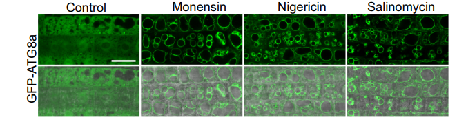
2025 Jul 18;16(1):6621. PMID: 40681515
Salinomycin purchased from MedChemExpress. Usage Cited in: Nat Commun. 2025 Jul 18;16(1):6621. [Abstract]
Salinomycin sodium salt (20 μM). The formation of membrane-like structures of GFP-ATG8a in response to three ionophores.
-
Adv Sci (Weinh)
Targeting Fibrotic Scarring by Mechanoregulation of Il11ra1+/Itga11+ Fibroblast Patterning Promotes Axon Growth after Spinal Cord Injury. [Abstract]2025 Sep 9:e13476. PMID: 40923460 -
Adv Sci (Weinh)
A Novel tRF, HCETSR, Derived From tRNA-Glu/TTC, Inhibits HCC Malignancy by Regulating the SPBTN1-catenin Complex Axis. [Abstract]2025 Feb 8:e2415229. PMID: 39921434 -
J Control Release
2020 Oct 10;326:387-395. PMID: 32702392 -
Cell Death Dis
PDIA4 confers resistance to ferroptosis via induction of ATF4/SLC7A11 in renal cell carcinoma. [Abstract]2023 Mar 11;14(3):193. PMID: 36906674 -
Pharmacol Res
Cardamonin retards progression of autosomal dominant polycystic kidney disease via inhibiting renal cyst growth and interstitial fibrosis. [Abstract]2020 May;155:104751. PMID: 32151678 -
Biosens Bioelectron
Proximity labeling-assisted click conjugation for electrochemical analysis of specific subpopulations in circulating extracellular vesicles. [Abstract]2024 Jul 1:255:116245. PMID: 38555770 -
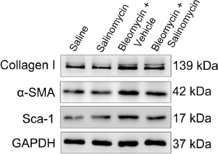
Cell Commun Signal
M2 macrophages promote myofibroblast differentiation of LR-MSCs and are associated with pulmonary fibrogenesis. [Abstract]2018 Nov 23;16(1):89. PMID: 30470231
Salinomycin purchased from MedChemExpress. Usage Cited in: Cell Commun Signal. 2018 Nov 23;16(1):89. [Abstract]
The expression of collagen I, α-smooth muscle actin (α-SMA), and Sca-1 in lung tissues is measured by western blotting in the treatment of Saline, Salinomycin, Bleomycin+Vehicle, and Bleomycin+ Salinomycin.
-
Acta Biomater
Reversal of epithelial-mesenchymal transition and inhibition of tumor stemness of breast cancer cells through advanced combined chemotherapy. [Abstract]2022 Oct 15:152:380-392. PMID: 36028199 -
Int J Nanomedicine
Salinomycin-Loaded Small-Molecule Nanoprodrugs Enhance Anticancer Activity in Hepatocellular Carcinoma. [Abstract]2020 Sep 15;15:6839-6854. PMID: 32982236 -
Mol Med
The mechanism of enterogenous toxin methylmalonic acid aggravating calcium-phosphorus metabolic disorder in uremic rats by regulating the Wnt/β-catenin pathway. [Abstract]2025 Jan 22;31(1):19. PMID: 39844078 -
EMBO Mol Med
A genome-wide RNAi screen reveals essential therapeutic targets of breast cancer stem cells. [Abstract]2019 Oct;11(10):e9930. PMID: 31476112 -
Cell Rep
Controlling mitochondrial membrane architecture via MIC60 determines viral replication to promote anti-viral immunity. [Abstract]2025 Jul 22;44(7):115922. PMID: 40628273 -
Anal Chem
Label-Free Isolation of Low-Adhesion Cells with Stem Properties for Cancer Stem Cell-Specific Drug Evaluation. [Abstract]2023 Apr 11;95(14):6191. PMID: 36122350 -
Talanta
Fluorescence-activated cell sorting-based efficient screening of monensin monoclonal antibodies and applications in lateral flow immunoassay. [Abstract]2025 Apr 10:293:128128. PMID: 40222095 -
Eur J Med Chem
A peptide-salinomycin conjugate with a bystander effect reduces the stemness characteristics of ovarian cancer cells and enhances drug sensitivity. [Abstract]2024 Oct 5:276:116701. PMID: 39067438 -
Cell Biosci
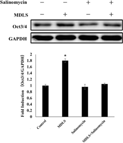
Modaline sulfate promotes Oct4 expression and maintains self-renewal and pluripotency of stem cells through JAK/STAT3 and Wnt signaling pathways. [Abstract]2021 Aug 4;11(1):156. PMID: 34348786
Salinomycin purchased from MedChemExpress. Usage Cited in: Cell Biosci. 2021 Aug 4;11(1):156. [Abstract]
Salinomycin (0.0078125 μg/ml; for 24 h) reverses the MDLS-induced up-regulation of Oct4. The expression of Oct4 is determined using Western blot analysis.
-
Biomed J
2020 Aug;43(4):368-374. PMID: 32563698 -
RSC Adv
BSA-MnO2-SAL multifunctional nanoparticle-mediated M1 macrophages polarization for glioblastoma therapy. [Abstract]2021 Nov 2;11(56):35331-35341. PMID: 35493189 -
J Mol Cell Biol
The inwardly rectifying potassium channel KCNJ12 regulates the stemness of hepatocellular carcinoma cells through the Wnt/β-catenin pathway. [Abstract]2025 Dec 2:mjaf048. PMID: 41328864 -
Hepatol Commun
Precision-cut tumor slices for modeling hepatocellular carcinoma enable at-scale drug screening. [Abstract]2025 May 16;9(6):e0706. PMID: 40377490 -
Int J Pharm
Polylactic Acid Based Biodegradable Hybrid Block Copolymeric Nanoparticle Mediated Co-delivery of Salinomycin and Doxorubicin for Cancer Therapy. [Abstract]2023 Mar 25:635:122779. PMID: 36842520 -
Eur J Pharmacol
To explore the potential combined treatment strategy for colorectal cancer: Inhibition of cancer stem cells and enhancement of intestinal immune microenvironment. [Abstract]2025 Jul 5:998:177533. PMID: 40120791 -
Cell Oncol (Dordr)
Inhibition of EREG/ErbB/ERK by Astragaloside IV reversed taxol-resistance of non-small cell lung cancer through attenuation of stemness via TGFβ and Hedgehog signal pathway. [Abstract]2024 Dec;47(6):2201-2215. PMID: 39373858 -
Int J Mol Sci
2023 Mar 21;24(6):5939. PMID: 36983012 -
Int J Mol Med
Simvastatin promotes endothelial dysfunction by activating the Wnt/β‑catenin pathway under oxidative stress. [Abstract]2019 Oct;44(4):1289-1298. PMID: 31432100 -
Int J Mol Sci
Sphingomyelin Synthase 2 Promotes Endothelial Dysfunction by Inducing Endoplasmic Reticulum Stress. [Abstract]2019 Jun 12;20(12):2861. PMID: 31212751 -
Nanoscale
Folic acid-targeted redox responsive polylactic acid-based nanoparticles co-delivering pirarubicin and salinomycin suppress breast cancer tumor growth in vivo. [Abstract]2024 Nov 7;16(43):20131-20146. PMID: 39420738 -
AAPS PharmSciTech
Concomitant Delivery of Pirarubicin and Salinomycin Synergistically Enhanced the Efficacy of Cancer Therapy and Reduced the Risk of Cancer Relapse. [Abstract]2024 Sep 7;25(7):211. PMID: 39242397 -
Sci Rep
PFDN2 stabilizes PYCR2 to activate Wnt/β-catenin signaling and promote colorectal cancer progression. [Abstract]2026 Feb 9;16(1):7909. PMID: 41656306 -
Cancers
Physical Cues in the Microenvironment Regulate Stemness-Dependent Homing of Breast Cancer Cells. [Abstract]2020 Aug 5;12(8):2176. PMID: 32764400 -
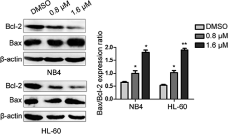
Oncol Rep
Salinomycin induces apoptosis and differentiation in human acute promyelocytic leukemia cells. [Abstract]2018 Aug;40(2):877-886. PMID: 29989650
Salinomycin purchased from MedChemExpress. Usage Cited in: Oncol Rep. 2018 Aug;40(2):877-886. [Abstract]
Salinomycin (SAL) has a pro-apoptotic effect on NB4 and HL-60 cells. The apoptosis-related protein levels of Bax and Bcl-2 are assessed by western blotting with the β-actin protein as an internal control.
-
ACS Infect Dis
Ex Vivo Phenotypic Screening of Two Small Repurposing Drug Collections Identifies Nifuratel as a Potential New Treatment against Visceral and Cutaneous Leishmaniasis. [Abstract]2021 Aug 13;7(8):2390-2401. PMID: 34114790 -
Antiviral Res
Salinomycin inhibits orthopoxvirus infection in vitro and in vivo by blocking endosomal acidification. [Abstract]2026 May:249:106403. PMID: 41941992 -
FASEB J
Stem Cells From the Apical Papilla Cultured in Spheroids Spontaneously Differentiate Into Odontoblasts via N-Cadherin Downregulation. [Abstract]2025 Dec 31;39(24):e71350. PMID: 41399894 -
Biochim Biophys Acta Mol Cell Res
Salinomycin inhibits SREBP1 to sensitize ferroptosis and ameliorate sorafenib resistance in clear cell renal cell carcinoma. [Abstract]2025 May 11:119989. PMID: 40360020 -
Viruses
2022 Aug 6;14(8):1734. PMID: 36016355 -
J Bioenerg Biomembr
Pre-clinical evidence that salinomycin is active against retinoblastoma via inducing mitochondrial dysfunction, oxidative damage and AMPK activation. [Abstract]2021 Oct;53(5):513-523. PMID: 34365583 -
Int J Hyperthermia
Eliminating cancer stem cells can inhibit progression of residual hepatocellular carcinoma after radiofrequency ablation. [Abstract]2026 Dec;43(1):2643510. PMID: 41883111 -
Exp Ther Med
Synthesis of chemical tools to improve water solubility and promote the delivery of salinomycin to cancer cells. [Abstract]2020 Mar;19(3):1835-1843. PMID: 32104239 -
Leuk Lymphoma
Salinomycin sodium exerts anti diffuse large B-cell lymphoma activity through inhibition of LRP6-mediated Wnt/β-catenin and mTORC1 signaling. [Abstract]2023 Jun;64(6):1151-1160. PMID: 37092573 -
-
-
-
-
-
Chemosphere
PFDA promotes cancer metastasis through macrophage M2 polarization mediated by Wnt/β-catenin signaling. [Abstract]2024 Jul 3:142758. PMID: 38969224 -
-
-
-
-


Solvent & Solubility
DMSO : ≥ 36.7 mg/mL (48.87 mM; Hygroscopic DMSO has a significant impact on the solubility of product, please use newly opened DMSO)
* "≥" means soluble, but saturation unknown.
Please refer to the solubility information to select the appropriate solvent. Once prepared, please aliquot and store the solution to prevent product inactivation from repeated freeze-thaw cycles.
Storage method and period of stock solution: -80°C, 6 months; -20°C, 1 month. When stored at -80°C, please use it within 6 months. When stored at -20°C, please use it within 1 month.
Concentration (start) × Volume (start) = Concentration (final) × Volume (final)
Select the appropriate dissolution method based on your experimental animal and administration route.
- For the following dissolution methods, please ensure to first prepare a clear stock solution using an In Vitro approach and then sequentially add co-solvents:
- To ensure reliable experimental results, the clarified stock solution can be appropriately stored based on storage conditions. As for the working solution for In Vivo experiments, it is recommended to prepare freshly and use it on the same day.
- The percentages shown for the solvents indicate their volumetric ratio in the final prepared solution. If precipitation or phase separation occurs during preparation, heat and/or sonication can be used to aid dissolution.
Add each solvent one by one: 10% DMSO 40% PEG300 5% Tween-80 45% Saline
Solubility: ≥ 2.5 mg/mL (3.33 mM); Clear solution
This protocol yields a clear solution of ≥ 2.5 mg/mL (saturation unknown).
Taking 1 mL working solution as an example, add 100 μL DMSO stock solution (25.0 mg/mL) to 400 μL PEG300, and mix evenly; then add 50 μL Tween-80 and mix evenly; then add 450 μL Saline to adjust the volume to 1 mL.
Preparation of Saline: Dissolve 0.9 g sodium chloride in ddH₂O and dilute to 100 mL to obtain a clear Saline solution.
Please enter the basic information of animal experiments:
-
-
-
-
Recommended: Prepare an additional quantity of animals to account for potential losses during experiments.
Please enter your animal formula composition:
-
%DMSO +
Recommended: Keep the proportion of DMSO in working solution below 2% if your animal is weak.
-
%+
-
+%Tween-80 + +
-
%Saline +
The co-solvents required include: DMSO, . All of co-solvents are available by MedChemExpress (MCE). , Tween 80. All of co-solvents are available by MedChemExpress (MCE).
Working solution concentration: 0.22 mg/mL
Method for preparing stock solution: mg drug dissolved in μL DMSO. Stock solution concentration: mg/mL.
1. Take μL DMSO stock solution;
2. Add μL .
μL , mix evenly;
3. Then add μL Tween 80, mix evenly;
4. Then add μL
Please ensure that the stock solution in the first step is dissolved to a clear state, and add co-solvents in sequence. You can use ultrasonic heating (ultrasonic cleaner, recommended frequency 20-40 kHz), vortexing, etc. to assist dissolution.

Protocol
For cisplatin or Salinomycin IC50 analysis in SW620 cells or Cisp-resistant SW620 cells, cells (1×10⁴/well) are cultured in 96-well plates and treated with different chemotherapeutics (cisplatin, Salinomycin) in different concentrations for 48 h. Then 20 μL of cell counting kit-8 (CCK-8) is added into each of the 96-wells. After 4 h incubation at 37°C, the optical density (OD) values are detected at 450 nm using the scan reader. Cell growth inhibiting rates are described as cell inhibiting curves and the IC50 parameters (inhibiting concentration of 50% cells) are evaluated by Xlfit 5.2 software. For cell proliferation analysis, SW620 cells or Cisp-resistant SW620 cells (5×10³/well) are also seeded in 96-well plates in serum-containing medium and treated with cisplatin (5 μM, according to the calculated IC50 values of cisplatin in SW620 cells) for 0, 12, 24, 48, 72 and 96 h. Then 20 μL cell counting kit-8 is added into each of the 96-wells. After 4-h incubation at 37°C, the coloring reactions are also quantified at 450 nm[2].
MedChemExpress (MCE) has not independently confirmed the accuracy of these methods. They are for reference only.
Mice[3]
Nude mice (nu/nu; 4-6 weeks of age) are used. HepG2 cells are suspended in 100 mL 1:1 serum-free DMEM and Matrigel. Mice are anesthetized with ketamine/xylazine and after surgically opening the abdomen, HepG2 cells are inoculated into the liver parenchyma and mice are monitored every 3 days for 35 days. Finally, 18 nude mice are divided into three groups that are intraperitoneally injected daily for 6 weeks: two Salinomycin-treated groups (4 mg/kg Salinomycin group, 8 mg/kg Salinomycin group) and the control group (saline water group).
Rats[4]
A total of 10 male rats are used in the experiment. After a routine anesthesia, the abdomen is opened. After a resuspension of high glucose medium not containing serum DMEM, and matrigel, the bladder transitional cancer cell line T24 is inoculated in the parenchyma of bladder in rats, and then the abdomen is sutured. After operation, the rats are randomized into the experiment group and the control group with five in each group. After operation, the rats in the experiment group are immediately given intraperitoneal injection of Salinomycin with a dosage of 8 mg/kg, while the rats in the control group are given intraperitoneal injection of normal saline. A close observation is paid during the drug administration period. After 15 d, the rats are sacrificed by cervical dislocation, and the complete tumor tissues are stripped to observe the tumor growth and metastasis.
MedChemExpress (MCE) has not independently confirmed the accuracy of these methods. They are for reference only.


Purity & Documentation
-
Data Sheet (286 KB)
-
SDS (479 KB)
- English - EN (479 KB)
- Français - FR (479 KB)
- Deutsch - DE (479 KB)
- Norwegian - NO (479 KB)
- Español - ES (479 KB)
- Swedish - SV (479 KB)
- Italian - IT (479 KB)
- Korean - KR (479 KB)
- Portuguese - PT (479 KB)
-
Handling Instructions (2659 KB)
References
[1]. Lu D, et al. Salinomycin inhibits Wnt signaling and selectively induces apoptosis in chronic lymphocytic leukemia cells. Proc Natl Acad Sci U S A. 2011 Aug 9;108(32):13253-7. [Content Brief]
[2]. Zhou J, et al. Salinomycin induces apoptosis in cisplatin-resistant colorectal cancer cells by accumulation of reactiveoxygen species. Toxicol Lett. 2013 Oct 24;222(2):139-45. [Content Brief]
[3]. Klose J, et al. Salinomycin: Anti-tumor activity in a pre-clinical colorectal cancer model. PLoS One. 2019 Feb 14;14(2):e0211916. [Content Brief]
[4]. Wang F, et al. Salinomycin Inhibits Proliferation and Induces Apoptosis of Human Hepatocellular Carcinoma Cells In Vitro and In Vivo. PLoS One. 2012; 7(12): e50638. [Content Brief]
[5]. Qu H, et al. Effect of salinomycin on metastasis and invasion of bladder cancer cell line T24. Asian Pac J Trop Med. 2015 Jul;8(7):578-82. [Content Brief]
[6]. Naujokat C, et al. Salinomycin as a drug for targeting human cancer stem cells. J Biomed Biotechnol. 2012;2012:950658. [Content Brief]


Complete Stock Solution Preparation Table
Please refer to the solubility information to select the appropriate solvent. Once prepared, please aliquot and store the solution to prevent product inactivation from repeated freeze-thaw cycles.
Storage method and period of stock solution: -80°C, 6 months; -20°C, 1 month. When stored at -80°C, please use it within 6 months. When stored at -20°C, please use it within 1 month.
| Optional Solvent | Concentration Solvent Mass | 1 mg | 5 mg | 10 mg | 25 mg |
|---|---|---|---|---|---|
| DMSO | 1 mM | 1.3316 mL | 6.6578 mL | 13.3156 mL | 33.2889 mL |
| 5 mM | 0.2663 mL | 1.3316 mL | 2.6631 mL | 6.6578 mL | |
| 10 mM | 0.1332 mL | 0.6658 mL | 1.3316 mL | 3.3289 mL | |
| 15 mM | 0.0888 mL | 0.4439 mL | 0.8877 mL | 2.2193 mL | |
| 20 mM | 0.0666 mL | 0.3329 mL | 0.6658 mL | 1.6644 mL | |
| 25 mM | 0.0533 mL | 0.2663 mL | 0.5326 mL | 1.3316 mL | |
| 30 mM | 0.0444 mL | 0.2219 mL | 0.4439 mL | 1.1096 mL | |
| 40 mM | 0.0333 mL | 0.1664 mL | 0.3329 mL | 0.8322 mL |


 Powered by Bioz