



β-Amyloid (31-35)

Based on 1 Customer Validation
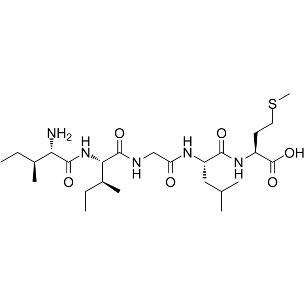
β-Amyloid (31-35) is the shortest sequence of native Amyloid-β peptide that retains neurotoxic activity.
For research use only. We do not sell to patients.
- Purity : 99.10%
- CAS No.: 149385-65-9
- Formula: C25H47N5O6S
- Molecular Weight:545.74
-
Storage:
Sealed storage, away from moisture.
Powder -80°C, 2 years , -20°C, 1 year* In solvent : -80°C, 6 months; -20°C, 1 month (sealed storage, away from moisture)

To place orders, for customer services and technical support, please contact: MedChemExpress USA
Tel: 609-228-6898 E-mail: [email protected] [email protected]


-
 Biological Activity
-
Chemical Information
-
Solvent & Solubility
- Protocols
- Purity & Documentation
- References
-
 Help & FAQs


-

HY-P1363A
β-Amyloid (1-42), humanAmyloid β Peptide -

HY-P1363B
β-Amyloid (1-42), human, HFIP-treatedβ-Amyloid (1-42) Peptide -
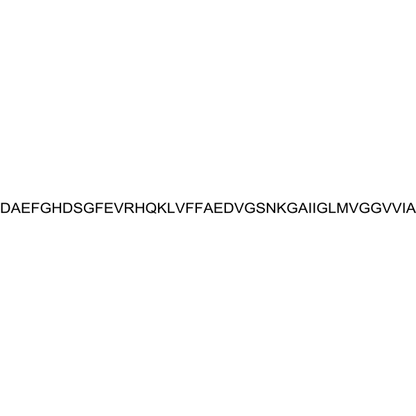
HY-P1388
β-Amyloid (1-42), (rat/mouse)β-Amyloid (1-42) Peptide -
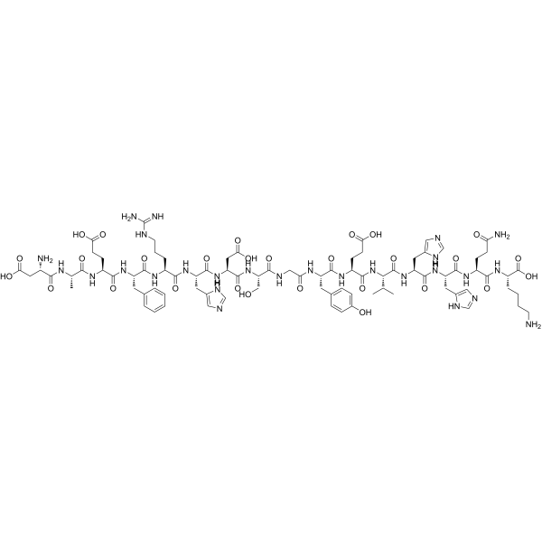
HY-P1466
β-Amyloid (1-16)β-Amyloid -
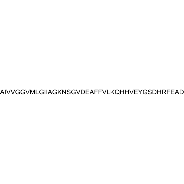
HY-P1362
β-Amyloid (42-1), humanInactive Form of Aβ(1-42) -
HY-P0128
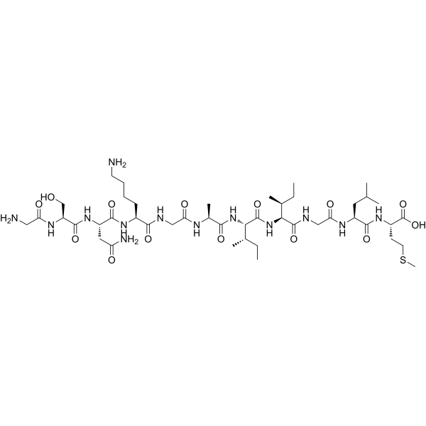
β-Amyloid (25-35)Amyloid β Peptide -

HY-P1468
β-Amyloid (1-28)Amyloid-β Peptide -

HY-P0128A
β-Amyloid (25-35), HFIP-treatedAmyloid β Peptide -
HY-P1046
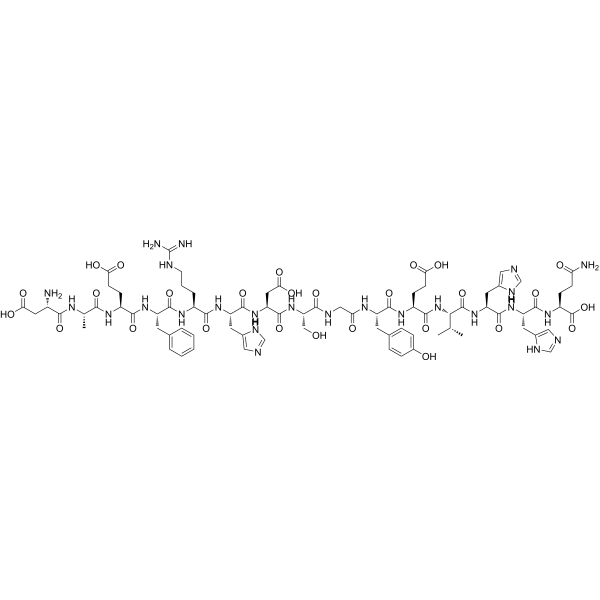
β-Amyloid (1-15)Peptide Fragment -

HY-P1474
β-Amyloid (22-35)β-Amyloid -

HY-P1522
β-Amyloid (29-40)Amyloid-β Peptide Fragment -
HY-N0615
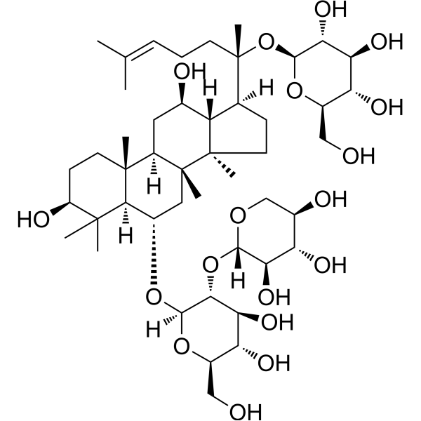
Notoginsenoside R1Apoptosis Inhibitor


Biological Activity
Description
In Vitro
β-Amyloid (31-35) is a functional cytotoxic domain of Aβ peptide. β-Amyloid (31-35) increases the phosphorylation of biotinylated Aβ(1-40), enhances CDK-1 activity, and also inhibits binding of Aβ to cyclin B1. β-Amyloid (31-35) is cytotoxic, and such an effect can be inhibited by olomoucine in differentiated human teratocarcinoma cell line, Ntera 2/cl-D1 (NT-2) neurons[1].
β-Amyloid Aggregation Guidelines (Following is our recommended protocol. This protocol only provides a guideline, and should be modified according to your specific needs).
1. Solid Aβ peptide was dissolved in cold hexafluoro-2-propanol (HFIP). The peptide was incubated at room temperature for at least 1h to establish monomerization and randomization of structure.
2. The HFIP was removed by evaporation, and the resulting peptide was stored as a film at -20 or -80 °C.
3. The resulting film was dissolved in anhydrous DMSO at 5 mM and then diluted into the appropriate concentration and buffer (serum- and phenol red-free culture medium) with vortexing.
4. Next, the solution was age 48h at 4-8 °C. The sample was then centrifuged at 14000g for 10 min at 4-8 °C; the soluble oligomers were in the supernatant. The supernatant was diluted 10-200-fold for experiments.
Methods vary depends on the downstream applications.
MedChemExpress (MCE) has not independently confirmed the accuracy of these methods. They are for reference only. Further protocols information, click here.


Chemical Information
-
CAS No. 149385-65-9
-
Appearance Solid
-
Molecular Weight 545.74
-
Formula C25H47N5O6S
-
Color White to off-white
-
Sequence
Ile-Ile-Gly-Leu-Met
-
Sequence Shortening
IIGLM
-
Shipping
Room temperature in continental US; may vary elsewhere.
-
Storage
Sealed storage, away from moisture
Powder -80°C 2 years -20°C 1 year * In solvent : -80°C, 6 months; -20°C, 1 month (sealed storage, away from moisture)


Solvent & Solubility
In Vitro:
DMSO : 5 mg/mL (9.16 mM; ultrasonic and adjust pH to 7 with HCl; Hygroscopic DMSO has a significant impact on the solubility of product, please use newly opened DMSO)
Please refer to the solubility information to select the appropriate solvent. Once prepared, please aliquot and store the solution to prevent product inactivation from repeated freeze-thaw cycles.
Storage method and period of stock solution: -80°C, 6 months; -20°C, 1 month (sealed storage, away from moisture). When stored at -80°C, please use it within 6 months. When stored at -20°C, please use it within 1 month.
Please refer to the solubility information to select the appropriate solvent. Once prepared, please aliquot and store the solution to prevent product inactivation from repeated freeze-thaw cycles.
Storage method and period of stock solution: -80°C, 6 months; -20°C, 1 month (sealed storage, away from moisture). When stored at -80°C, please use it within 6 months. When stored at -20°C, please use it within 1 month.
Concentration (start) × Volume (start) = Concentration (final) × Volume (final)
In Vivo:
Select the appropriate dissolution method based on your experimental animal and administration route.
- For the following dissolution methods, please ensure to first prepare a clear stock solution using an In Vitro approach and then sequentially add co-solvents:
- To ensure reliable experimental results, the clarified stock solution can be appropriately stored based on storage conditions. As for the working solution for In Vivo experiments, it is recommended to prepare freshly and use it on the same day.
- The percentages shown for the solvents indicate their volumetric ratio in the final prepared solution. If precipitation or phase separation occurs during preparation, heat and/or sonication can be used to aid dissolution.
Add each solvent one by one: 10% DMSO 90% (20% SBE-β-CD in Saline)
Solubility: ≥ 2.08 mg/mL (3.81 mM); Clear solution
This protocol yields a clear solution of ≥ 2.08 mg/mL (saturation unknown).
Taking 1 mL working solution as an example, add 100 μL DMSO stock solution (20.8 mg/mL) to 900 μL 20% SBE-β-CD in Saline, and mix evenly.
Preparation of 20% SBE-β-CD in Saline (4°C, storage for one week): 2 g SBE-β-CD powder is dissolved in 10 mL Saline, completely dissolve until clear.
In Vivo Dissolution Calculator
Please enter the basic information of animal experiments:
-
-
-
-
Recommended: Prepare an additional quantity of animals to account for potential losses during experiments.
Please enter your animal formula composition:
-
%DMSO +
Recommended: Keep the proportion of DMSO in working solution below 2% if your animal is weak.
-
%+
-
+%Tween-80 + +
-
%Saline +
The co-solvents required include: DMSO, . All of co-solvents are available by MedChemExpress (MCE). , Tween 80. All of co-solvents are available by MedChemExpress (MCE).
Working solution concentration: 0.22 mg/mL
Method for preparing stock solution: mg drug dissolved in μL DMSO. Stock solution concentration: mg/mL. * In solvent : -80°C, 6 months; -20°C, 1 month (sealed storage, away from moisture)
1. Take μL DMSO stock solution;
2. Add μL .
μL , mix evenly;
3. Then add μL Tween 80, mix evenly;
4. Then add μL
Please ensure that the stock solution in the first step is dissolved to a clear state, and add co-solvents in sequence. You can use ultrasonic heating (ultrasonic cleaner, recommended frequency 20-40 kHz), vortexing, etc. to assist dissolution.

Protocols
-
Neurotoxicity Study
This protocol assesses in vitro neurotoxicity by combining neuronal viability, mitochondrial/metabolic activity, neurite outgrowth, and optional neuronal network function readouts. Calcein-AM or resazurin/PrestoBlue readouts estimate viable or metabolically active cells; βIII-tubulin immunofluorescence detects neuronal morphology and neurite networks; TMRE detects mitochondrial membrane potential; and MEA recordings detect functional changes in neuronal network activity.
-
Amyloid: Congo Red Amyloid Staining
Congo red amyloid staining is a histochemical method used to detect extracellular amyloid deposits in tissue sections based on the affinity of Congo red dye for β-pleated sheet-rich protein aggregates. When bound to amyloid, Congo red produces characteristic apple-green birefringence under polarized light microscopy, which is widely regarded as a diagnostic feature of amyloid deposition in histopathology. The diagnostic principle relies on the combination of dye binding (congophilia) and optical anisotropy under polarized illumination, which distinguishes amyloid from most non-amyloid eosinophilic extracellular deposits in routine histological evaluation. Amyloid identification by Congo red staining remains a cornerstone in diagnostic pathology despite the availability of adjunct methods such as immunohistochemistry and mass spectrometry, particularly because of its ability to localize deposits directly within tissue architecture. The specificity of Congo red-positive deposits is incre
-
Alzheimer’s Disease Modeling
Alzheimer’s Disease (AD) is a neurodegenerative disorder characterized by a progressive decline in cognitive functions and loss of specific types of neurons and synapses. Alzheimer's symptoms can be simulated in mice by injecting drugs (such as Aβ) or genetically modified.


Purity & Documentation
-
Data Sheet (273 KB)
-
SDS (252 KB)
- English - EN (252 KB)
- Français - FR (252 KB)
- Deutsch - DE (252 KB)
- Norwegian - NO (252 KB)
- Español - ES (252 KB)
- Swedish - SV (252 KB)
- Italian - IT (252 KB)
- Korean - KR (252 KB)
- Portuguese - PT (252 KB)
-
Handling Instructions (2659 KB)
References
[1]. Milton NG, et al. The amyloid-beta peptide binds to cyclin B1 and increases human cyclin-dependent kinase-1 activity. Neurosci Lett. 2002 Apr 5;322(2):131-3. [Content Brief]
[2]. Misiti F, et al. Fragment 31-35 of beta-amyloid peptide induces neurodegeneration in rat cerebellar granule cells via bax gene expression and caspase-3 activation. A crucial role for the redox state of methionine-35 residue. Neurochem Int. 2006 Oct;49(5):525-32. [Content Brief]
[3]. M Jesús Pérez de Vega, et al. Synthesis and biological properties of beta-turned Abeta(31-35) constrained analogues. Bioorg Med Chem Lett. 2008 Mar 15;18(6):2078-82. [Content Brief]


Complete Stock Solution Preparation Table
Please refer to the solubility information to select the appropriate solvent. Once prepared, please aliquot and store the solution to prevent product inactivation from repeated freeze-thaw cycles.
Storage method and period of stock solution: -80°C, 6 months; -20°C, 1 month (sealed storage, away from moisture). When stored at -80°C, please use it within 6 months. When stored at -20°C, please use it within 1 month.
| Optional Solvent | Concentration Solvent Mass | 1 mg | 5 mg | 10 mg | 25 mg |
|---|---|---|---|---|---|
| DMSO | 1 mM | 1.8324 mL | 9.1619 mL | 18.3237 mL | 45.8094 mL |
| 5 mM | 0.3665 mL | 1.8324 mL | 3.6647 mL | 9.1619 mL |


β-Amyloid (31-35) Related Classifications
HY-P1517 Related Classifications
 Powered by Bioz