



Anti-Mouse Ly6C Antibody (Monts 1)

Based on 1 Customer Validation
Anti-Mouse Ly6C Antibody (Monts 1) is a rat-derived anti-mouse Ly6C IgG2a type antibody inhibitor. Anti-Mouse Ly6C Antibody (Monts 1) depletes monocytes, neutrophils, and some other granulocyte populations. Anti-Mouse Ly6C Antibody (Monts 1) can be used for the researches of cancer, immunology and infection, such as chikungunya (CHIKV) and aggressive tumor.
For research use only. We do not sell to patients.
- Purity : 95.00%
- Molecular Weight:150 kDa
-
Storage:
Please store the product under the recommended conditions in the Certificate of Analysis.

To place orders, for customer services and technical support, please contact: MedChemExpress USA
Tel: 609-228-6898 E-mail: [email protected] [email protected]


-
 Biological Activity
-
Chemical Information
- Protocols
- Purity & Documentation
- References
-
 Help & FAQs




Biological Activity
Description
Isotype
Rat IgG2a kappa
Recommend Isotype Controls
Species Reactivity
Mouse
IC50 & Target
Ly6C
In Vitro
The antibody framework is stable, specific and adaptable, and has the ability to bind both antigens and endogenous immune receptors. Monoclonal antibodies have several derivatives, including bispecific antibodies, antibody-drug conjugates, and antibody fragments, and have significant effects in fields such as immunology and oncology. When designing inhibitory antibodies, considerations include identification of antigen-specific variable regions, choice of expression system, use of multispecific formats, and antibody derivatives based on fragmentation, oligomerization, or conjugation with other functional moieties[1].
MedChemExpress (MCE) has not independently confirmed the accuracy of these methods. They are for reference only. Further protocols information, click here.
In Vivo
Anti-Mouse Ly6C Antibody (Monts 1) (700 μg, i.p., 24 h prior to infection) reduces chikungunya (CHIKV) infection in the skin and subsequent dissemination in CHIKV mice models[2].
Anti-Mouse Ly6C Antibody (Monts 1) (70 μg, Subconjuctivally administration, 7 days before infection) depletes macrophages in Herpes Simplex Virus-1-induced subclinical corneal inflammation[3].
MedChemExpress (MCE) has not independently confirmed the accuracy of these methods. They are for reference only.
-
Animal Model:Aggressive KP 344SQ tumor mice models treated with PD-1/CTLA-4[1]
-
Dosage:200 μg
-
Administration:Intraperitoneally injection, once a week for 4 weeks
-
Result:Reversed the resistance to anti-PD-1/CTLA-4 treatment.
Restored the levels of classical, non-classical monocytes and immune cell responses.
-
Animal Model:Mice were inoculated with chikungunya-LR in footpad[2]
-
Dosage:700 μg
-
Administration:Intraperitoneally injection, 24 h prior to inoculation
-
Result:Reduced viral RNA levels.
Depleted monocytes, neutrophils, and some other granulocyte populations.
Significantly reduced in viral RNA levels in the skin, DLN, serum, spleen, and contralateral foot.
-
Animal Model:Mice with HSV-1(Herpes Simplex Virus-1)-induced subclinical corneal inflammation[3]
-
Dosage:70 μg
-
Administration:Subconjuctivally administration, 7 days before infection
-
Result:Effectively depleted macrophages from HSV-1 infected.
Gene ID
17067 [NCBI]
Accession
Conjugated
Unconjugated
Reconsititution
The product can be reconstituted/diluted with sterile PBS or saline.
Format
-
Product Image
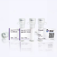
Application
in vivo macrophage depletion (in combination with clodronate liposomes); Flow cytometry


Chemical Information
-
Appearance Liquid
-
Molecular Weight 150 kDa
-
Color Colorless to light yellow
-
SMILES
[Anti-Mouse Ly6C Antibody (Monts 1)]
-
Formulation
Please refer to the lot-specific COA for specific buffer information.
-
Storage
Please store the product under the recommended conditions in the Certificate of Analysis.

Protocols
-
Research Protocol for Infectious Diseases
Infectious-disease experiments test how pathogens interact with host barriers, innate immune receptors, inflammatory signaling, pathogen replication, and tissue injury; pattern-recognition receptors such as TLRs, RIG-I-like receptors, NOD-like receptors, and inflammasomes detect microbial molecules and activate NF-κB, interferon, and cytokine responses. The central hypothesis is that infection severity reflects the balance between pathogen burden and host response: protective inflammation restricts pathogen growth, whereas excessive or mislocalized inflammation contributes to tissue damage and disease phenotype. Unresolved questions include which host pathways are protective versus pathogenic, why some infection models fail to translate to human disease, and which combined readouts best predict clinically relevant infection outcomes.


Purity & Documentation
-
Data Sheet (262 KB)
-
SDS (251 KB)
- English - EN (251 KB)
- Français - FR (251 KB)
- Deutsch - DE (251 KB)
- Norwegian - NO (251 KB)
- Español - ES (251 KB)
- Swedish - SV (251 KB)
- Italian - IT (251 KB)
- Korean - KR (251 KB)
- Portuguese - PT (251 KB)
-
Inhibitory Antibodies User Guide (603 KB)
References
[1]. Rodriguez BL, et al. Targeting immunosuppressive Ly6C+ classical monocytes reverses anti-PD-1/CTLA-4 immunotherapy resistance. Front Immunol. 2023 Jun 28;14:1161869. [Content Brief]
[2]. Holmes AC, et al. Ly6C+ monocytes in the skin promote systemic alphavirus dissemination. Cell Rep. 2024 Mar 26;43(3):113876. [Content Brief]
[3]. Rowe AM, et al. Subclinical Herpes Simplex Virus Type 1 Infections Provide Site-Specific Resistance to an Unrelated Pathogen. J Immunol. 2017 Feb 15;198(4):1706-1717. [Content Brief]

Calculators
Concentration (start) × Volume (start) = Concentration (final) × Volume (final)


Anti-Mouse Ly6C Antibody (Monts 1) Related Classifications
HY-P990200 Related Classifications
 Powered by Bioz