



Atg12 Antibody (YA830)

Based on 1 Customer Validation
Atg12 Antibody (YA830) is a Mouse-derived and non-conjugated IgE monoclonal antibody, targeting to Atg12.
-
Host:
Mouse
-
Isotype:
IgE
-
Application:
WB, IHC-P, ICC/IF
-
Reactivity :
Human, Mouse, Rat
-
Formulation:
Supplied in 1*PBS (pH7.4), 0.2% BSA and 40% Glycerol. Preservative: 0.05% Sodium Azide.
-
Conjugation:
Non-conjugated


To place orders, for customer services and technical support, please contact: MedChemExpress USA
Tel: 609-228-6898 E-mail: [email protected] [email protected]





Applications
| Application |
WB

WB: Western Blot
|
ICC/IF
ICC/IF: Immunocytochemistry/
Immunofluorescence |
IHC-P
IHC-P: Immunohistochemistry-Paraffin
|
|---|---|---|---|
| Dilution Ratio | 1:500-1:1000 | 1:50-1:200 | 1:50-1:200 |


Product Details
Atg12 Antibody (YA830) is a Mouse-derived and non-conjugated IgE monoclonal antibody, targeting to Atg12.
-
Host Mouse

-
Clonality Monoclonal

-
Species ReactivityHuman, Mouse, Rat
-
Observed Molecular WeightObserved band size: 14 kDa
Note: Due to possible protein modifications or aggregation, the molecular weight should be confirmed by actual measurement, and the predicted value is for reference only.

-
Calculated Molecular Weight Predicted band size: 15 kDa

Entrez Gene: 9140 Human ; 67526 Mouse ; 361321 Rat
SwissProt: O94817 Human ; Q9CQY1 Mouse ; Q2TBJ5 Rat
OMIM: 609608 Human
Synthetic peptide corresponding to Human Atg12.AA range:91-140.
Endogenous
Protein A affinity purified.
Non-conjugated
Unmodified
IgE


Product Properties
-
Appearance
Liquid
-
Formulation
Supplied in 1*PBS (pH7.4), 0.2% BSA and 40% Glycerol. Preservative: 0.05% Sodium Azide.
-
Concentration
Batch-dependent, Please check the COA for the concentration of each lot. Check Lot Concentration
-
Storage & Stability
Stored at -20°C for 1 year. Avoid repeated freeze / thaw cycles.
-
Livraison
Shipping with blue ice.


Verification Images
-
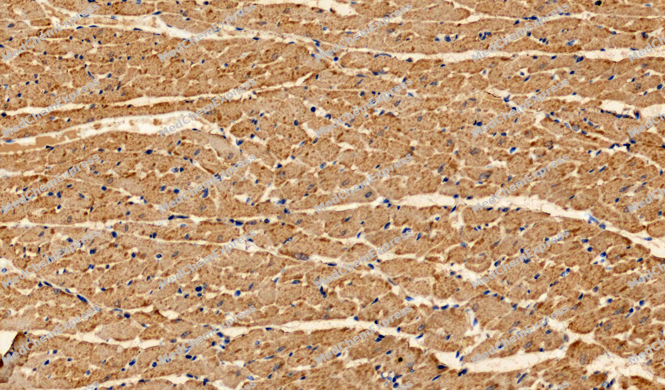 Immunohistochemical analysis of paraffin-embedded Rat heart tissue using Atg12 Antibody. The section was pre-treated using heat mediated antigen retrieval with sodium citrate buffer (pH 6.0) for 8 minutes. The tissues were blocked in QuickBlock for 20 minutes at room temperature, washed with ddH2O and PBS, and then probed with the primary antibody (HY-P80021, 1/200) in 4℃ overnight. The detection was performed using an HRP conjugated compact polymer system. DAB was used as the chromogen. Tissues were counterstained with hematoxylin and mounted with DPX.
 -
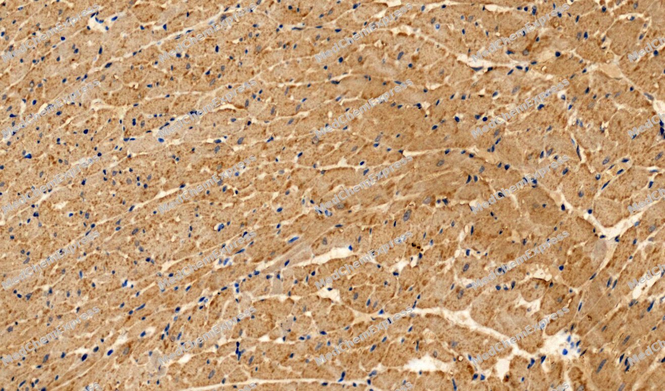 Immunohistochemical analysis of paraffin-embedded Rat heart tissue using Atg12 Antibody. The section was pre-treated using heat mediated antigen retrieval with sodium citrate buffer (pH 6.0) for 8 minutes. The tissues were blocked in QuickBlock for 20 minutes at room temperature, washed with ddH2O and PBS, and then probed with the primary antibody (HY-P80021, 1/200) in 4℃ overnight. The detection was performed using an HRP conjugated compact polymer system. DAB was used as the chromogen. Tissues were counterstained with hematoxylin and mounted with DPX.
 -
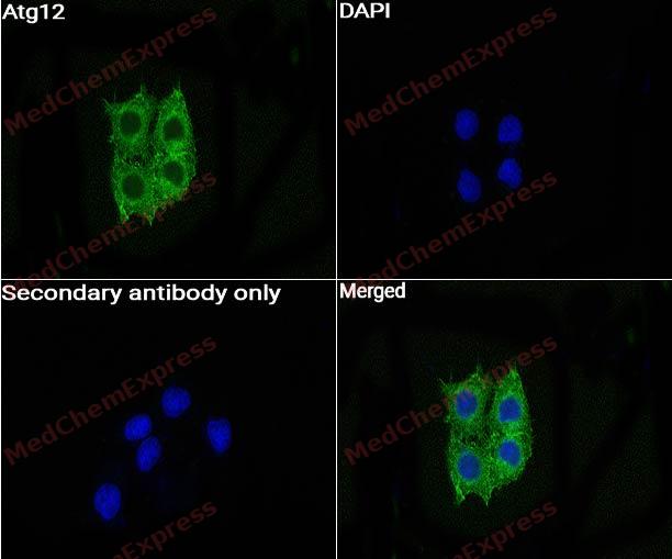 Immunocytochemistry analysis of Hela cells labeling Atg12 with Atg12 Antibody (HY-P80021) at 1:50 dilution. Cells were fixed in 4% paraformaldehyde for 15 minutes at room temperature, permeabilized with 0.1% Triton X-100 in PBS for 15 minutes at room temperature, then blocked with quick block buffer for 10 minutes at room temperature. Cells were then incubated with Atg12 Antibody (HY-P80021) at 1:50 dilution in quick block buffer overnight at 4 ℃. AF488-conjugated Goat Anti-Rabbit IgG H&L(HY-P8002, Green) was used as the secondary antibody at 1/1,000 dilution. PBS instead of the primary antibody was used as the secondary antibody only control. The Nuclear counterstain was DAPI (Blue).
 -
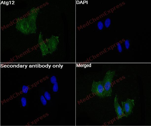 Immunocytochemistry analysis of Hela cells labeling Atg12 with Atg12 Antibody (HY-P80021) at 1:200 dilution. Cells were fixed in 4% paraformaldehyde for 15 minutes at room temperature, permeabilized with 0.1% Triton X-100 in PBS for 15 minutes at room temperature, then blocked with quick block buffer for 10 minutes at room temperature. Cells were then incubated with Atg12 Antibody (HY-P80021) at 1:200 dilution in quick block buffer overnight at 4 ℃. AF488-conjugated Goat Anti-Rabbit IgG H&L(HY-P8002, Green) was used as the secondary antibody at 1/1,000 dilution. PBS instead of the primary antibody was used as the secondary antibody only control. The Nuclear counterstain was DAPI (Blue).


Background
-
Function
Atg12 is an Ubiquitin-like protein involved in autophagy vesicles formation. Conjugation with ATG5 through a ubiquitin-like conjugating system involving also ATG7 as an E1-like activating enzyme and ATG10 as an E2-like conjugating enzyme, is essential for its function. The ATG12-ATG5 conjugate acts as an E3-like enzyme which is required for lipidation of ATG8 family proteins and their association to the vesicle membranes. As part of the ATG8 conjugation system with ATG5 and ATG16L1, required for recruitment of LRRK2 to stressed lysosomes and induction of LRRK2 kinase activity in response to lysosomal stress; (Microbial infection) May act as a proviral factor. In association with ATG5, negatively regulates the innate antiviral immune response by impairing the type I IFN production pathway upon vesicular stomatitis virus (VSV) infection. Required for the translation of incoming hepatitis C virus (HCV) RNA and, thereby, for the initiation of HCV replication, but not required once infection is established[1][2].
-
Subcellular Localization
Cytoplasm; Preautophagosomal structure membrane; Peripheral membrane protein
-
Expression
Tissue_specificity:general expression
Induction:Expression is induced by mitochondrial DNA deletions, chloramphenicol and nicotinamide -
Subunit
Forms a conjugate with ATG5 (PubMed:11096062, PubMed:11825910, PubMed:12207896, PubMed:17709747, PubMed:23202584, PubMed:24191030, PubMed:26812546).
-
SwissProt ID
-
Research Field
Cell Biology


Documentation
-
Fiche technique (235 KB)
-
SDS (251 KB)
- English - EN (251 KB)
- Français - FR (251 KB)
- Deutsch - DE (251 KB)
- Norwegian - NO (251 KB)
- Español - ES (251 KB)
- Swedish - SV (251 KB)
- Italian - IT (251 KB)
- Korean - KR (251 KB)
- Portuguese - PT (251 KB)
-
User Guide for Antibodies (1077 KB)

 Powered by Bioz
