



Bmi-1 Antibody (YA821)
(Synonyms: PCGF4, RNF51, BMI1, Polycomb complex protein BMI-1, Polycomb group RING finger protein 4, RING finger protein 51)Based on 1 publication(s) in Google Scholar
Bmi-1 Antibody (YA821) is a Mouse-derived and non-conjugated IgG2a monoclonal antibody, targeting to Bmi-1.
-
Host:
Mouse
-
Isotype:
IgG
-
Application:
WB, ICC/IF, IHC-P, FC
-
Reactivity :
Human, Mouse, Rat
-
Formulation:
Supplied in 1*PBS (pH7.4), 0.2% BSA and 40% Glycerol. Preservative: 0.05% Sodium Azide.
-
Conjugation:
Non-conjugated


To place orders, for customer services and technical support, please contact: MedChemExpress USA
Tel: 609-228-6898 E-mail: [email protected] [email protected]



Publications Citing Use of MedChemExpress (MCE) Bmi-1 Antibody (YA821)
More

Applications
| Application |
WB

WB: Western Blot
|
IHC-P
IHC-P: Immunohistochemistry-Paraffin
|
ICC/IF
ICC/IF: Immunocytochemistry/
Immunofluorescence |
FC
FC: Flow Cytometry
|
|---|---|---|---|---|
| Dilution Ratio | 1:1000 | 1:200 | 1:50 | 1:100-1:200 |


Product Details
Bmi-1 Antibody (YA821) is a Mouse-derived and non-conjugated IgG2a monoclonal antibody, targeting to Bmi-1.
-
Host Mouse

-
Clonality Monoclonal

-
Species ReactivityHuman, Mouse, Rat
-
Observed Molecular WeightObserved band size: 40 kDa
Note: Due to possible protein modifications or aggregation, the molecular weight should be confirmed by actual measurement, and the predicted value is for reference only.

-
Calculated Molecular Weight Predicted band size: 37 kDa

Entrez Gene: 100532731 Human ; 648 Human ; 12151 Mouse ; 307151 Rat
SwissProt: P35226 Human ; P25916 Mouse ;
OMIM: 164831 Human
Synthetic peptide corresponding to Human Bmi1.AA range: full length.
Endogenous
Protein A affinity purified.
Non-conjugated
Unmodified
IgG


Product Properties
-
Appearance
Solution
-
Formulation
Supplied in 1*PBS (pH7.4), 0.2% BSA and 40% Glycerol. Preservative: 0.05% Sodium Azide.
-
Concentration
Batch-dependent, Please check the COA for the concentration of each lot. Check Lot Concentration
-
Storage & Stability
Stored at -20°C for 1 year. Avoid repeated freeze / thaw cycles.
-
Shipping
Shipping with blue ice.


Publications (1)
-
Journal Impact Factor
-
Most Recent
-
Toxics
Triptolide Causes Spermatogenic Disorders by Inducing Apoptosis in the Mitochondrial Pathway of Mouse Testicular Spermatocytes. [Abstract]2024 Dec 10;12(12):896. PMID: 39771111


Verification Images
-
Western blot analysis of extracts from K562 (lane 1) and Hela (lane 2) and Raji (lane 3) using Bmil antibody. Proteins were transferred to a PVDF membrane and blocked with 5% nonfat powdered milk in PBST for 2 hour at room temperature. The primary antibody (1/1000) and Loading control antibody (GAPDH, 1/3000) was diluted with 5% nonfat powdered milk in PBST at 4°C overnight. Goat Anti-mouse IgG-HRP Secondary Antibody (1/8,000) was incubated for 45min at room temperature.
-
 Immunohistochemical analysis of paraffin-embedded human colon cancer using Bmi-1 antibody. The section was pre-treated using heat mediated antigen retrieval with EDTA (pH 9.0) for 14 minutes. The tissues were blocked in 5% BSA for 30 minutes at room temperature, washed with TBST, and then probed with the primary antibody (HY-P80034, 1/200) for 30 minutes at room temperature. The detection was performed using Polymer HRP-conjugated Goat Anti-Mouse/Rabbit lgG(H&L) secondary antibody (HY-P83652). DAB was used as the chromogen. Tissues were counterstained with hematoxylin and mounted with DPX.
 -
 Immunohistochemical analysis of paraffin-embedded human breast cancer using Bmi-1 antibody. The section was pre-treated using heat mediated antigen retrieval with EDTA (pH 9.0) for 14 minutes. The tissues were blocked in 5% BSA for 30 minutes at room temperature, washed with TBST, and then probed with the primary antibody (HY-P80034, 1/200) for 30 minutes at room temperature. The detection was performed using Polymer HRP-conjugated Goat Anti-Mouse/Rabbit lgG(H&L) secondary antibody (HY-P83652). DAB was used as the chromogen. Tissues were counterstained with hematoxylin and mounted with DPX.
 -
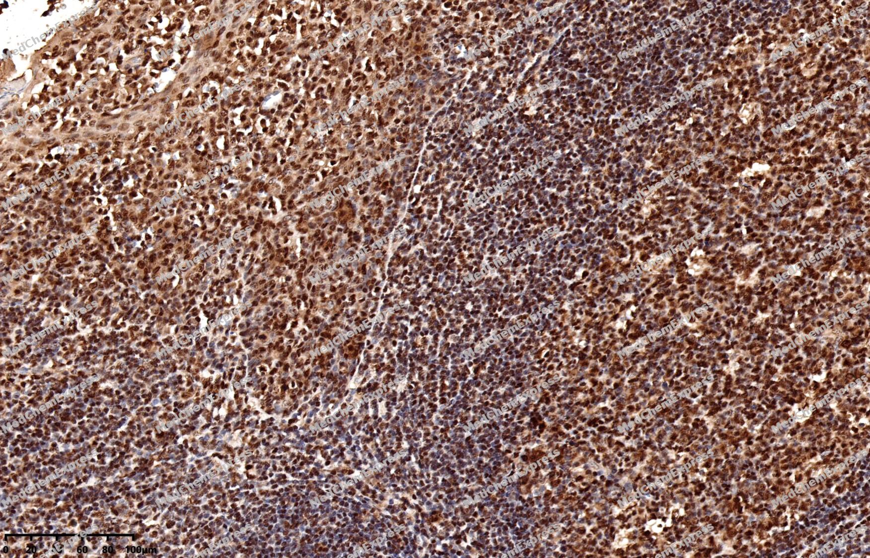 Immunohistochemical analysis of paraffin-embedded human tonsil using Bmi-1 antibody. The section was pre-treated using heat mediated antigen retrieval with EDTA (pH 9.0) for 14 minutes. The tissues were blocked in 5% BSA for 30 minutes at room temperature, washed with TBST, and then probed with the primary antibody (HY-P80034, 1/200) for 30 minutes at room temperature. The detection was performed using Polymer HRP-conjugated Goat Anti-Mouse/Rabbit lgG(H&L) secondary antibody (HY-P83652). DAB was used as the chromogen. Tissues were counterstained with hematoxylin and mounted with DPX.
 -
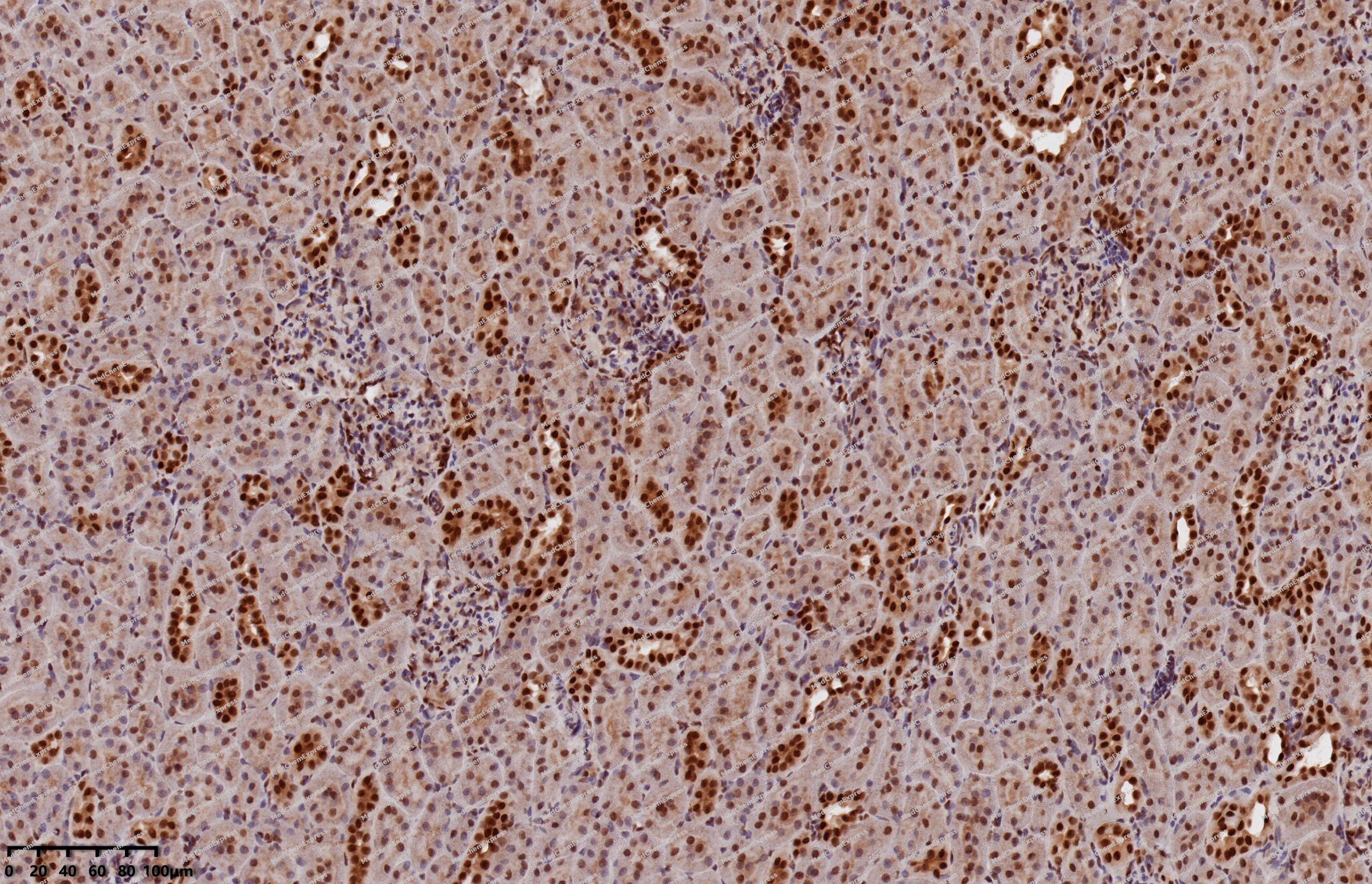 Immunohistochemical analysis of paraffin-embedded rat kidney using Bmi-1 antibody. The section was pre-treated using heat mediated antigen retrieval with EDTA (pH 9.0) for 14 minutes. The tissues were blocked in 5% BSA for 30 minutes at room temperature, washed with TBST, and then probed with the primary antibody (HY-P80034, 1/200) for 30 minutes at room temperature. The detection was performed using Polymer HRP-conjugated Goat Anti-Mouse/Rabbit lgG(H&L) secondary antibody (HY-P83652). DAB was used as the chromogen. Tissues were counterstained with hematoxylin and mounted with DPX.
 -
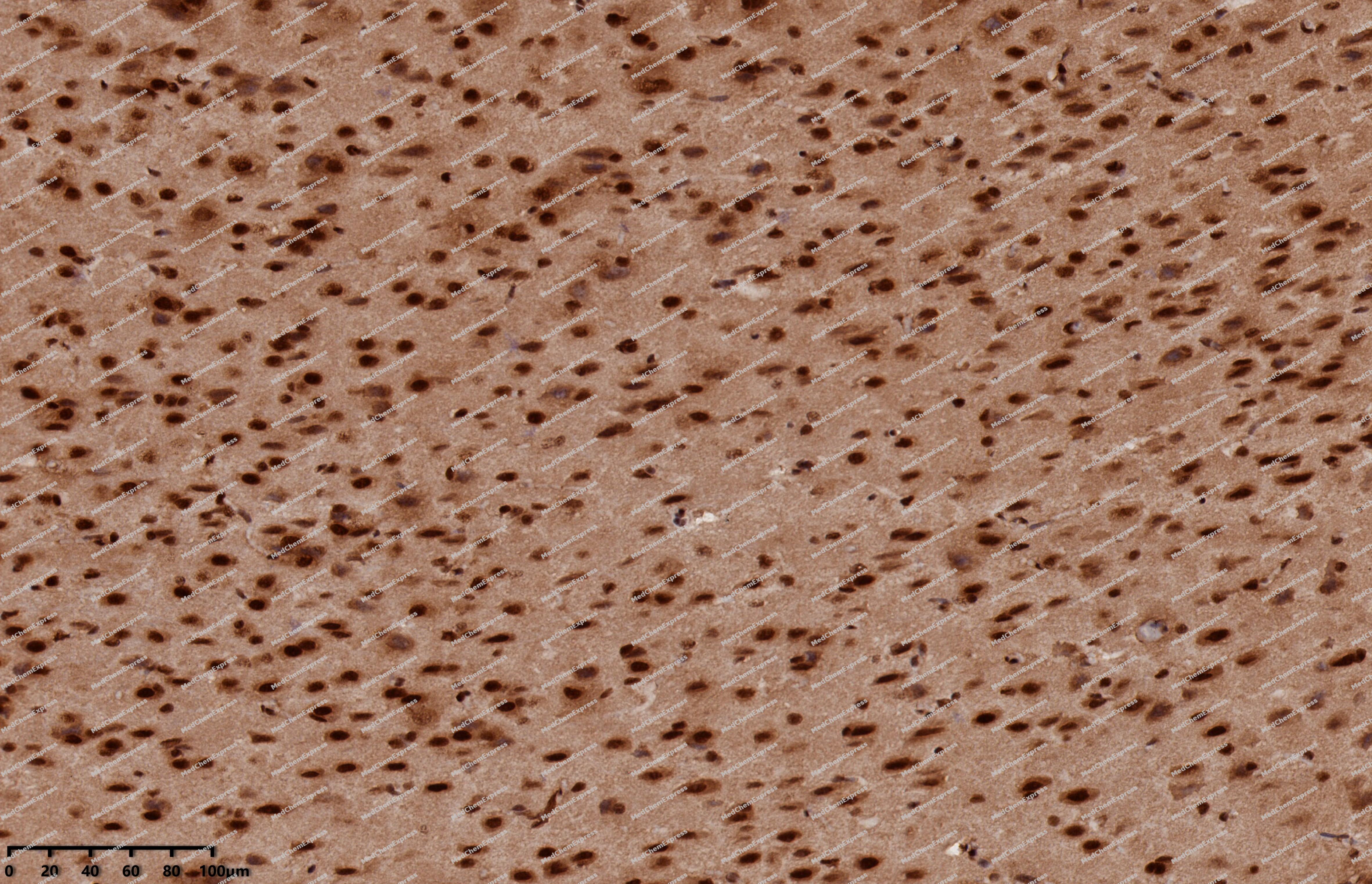 Immunohistochemical analysis of paraffin-embedded rat brain using Bmi-1 antibody. The section was pre-treated using heat mediated antigen retrieval with EDTA (pH 9.0) for 14 minutes. The tissues were blocked in 5% BSA for 30 minutes at room temperature, washed with TBST, and then probed with the primary antibody (HY-P80034, 1/200) for 30 minutes at room temperature. The detection was performed using Polymer HRP-conjugated Goat Anti-Mouse/Rabbit lgG(H&L) secondary antibody (HY-P83652). DAB was used as the chromogen. Tissues were counterstained with hematoxylin and mounted with DPX.
 -
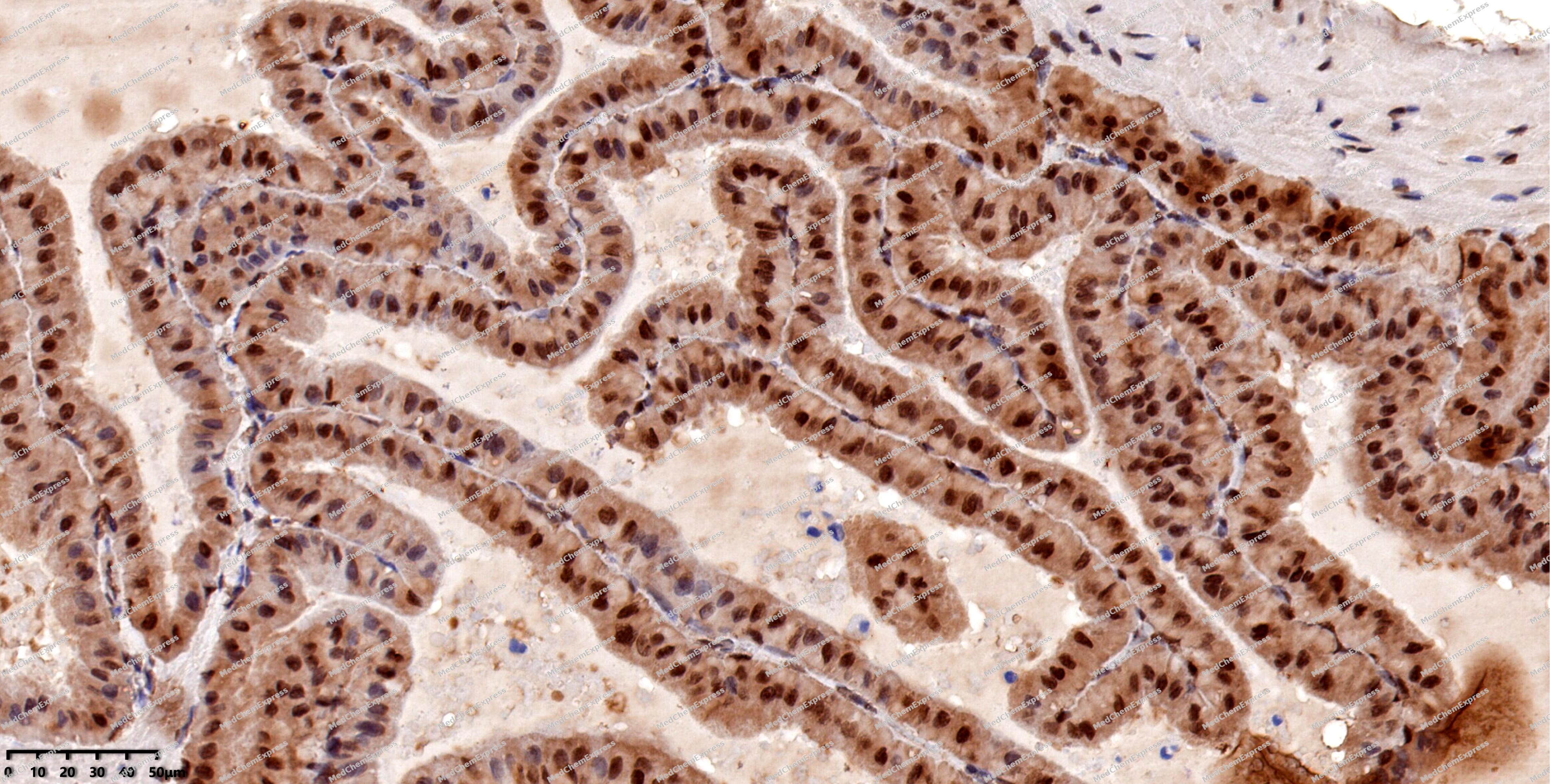 Immunohistochemical analysis of paraffin-embedded rat prostate using Bmi-1 antibody. The section was pre-treated using heat mediated antigen retrieval with EDTA (pH 9.0) for 14 minutes. The tissues were blocked in 5% BSA for 30 minutes at room temperature, washed with TBST, and then probed with the primary antibody (HY-P80034, 1/200) for 30 minutes at room temperature. The detection was performed using Polymer HRP-conjugated Goat Anti-Mouse/Rabbit lgG(H&L) secondary antibody (HY-P83652). DAB was used as the chromogen. Tissues were counterstained with hematoxylin and mounted with DPX.
 -
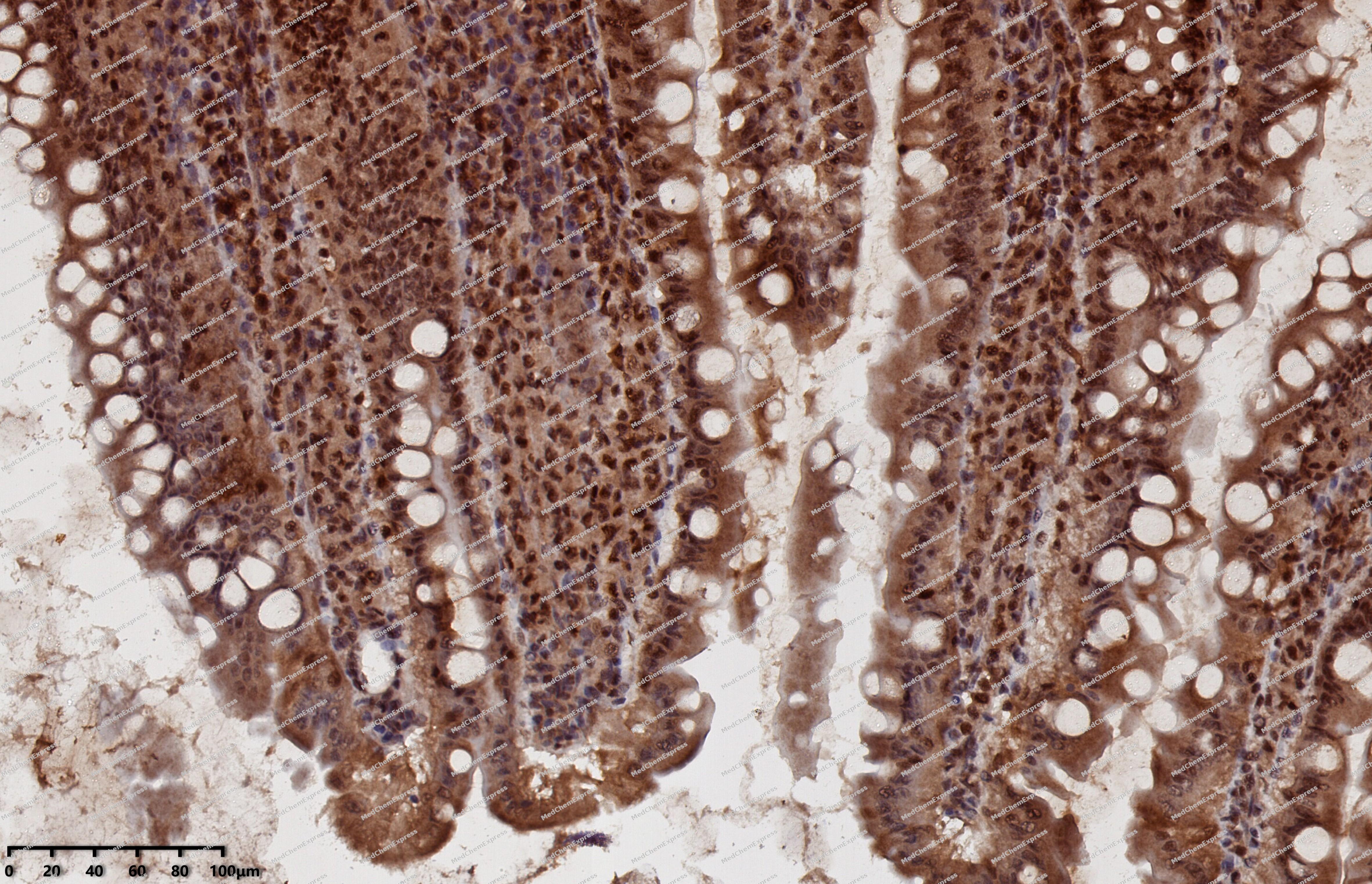 Immunohistochemical analysis of paraffin-embedded rat small intestine using Bmi-1 antibody. The section was pre-treated using heat mediated antigen retrieval with EDTA (pH 9.0) for 14 minutes. The tissues were blocked in 5% BSA for 30 minutes at room temperature, washed with TBST, and then probed with the primary antibody (HY-P80034, 1/200) for 30 minutes at room temperature. The detection was performed using Polymer HRP-conjugated Goat Anti-Mouse/Rabbit lgG(H&L) secondary antibody (HY-P83652). DAB was used as the chromogen. Tissues were counterstained with hematoxylin and mounted with DPX.
 -
 Flow cytometric analysis of 1X10⁶ HeLa cells labeling Bmi-1 Antibody (HY-P80034, red). Cells were fixed with 4% paraformaldehyde and permeabilised with 90% methanol. Then stained with the primary antibody at 1/200 dilution for an hour at 4℃. AF488-conjugated Goat Anti-Mouse IgG H&L (HY-P8005) was used as the secondary antibody at 1/1,000 dilution for 30 minutes at 4℃. Mouse IgG Isotype Control (HY-P80757, blue) was used as the isotype control, cells without incubation with primary antibody were used as the unlabeled control (black).
 -
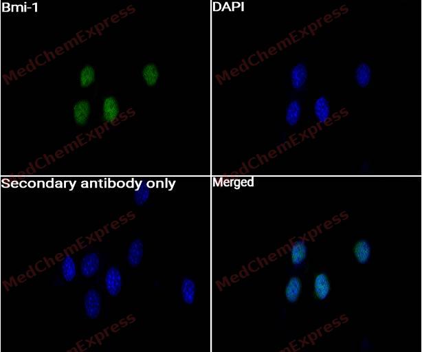 Immunocytochemistry analysis of NIH-3T3 cells labeling Bmi-1 with Bmi-1 Antibody (HY-P80034) at 1:50 dilution. Cells were fixed in 4% paraformaldehyde for 15 minutes at room temperature, permeabilized with 0.1% Triton X-100 in PBS for 15 minutes at room temperature, then blocked with quick block buffer for 10 minutes at room temperature. Cells were then incubated with Bmi-1 Antibody (HY-P80034) at 1:50 dilution in quick block buffer overnight at 4 ℃. AF488-conjugated Goat Anti-Rabbit IgG H&L(HY-P8002, Green) was used as the secondary antibody at 1/1,000 dilution. PBS instead of the primary antibody was used as the secondary antibody only control. The Nuclear counterstain was DAPI (Blue).
 -
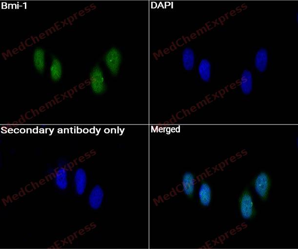 Immunocytochemistry analysis of Hela cells labeling Bmi-1 with Bmi-1 Antibody (HY-P80034) at 1:50 dilution. Cells were fixed in 4% paraformaldehyde for 15 minutes at room temperature, permeabilized with 0.1% Triton X-100 in PBS for 15 minutes at room temperature, then blocked with quick block buffer for 10 minutes at room temperature. Cells were then incubated with Bmi-1 Antibody (HY-P80034) at 1:50 dilution in quick block buffer overnight at 4 ℃. AF488-conjugated Goat Anti-Rabbit IgG H&L(HY-P8002, Green) was used as the secondary antibody at 1/1,000 dilution. PBS instead of the primary antibody was used as the secondary antibody only control. The Nuclear counterstain was DAPI (Blue).


Background
-
Function
B cell-specific Moloney murine leukemia virus integration site 1 (BMI1), a component of the polycomb repressive complex 1 (PRC1), is recognized as a polycomb protein that prevents premature senescence and maintains the self-renewal capacity of stem cells.
-
Subcellular Localization
Nucleus; Cytoplasm
-
Expression
Induction:Down-regulated by oxidative stress -
Subunit
Component of a PRC1-like complex (PubMed:12167701, PubMed:15386022, PubMed:19636380, PubMed:21282530, PubMed:21772249, PubMed:25355358, PubMed:26151332). Identified in a PRC1-like HPRC-H complex with CBX2, CBX4, CBX8, PHC1, PHC2, PHC3 RING1 and RNF2 (PubMed:12167701). Interacts with RNF2/RING2 (PubMed:16714294, PubMed:21772249, PubMed:25355358). Interacts with RING1 (By similarity). Part of a complex that contains RNF2, UB2D3 and BMI1, where RNF2 and BMI1 form a tight heterodimer, and UB2D3 interacts only with RNF2 (PubMed:21772249, PubMed:25355358). The complex composed of RNF2, UB2D3 and BMI1 binds nucleosomes, and has activity only with nucleosomal histone H2A (PubMed:21772249, PubMed:25355358). Interacts with CBX7 and CBX8 (PubMed:19636380). Interacts with SPOP (PubMed:15897469). Part of a complex consisting of BMI1, CUL3 and SPOP (PubMed:15897469). Interacts with E4F1 (PubMed:16882984). Interacts with PHC2 (PubMed:27827373, PubMed:9121482, PubMed:9199346). Interacts with zinc finger protein ZNF277 (By similarity). May be part of a complex including at least ZNF277, BMI1 and RNF2/RING2 (By similarity)
-
SwissProt ID
-
Synonyms
PCGF4, RNF51, BMI1, Polycomb complex protein BMI-1, Polycomb group RING finger protein 4, RING finger protein 51
-
Research Field
Cell Biology
Documentation
-
Data Sheet (235 KB)
-
SDS (252 KB)
- English - EN (252 KB)
- Français - FR (252 KB)
- Deutsch - DE (252 KB)
- Norwegian - NO (252 KB)
- Español - ES (252 KB)
- Swedish - SV (252 KB)
- Italian - IT (252 KB)
- Korean - KR (252 KB)
- Portuguese - PT (252 KB)
-
User Guide for Antibodies (1077 KB)

 Powered by Bioz
