



FGR Antibody (YA4792)
(Synonyms: FGR; SRC2; Tyrosine-protein kinase Fgr; Gardner-Rasheed feline sarcoma viral; v-fgr; oncogene homolog; Proto-oncogene c-Fgr; p55-Fgr; p58-Fgr; p58c-Fgr)
Based on 1 Customer Validation
FGR Antibody (YA4792) is a Mouse-derived and non-conjugated monoclonal antibody, targeting to FGR.
-
Host:
Mouse
-
Application:
WB, IHC-P, ICC/IF, ELISA
-
Reactivity :
Human, Mouse
-
Formulation:
Supplied in PBS containing 50% glycerol, 0.5% BSA and 0.02% sodium azide.
-
Conjugation:
Non-conjugated


To place orders, for customer services and technical support, please contact: MedChemExpress USA
Tel: 609-228-6898 E-mail: [email protected] [email protected]





Applications
| Application |
WB

WB: Western Blot
|
IHC-P
IHC-P: Immunohistochemistry-Paraffin
|
ELISA
ELISA: Enzyme Linked Immunosorbent Assay
|
ICC/IF
ICC/IF: Immunocytochemistry/
Immunofluorescence |
|---|---|---|---|---|
| Dilution Ratio | 1:500-1:2000 | 1:200-1:1000 | 1:10000 | 1:50-200 |


Product Details
FGR Antibody (YA4792) is a Mouse-derived and non-conjugated monoclonal antibody, targeting to FGR.
-
Host Mouse

-
Clonality Monoclonal

-
Species ReactivityHuman, Mouse
-
Calculated Molecular Weight Predicted band size: 59 kDa

Purified recombinant fragment of human c-Fgr expressed in E. Coli.
affinity purified.
Non-conjugated
Unmodified


Product Properties
-
Appearance
Solution
-
Formulation
Supplied in PBS containing 50% glycerol, 0.5% BSA and 0.02% sodium azide.
-
Concentration
Batch-dependent, Please check the COA for the concentration of each lot. Check Lot Concentration
-
Storage & Stability
Stored at -20°C for 1 year. Avoid repeated freeze / thaw cycles.
-
Shipping
Shipping with blue ice.


Verification Images
-
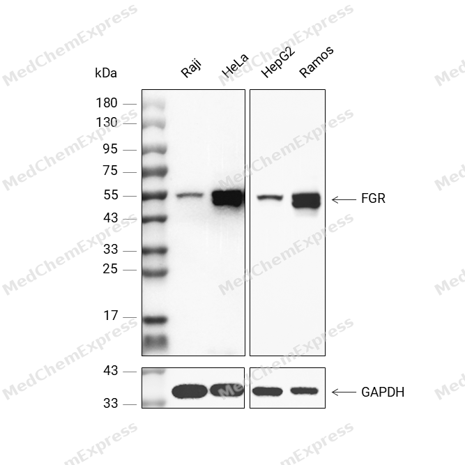 Western blot analysis was performed on protein extracts (25 μg) from Raji (lane 2), HeLa (lane 3), HepG2 (lane 4) and Ramos (lane 5) using FGR antibody. Proteins were transferred onto a 0.45 μm PVDF membrane using the Trans-Blot® Turbo™ system for 13 min. The membrane was then blocked with 5% nonfat milk in TBST (HY-K1025) for 1 h at room temperature. The primary antibody (1:1000) and loading control antibody GAPDH Antibody (HRP) (HY-P80954A) (1:5000) were diluted in 5% nonfat milk in TBST and incubated with the membrane overnight at 4°C. After washing, the membrane of primary antibody was incubated with HRP-conjugated goat anti-rabbit/mouse IgG secondary antibody (HY-P8001/HY-P8004) (1:5000) diluted in 5% nonfat milk in TBST for 1 h at room temperature. Protein bands were visualized using an Ultra High Sensitivity ECL detection kit (HY-K1005).


Background
-
Function
Fgr is a Src family tyrosine kinase activated during β2 integrin-dependent signaling in human neutrophils, linking adhesion to protein tyrosine phosphorylation[1]. Mechanistically, Fgr and Hck support adhesion-dependent neutrophil respiratory burst, spreading, and degranulation, showing that Fgr participates in integrin-driven inflammatory effector functions[2][3]. Fgr also acts with Hck to negatively regulate neutrophil and dendritic-cell chemokine signaling through PIR-B, indicating a context-dependent role in myeloid signal control[4]. In disease models, hematopoietic loss of Hck, Fgr, and Lyn protected mice from autoantibody-induced arthritis, blistering skin inflammation, and reverse passive Arthus reaction by impairing inflammatory-environment generation rather than intrinsic leukocyte recruitment[5]. Compared with related Src family isoforms, Fgr shows substantial functional overlap with Hck and Lyn, because single Fgr deficiency did not block arthritis development, whereas combined Hck/Fgr/Lyn loss produced complete protection[5]. For experimental applications, an Fgr kinase inhibitor attenuated sepsis-associated encephalopathy by reducing mitochondrial dysfunction, oxidative stress, and neuroinflammation through the SIRT1/PGC-1α pathway[6].
-
Subcellular Localization
Cell membrane; Lipid-anchor; Cytoplasmic side; Cell membrane; Peripheral membrane protein; Cytoplasmic side; Cell projection, ruffle membrane; Cytoplasm, cytosol; Cytoplasm, cytoskeleton; Mitochondrion inner membrane; Mitochondrion intermembrane space
-
Expression
Tissue_specificity:Detected (protein levels) in neutrophils, monocytes, and natural killer cells. Detected in monocytes and large lymphocytes. -
Subunit
Interacts with ITGB1, ITGB2, MS4A2/FCER1B, FCER1G, FCGR2A and/or FCGR2B. Interacts (via SH2 domain) with SYK (tyrosine phosphorylated). Interacts (via SH2 domain) with FLT3 (tyrosine phosphorylated).
-
SwissProt ID
-
Synonyms
FGR; SRC2; Tyrosine-protein kinase Fgr; Gardner-Rasheed feline sarcoma viral; v-fgr; oncogene homolog; Proto-oncogene c-Fgr; p55-Fgr; p58-Fgr; p58c-Fgr


Documentation
-
Data Sheet (262 KB)
-
SDS (251 KB)
- English - EN (251 KB)
- Français - FR (251 KB)
- Deutsch - DE (251 KB)
- Norwegian - NO (251 KB)
- Español - ES (251 KB)
- Swedish - SV (251 KB)
- Italian - IT (251 KB)
- Korean - KR (251 KB)
- Portuguese - PT (251 KB)
-
User Guide for Antibodies (1077 KB)
References
[1]. Berton G, et al. Beta 2 integrin-dependent protein tyrosine phosphorylation and activation of the FGR protein tyrosine kinase in human neutrophils. J Cell Biol. 1994 Aug;126(4):1111-21. [Content Brief]
[2]. Lowell CA, et al. Deficiency of Src family kinases p59/61hck and p58c-fgr results in defective adhesion-dependent neutrophil functions. J Cell Biol. 1996 May;133(4):895-910. [Content Brief]
[3]. Sibley CD, et al. Discovery of a Small Side Cavity in Sphingosine Kinase 2 that Enhances Inhibitor Potency and Selectivity. J Med Chem. 2020 Feb 13;63(3):1178-1198. [Content Brief]
[4]. Zhang H, et al. The Src family kinases Hck and Fgr negatively regulate neutrophil and dendritic cell chemokine signaling via PIR-B. Immunity. 2005 Feb;22(2):235-46. [Content Brief]
[5]. Kovács M, et al. The Src family kinases Hck, Fgr, and Lyn are critical for the generation of the in vivo inflammatory environment without a direct role in leukocyte recruitment. J Exp Med. 2014 Sep 22;211(10):1993-2011. [Content Brief]
[6]. Liu Y, et al. An Fgr kinase inhibitor attenuates sepsis-associated encephalopathy by ameliorating mitochondrial dysfunction, oxidative stress, and neuroinflammation via the SIRT1/PGC-1α signaling pathway. J Transl Med. 2023 Jul 20;21(1):486. [Content Brief]

 Powered by Bioz
