



MCP1 Antibody (YA3779)
(Synonyms: HC11; MCAF; MCP1; MCP-1; SCYA2; GDCF-2; SMC-CF; HSMCR30)
Based on 1 Customer Validation
MCP1 Antibody (YA3779) is a Mouse-derived and non-conjugated IgG1 monoclonal antibody, targeting to MCP1.
-
Host:
Mouse
-
Isotype:
IgG1
-
Application:
WB, IHC-P, ICC/IF, FC, ELISA
-
Reactivity :
Human, Mouse, Rat, Monkey
-
Formulation:
Supplied in PBS with 0.05% sodium azide
-
Conjugation:
Non-conjugated


To place orders, for customer services and technical support, please contact: MedChemExpress USA
Tel: 609-228-6898 E-mail: [email protected] [email protected]





Applications
| Application |
WB

WB: Western Blot
|
IHC-P
IHC-P: Immunohistochemistry-Paraffin
|
ICC/IF
ICC/IF: Immunocytochemistry/
Immunofluorescence |
FC
FC: Flow Cytometry
|
ELISA
ELISA: Enzyme Linked Immunosorbent Assay
|
|---|---|---|---|---|---|
| Dilution Ratio | 1:500-1:2000 | 1:200-1:1000 | 1:200-1:1000 | 1:200-1:400 | 1:10000 |


Product Details
MCP1 Antibody (YA3779) is a Mouse-derived and non-conjugated IgG1 monoclonal antibody, targeting to MCP1.
-
Host Mouse

-
Clonality Monoclonal

-
Species ReactivityHuman, Mouse, Rat, Monkey
-
Observed Molecular WeightObserved band size: 11 kDa
Note: Due to possible protein modifications or aggregation, the molecular weight should be confirmed by actual measurement, and the predicted value is for reference only.

-
Calculated Molecular Weight Predicted band size: 11 kDa

Purified recombinant fragment of human CCL2 full(aa 1-99).
affinity purified.
Non-conjugated
Unmodified
IgG1


Product Properties
-
Appearance
Solution
-
Formulation
Supplied in PBS with 0.05% sodium azide
-
Concentration
Batch-dependent, Please check the COA for the concentration of each lot. Check Lot Concentration
-
Storage & Stability
Stored at -20°C for 1 year. Avoid repeated freeze / thaw cycles.
-
Shipping
Shipping with blue ice.


Verification Images
-
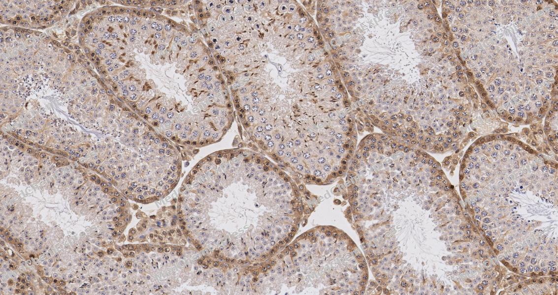 Immunohistochemical analysis of paraffin-embedded human testis tissue using MCP1 antibody. The section was pre-treated using heat mediated antigen retrieval with sodium citrate buffer (pH 6.0) for 2 minutes. The tissues were blocked with QuickBlock at room temperature for 30 minutes, washed with PBSand PBST, and then incubated with the primary antibody (HY-P84082, 1/500) overnight at 4℃. The detection was performed using an HRP conjugated compact polymer system. DAB was used as the chromogen. Tissues were counterstained with hematoxylin and mounted with DPX.
 -
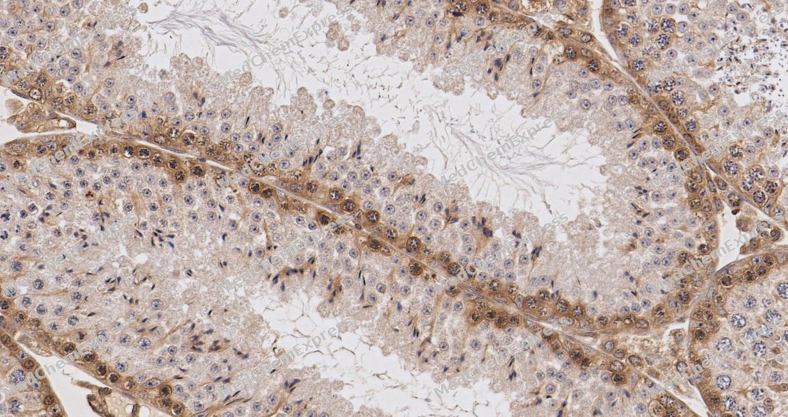 Immunohistochemical analysis of paraffin-embedded rat testis tissue using MCP1 antibody. The section was pre-treated using heat mediated antigen retrieval with sodium citrate buffer (pH 6.0) for 2 minutes. The tissues were blocked with QuickBlock at room temperature for 30 minutes, washed with PBSand PBST, and then incubated with the primary antibody (HY-P84082, 1/500) overnight at 4℃. The detection was performed using an HRP conjugated compact polymer system. DAB was used as the chromogen. Tissues were counterstained with hematoxylin and mounted with DPX.
 -
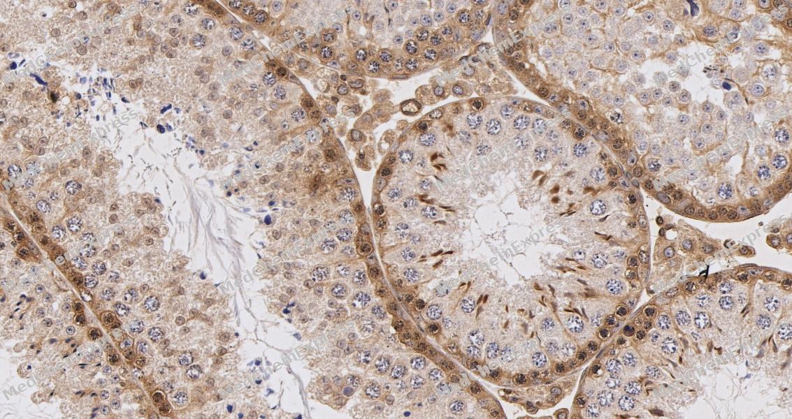 Immunohistochemical analysis of paraffin-embedded mouse testis tissue using MCP1 antibody. The section was pre-treated using heat mediated antigen retrieval with sodium citrate buffer (pH 6.0) for 2 minutes. The tissues were blocked with QuickBlock at room temperature for 30 minutes, washed with PBSand PBST, and then incubated with the primary antibody (HY-P84082, 1/500) overnight at 4℃. The detection was performed using an HRP conjugated compact polymer system. DAB was used as the chromogen. Tissues were counterstained with hematoxylin and mounted with DPX.
 -
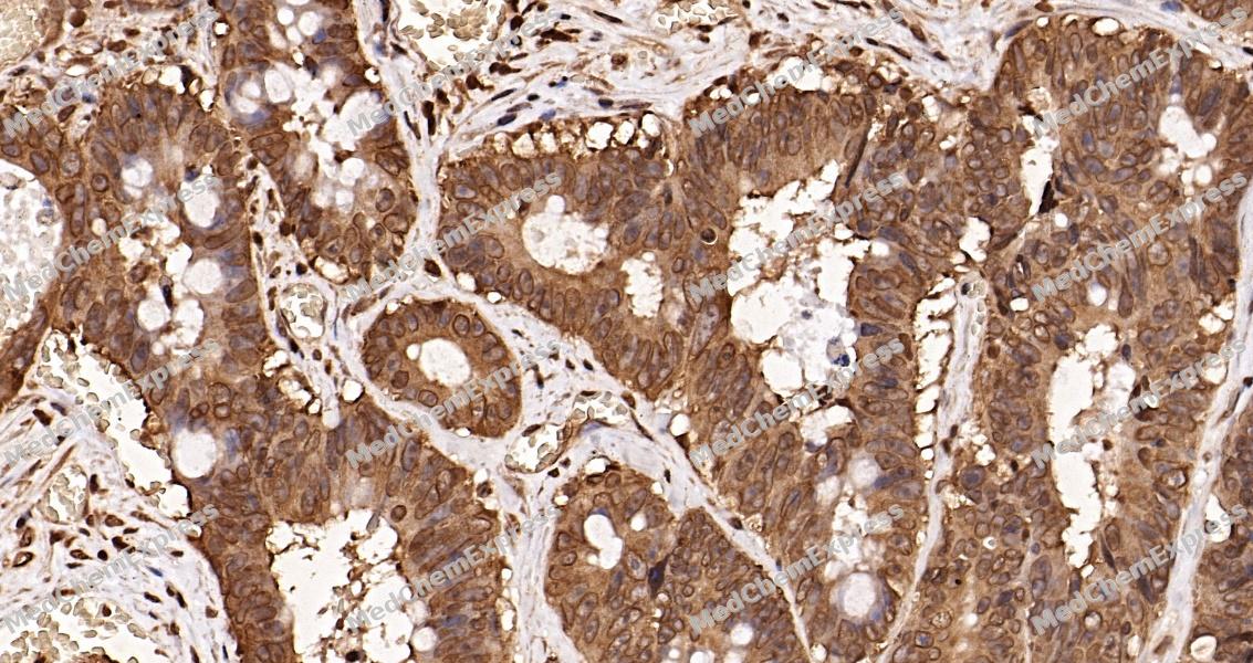 Immunohistochemical analysis of paraffin-embedded human Colorectal cancer 1 tissue using MCP1 antibody. The section was pre-treated using heat mediated antigen retrieval with sodium citrate buffer (pH 6.0) for 2 minutes. The tissues were blocked with QuickBlock at room temperature for 30 minutes, washed with PBSand PBST, and then incubated with the primary antibody (HY-P84082, 1/500) overnight at 4℃. The detection was performed using an HRP conjugated compact polymer system. DAB was used as the chromogen. Tissues were counterstained with hematoxylin and mounted with DPX.
 -
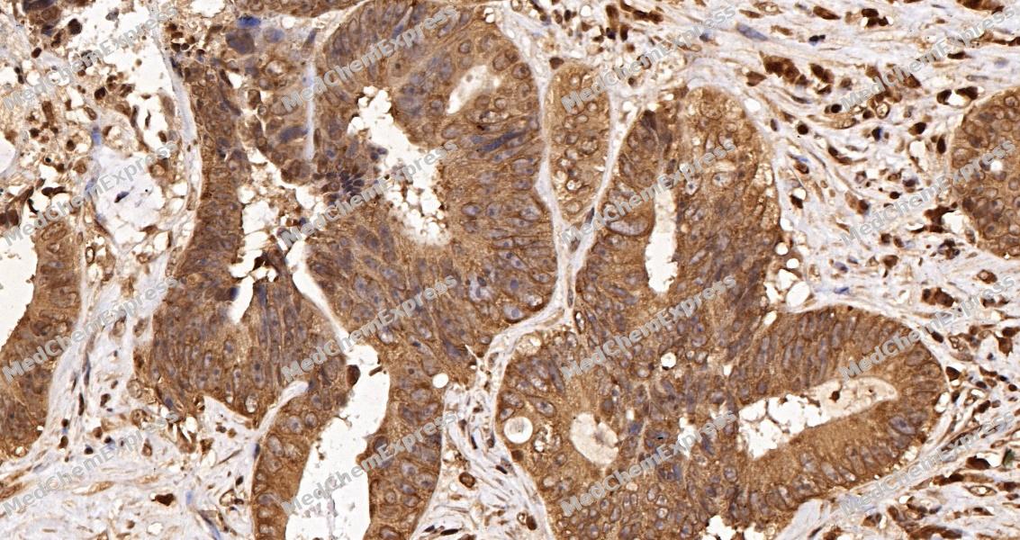 Immunohistochemical analysis of paraffin-embedded human Colorectal cancer 2 tissue using MCP1 antibody. The section was pre-treated using heat mediated antigen retrieval with sodium citrate buffer (pH 6.0) for 2 minutes. The tissues were blocked with QuickBlock at room temperature for 30 minutes, washed with PBSand PBST, and then incubated with the primary antibody (HY-P84082, 1/500) overnight at 4℃. The detection was performed using an HRP conjugated compact polymer system. DAB was used as the chromogen. Tissues were counterstained with hematoxylin and mounted with DPX.
 -
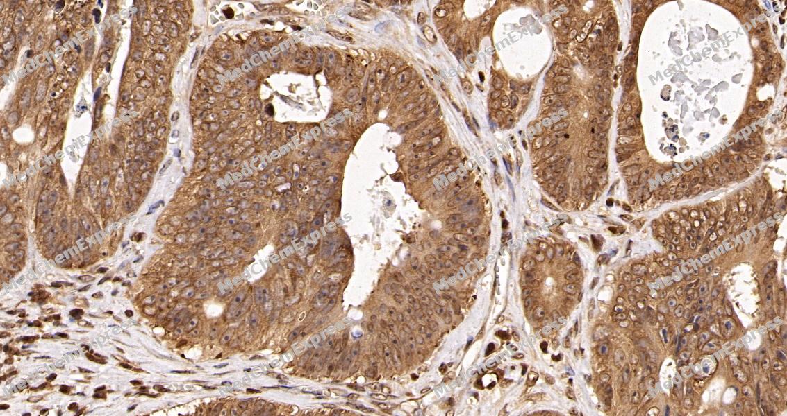 Immunohistochemical analysis of paraffin-embedded human Colorectal cancer 3 tissue using MCP1 antibody. The section was pre-treated using heat mediated antigen retrieval with sodium citrate buffer (pH 6.0) for 2 minutes. The tissues were blocked with QuickBlock at room temperature for 30 minutes, washed with PBSand PBST, and then incubated with the primary antibody (HY-P84082, 1/500) overnight at 4℃. The detection was performed using an HRP conjugated compact polymer system. DAB was used as the chromogen. Tissues were counterstained with hematoxylin and mounted with DPX.
 -
 Immunocytochemistry analysis of HepG2 cells labeling MCP1 with MCP1 Antibody (HY-P84082) at 1/200 dilution. Cells were fixed in 4% paraformaldehyde for 15 minutes at room temperature, permeabilized with 0.1% Triton X-100 in PBS for 15 minutes at room temperature, then blocked with quick block buffer for 10 minutes at room temperature. Cells were then incubated with MCP1 Antibody (HY-P84082) at 1/200 dilution in quick block buffer overnight at 4 ℃. AF488-conjugated Goat Anti-Mouse IgG H&L(HY-P8005, Green) was used as the secondary antibody at 1/1,000 dilution. PBS instead of the primary antibody was used as the secondary antibody only control. The Nuclear counterstain was DAPI (Blue).
 -
 Immunocytochemistry analysis of U2OS cells labeling MCP1 with MCP1 Antibody (HY-P84082) at 1/200 dilution. Cells were fixed in 4% paraformaldehyde for 15 minutes at room temperature, permeabilized with 0.1% Triton X-100 in PBS for 15 minutes at room temperature, then blocked with quick block buffer for 10 minutes at room temperature. Cells were then incubated with MCP1 Antibody (HY-P84082) at 1/200 dilution in quick block buffer overnight at 4 ℃. AF488-conjugated Goat Anti-Mouse IgG H&L(HY-P8005, Green) was used as the secondary antibody at 1/1,000 dilution. PBS instead of the primary antibody was used as the secondary antibody only control. The Nuclear counterstain was DAPI (Blue).


Background
-
Function
MCP1 acts as a ligand for C-C chemokine receptor CCR2. Signals through binding and activation of CCR2 and induces a strong chemotactic response and mobilization of intracellular calcium ions. Exhibits a chemotactic activity for monocytes and basophils but not neutrophils or eosinophils. May be involved in the recruitment of monocytes into the arterial wall during the disease process of atherosclerosis[1][2][3][4][5][6][7].
-
Subcellular Localization
Secreted
-
Expression
Tissue_specificity:Expression (protein level) in seminal plasma, endometrial fluid, and follicular fluid (PubMed: 23765988) . Expression in monocytes (PubMed: 2513477) .
Induction:Up-regulated upon hypertonic conditions (PubMed:23233732) . In pancreatic islets, secretion is stimulated by IL1B (PubMed:23955712) . Up-regulated by coagulation factor Xa (F10) in PAR-1 (F2R) -dependent manner in cardiac fibroblasts and endothelial cells (PubMed:30568593, PubMed:34831181) . Up-regulated by thrombin (F2) in endothelial cells (PubMed:30568593) -
Subunit
Monomer or homodimer; in equilibrium (PubMed:15033992, PubMed:8898111, PubMed:8989326, PubMed:9837883). Is tethered on endothelial cells by glycosaminoglycan (GAG) side chains of proteoglycans (PubMed:9792674). Interacts with TNFAIP6 (via Link domain)
-
SwissProt ID
-
Synonyms
HC11; MCAF; MCP1; MCP-1; SCYA2; GDCF-2; SMC-CF; HSMCR30


Documentation
-
Data Sheet (262 KB)
-
SDS (251 KB)
- English - EN (251 KB)
- Français - FR (251 KB)
- Deutsch - DE (251 KB)
- Norwegian - NO (251 KB)
- Español - ES (251 KB)
- Swedish - SV (251 KB)
- Italian - IT (251 KB)
- Korean - KR (251 KB)
- Portuguese - PT (251 KB)
-
User Guide for Antibodies (1077 KB)
References
[1]. Hemmerich S, et al. Identification of residues in the monocyte chemotactic protein-1 that contact the MCP-1 receptor, CCR2. Biochemistry. 1999 Oct 5;38(40):13013-25. [Content Brief]
[2]. Jarnagin K, et al. Identification of surface residues of the monocyte chemotactic protein 1 that affect signaling through the receptor CCR2. Biochemistry. 1999 Dec 7;38(49):16167-77. [Content Brief]
[3]. Paavola CD, et al. Monomeric monocyte chemoattractant protein-1 (MCP-1) binds and activates the MCP-1 receptor CCR2B. J Biol Chem. 1998 Dec 11;273(50):33157-65. [Content Brief]
[4]. Zhang YJ, et al. Structure/activity analysis of human monocyte chemoattractant protein-1 (MCP-1) by mutagenesis. Identification of a mutated protein that inhibits MCP-1-mediated monocyte chemotaxis. J Biol Chem. 1994 Jun 3;269(22):15918-24. [Content Brief]
[5]. Weber M, et al. Deletion of the NH2-terminal residue converts monocyte chemotactic protein 1 from an activator of basophil mediator release to an eosinophil chemoattractant. J Exp Med. 1996 Feb 1;183(2):681-5. [Content Brief]
[6]. Chakravarty L, et al. Lysine 58 and histidine 66 at the C-terminal alpha-helix of monocyte chemoattractant protein-1 are essential for glycosaminoglycan binding. J Biol Chem. 1998 Nov 6;273(45):29641-7. [Content Brief]
[7]. Li YS, et al. The expression of monocyte chemotactic protein (MCP-1) in human vascular endothelium in vitro and in vivo. Mol Cell Biochem. 1993 Sep 8;126(1):61-8. [Content Brief]

 Powered by Bioz
