



CD19 Antibody (YA543)
(Synonyms: CD19_HUMAN; B-lymphocyte antigen CD19; B-lymphocyte surface antigen B4; Differentiation antigen CD19; T-cell surface antigen Leu-12; CD_antigen: CD19; B-lymphocyte surface antigen B4; Leu 12; Leu12; )
Based on 1 Customer Validation
CD19 Antibody (YA543) is a Rabbit-derived and non-conjugated IgG monoclonal antibody, targeting to CD19.
-
Host:
Rabbit
-
Isotype:
IgG
-
Application:
WB, ICC/IF, IHC-P, FC
-
Reactivity :
Human
-
Formulation:
Supplied in 1*TBS (pH7.4), 0.05% BSA and 40% Glycerol. Preservative: 0.05% Sodium Azide.
-
Conjugation:
Non-conjugated


To place orders, for customer services and technical support, please contact: MedChemExpress USA
Tel: 609-228-6898 E-mail: [email protected] [email protected]





Applications
| Application |
WB

WB: Western Blot
|
ICC/IF
ICC/IF: Immunocytochemistry/
Immunofluorescence |
IHC-P
IHC-P: Immunohistochemistry-Paraffin
|
FC
FC: Flow Cytometry
|
|---|---|---|---|---|
| Dilution Ratio | 1:1000-1:2000 | 1:50-1:200 | 1:50-1:200 | 1:50-1:1000 |


Product Details
CD19 Antibody (YA543) is a Rabbit-derived and non-conjugated IgG monoclonal antibody, targeting to CD19.
-
Host Rabbit

-
Clonality Recombinant,Monoclonal

-
Species ReactivityHuman
-
Observed Molecular WeightObserved band size: 90 kDa
Note: Due to possible protein modifications or aggregation, the molecular weight should be confirmed by actual measurement, and the predicted value is for reference only.

-
Calculated Molecular Weight Predicted band size: 61 kDa

Synthetic peptide corresponding to Human CD19.AA range:294-556.
Endogenous
Protein A affinity purified.
Non-conjugated
Unmodified
IgG


Product Properties
-
Appearance
Solution
-
Formulation
Supplied in 1*TBS (pH7.4), 0.05% BSA and 40% Glycerol. Preservative: 0.05% Sodium Azide.
-
Concentration
Batch-dependent, Please check the COA for the concentration of each lot. Check Lot Concentration
-
Storage & Stability
Stored at -20°C for 1 year. Avoid repeated freeze / thaw cycles.
-
Shipping
Shipping with blue ice.


Verification Images
-
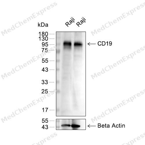 Western blot analysis of extracts from Raji(lane 2(20μg)) or (lane 4(40μg)) using CD19 (HY-P80446) Rabbit mAb. Proteins were transferred to a PVDF membrane and blocked with 5% BSA in TBST for 2 hour at room temperature. The primary antibody (1/1000) and Loading control antibody (Beta Actin, HY-P80438, 1/10000) was used in 5% BSA in TBST at 4°C overnight. Goat Anti-Rabbit IgG-HRP Secondary Antibody (1/10,000) was used for 1 hour at room temperature.
 -
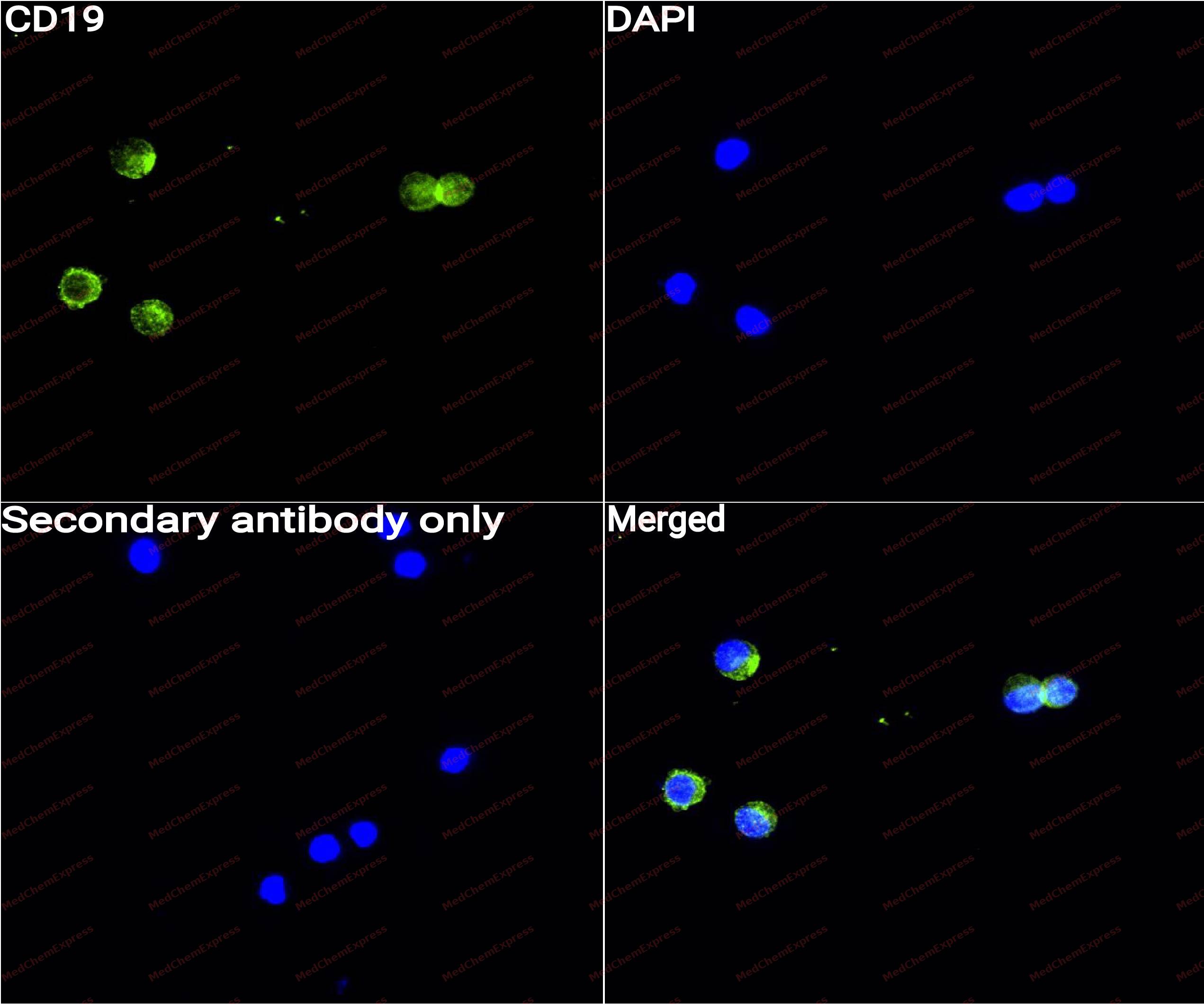 Immunocytochemistry analysis of Raji cells labeling CD19 with CD19 Antibody (HY-P80446) at 1/50 dilution. Cells were fixed in 4% paraformaldehyde for 15 minutes at room temperature, permeabilized with 0.1% Triton X-100 for 10 minutes at room temperature, then blocked with QuickBlock™ Blocking Buffer for Immunol Staining for 10 min at room temperature. Cells were then incubated with CD19 Antibody (HY-P80446) at 1/50 dilution in QuickBlock™ Blocking Buffer for Immunol Staining at 4 ℃. Alexa Fluor® 488-conjugated AffiniPure Goat Anti-Rabbit IgG H&L(HY-P8002, Green) was used as the secondary antibody at 1/1,000 dilution. PBS instead of the primary antibody was used as the secondary antibody only control. The Nuclear counterstain was DAPI (Blue).
 -
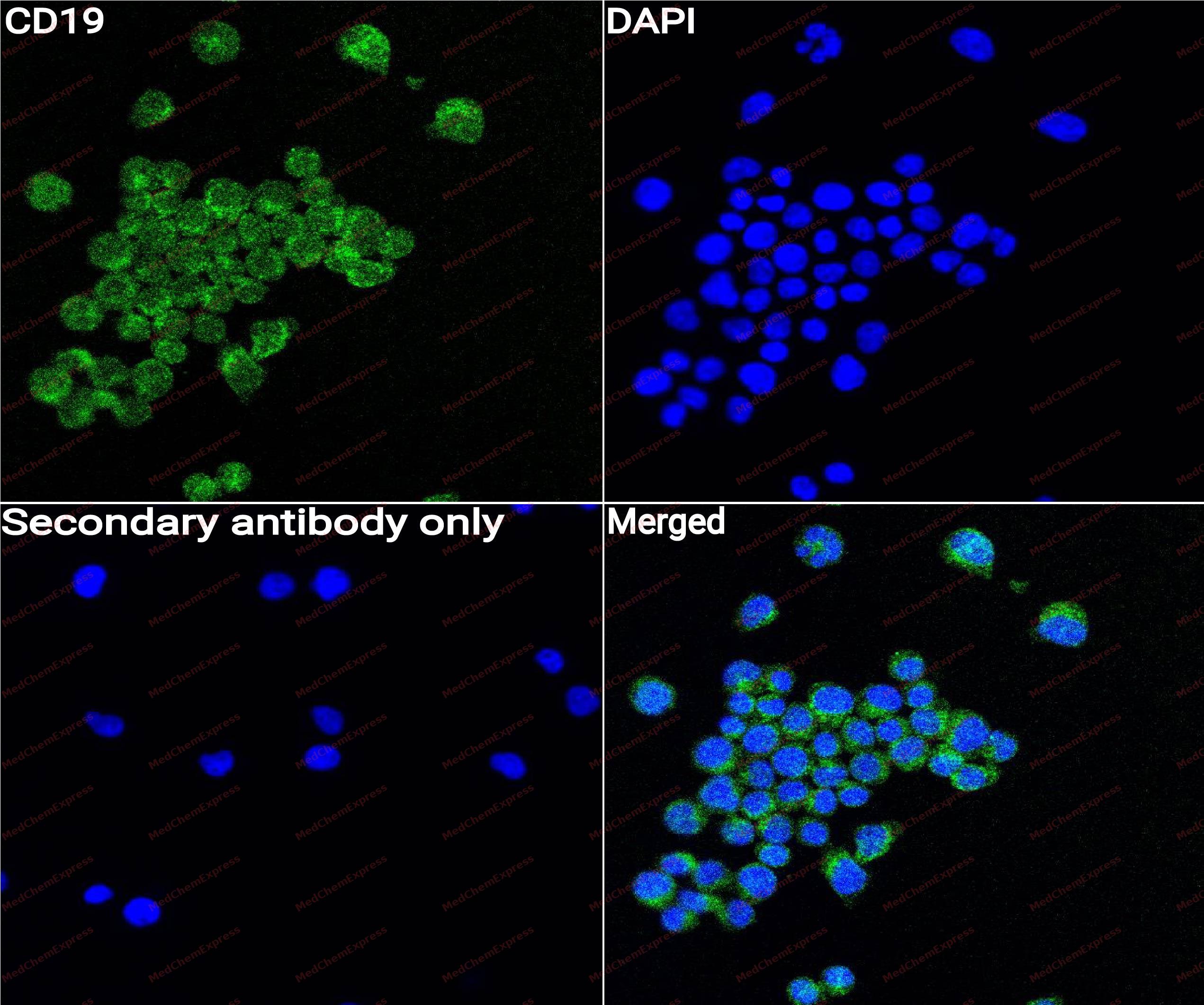 Immunocytochemistry analysis of Ramos cells labeling CD19 with CD19 Antibody (HY-P80446) at 1/50 dilution. Cells were fixed in 4% paraformaldehyde for 15 minutes at room temperature, permeabilized with 0.1% Triton X-100 for 10 minutes at room temperature, then blocked with QuickBlock™ Blocking Buffer for Immunol Staining for 10 min at room temperature. Cells were then incubated with CD19 Antibody (HY-P80446)at 1/50 dilution in QuickBlock™ Blocking Buffer for Immunol Staining at 4 ℃. Alexa Fluor® 488-conjugated AffiniPure Goat Anti-Rabbit IgG H&L(HY-P8002,Green) was used as the secondary antibody at 1/1,000 dilution. PBS instead of the primary antibody was used as the secondary antibody only control. The Nuclear counterstain was DAPI (Blue).
 -
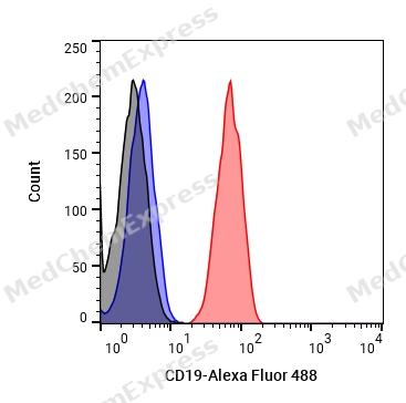 Flow cytometric analysis of 1X10^6 Raji cells labeling CD19 Antibody (HY-P80446, red). Cells were fixed with 4% paraformaldehyde and permeabilised with 90% methanol. Then stained with the primary antibody at 1μg/mL dilution for an hour at 4℃. Alexa Fluor® 488-conjugated AffiniPure Goat Anti-Rabbit IgG H&L (HY-P8002) was used as the secondary antibody at 1/1,000 dilution for 30 minutes at 4℃. Rabbit IgG Isotype Control (HY-P80879, blue) was used as the isotype control, cells without incubation with primary antibody were used as the unlabeled control (black).


Background
-
Function
CD19 is a B cell-specific transmembrane glycoprotein and a core member of the B cell receptor (BCR) co-receptor complex. It can significantly enhance BCR signaling by activating signaling pathways such as PI3K and BTK, promoting B cell development, activation, and antibody production. CD19 is present throughout almost the entire B cell development process and is a biomarker for all B cells. CD19 is an important marker for immunophenotyping of B-cell leukemia and lymphoma, and is also one of the targets for CAR-T cell therapy, bispecific antibodies, and other B cell-targeted therapies.
-
Subcellular Localization
Cell membrane; Single-pass type I membrane protein; Membrane raft; Single-pass type I membrane protein
-
Expression
Tissue_specificity:Detected in B cells in the marginal zone of lymph nodes and germinal centers (PubMed:2463100) . Detected in blood B cells (protein levels) (PubMed:16672701, PubMed:2463100) . -
Isoforms & Post-Translational Modification
CD19 has 2 isomers: P15391-1: 61128 Da (predicted); P15391-2: 61200 Da (predicted).
Phosphorylated on tyrosine following B-cell activation. Phosphorylated on tyrosine residues by LYN. Tyrosine residues are phosphorylated sequentially after activation of the B cell receptor. Phosphorylation of Tyr-531 is extremely rapid, followed by phosphorylation at Tyr-409. -
Subunit
Interacts with CR2/CD21 (PubMed:1702139). Part of a complex composed of CD19, CR2/CD21, CD81 and IFITM1/CD225 in the membrane of mature B-cells (PubMed:1383329). Interacts directly with CD81 (via the second extracellular domain); this interaction is initiated early during biosynthesis in the ER/pre-Golgi compartments and is essential for trafficking and compartmentalization of CD19 receptor on the cell surface of activated B cells (PubMed:16449649). Interacts with VAV (PubMed:10706702). Interacts with GRB2 and SOS when phosphorylated on Tyr-348 and/or Tyr-378. Interacts with PLCG2 when phosphorylated on Tyr-409 (PubMed:10706702). Interacts with LYN (PubMed:7687428). Interacts (when tyrosine phosphorylated) with the regulatory p85 subunit of phosphatidylinositol 3-kinase (PIK3R1 or PIK3R2) (PubMed:7684160). Interacts with GRB2 (PubMed:29523808)
-
SwissProt ID
-
Synonyms
CD19_HUMAN; B-lymphocyte antigen CD19; B-lymphocyte surface antigen B4; Differentiation antigen CD19; T-cell surface antigen Leu-12; CD_antigen: CD19; B-lymphocyte surface antigen B4; Leu 12; Leu12;
-
Research Field
Immunology


Documentation
-
Data Sheet (261 KB)
-
SDS (252 KB)
- English - EN (252 KB)
- Français - FR (252 KB)
- Deutsch - DE (252 KB)
- Norwegian - NO (252 KB)
- Español - ES (252 KB)
- Swedish - SV (252 KB)
- Italian - IT (252 KB)
- Korean - KR (252 KB)
- Portuguese - PT (252 KB)
-
User Guide for Antibodies (1077 KB)
References

 Powered by Bioz
