



CDK5 Antibody (YA796)
(Synonyms: Cdk 5; Cdk5; CDK5_HUMAN; Cell division protein kinase 5; Crk6; Cyclin dependent kinase 5; Cyclin-dependent kinase 5; Protein kinase CDK5 splicing; PSSALRE; Serine threonine protein kinase PSSALRE; Serine/threonine-protein kinase PSSALRE; Tau protein kinas)
Based on 1 Customer Validation
CDK5 Antibody (YA796) is a Mouse-derived and non-conjugated IgG1 monoclonal antibody, targeting to CDK5.
-
Host:
Mouse
-
Isotype:
IgG
-
Application:
WB, ICC/IF
-
Reactivity :
Human, Mouse, Rat, Monkey
-
Formulation:
Supplied in 1*PBS (pH 7.3), 50% glycerol and 0.5% BSA. Preservative: 0.02% sodium azide.
-
Conjugation:
Non-conjugated


To place orders, for customer services and technical support, please contact: MedChemExpress USA
Tel: 609-228-6898 E-mail: [email protected] [email protected]





Applications
| Application |
WB

WB: Western Blot
|
ICC/IF
ICC/IF: Immunocytochemistry/
Immunofluorescence |
|---|---|---|
| Dilution Ratio | 1:500-1:1000 | 1:50-1:200 |


Product Details
CDK5 Antibody (YA796) is a Mouse-derived and non-conjugated IgG1 monoclonal antibody, targeting to CDK5.
-
Host Mouse

-
Clonality Monoclonal

-
Species ReactivityHuman, Mouse, Rat, Monkey
-
Observed Molecular WeightObserved band size: 36 kDa
Note: Due to possible protein modifications or aggregation, the molecular weight should be confirmed by actual measurement, and the predicted value is for reference only.

-
Calculated Molecular Weight Predicted band size: 33 kDa

Entrez Gene: 1020 Human ; 12568 Mouse ; 140908 Rat
SwissProt: Q00535 Human ; P49615 Mouse ; Q03114 Rat
OMIM: 616342 Human
Synthetic peptide corresponding to Human CDK5.The exact sequence is proprietary to MCE.
Endogenous
affinity purified
Non-conjugated
Unmodified
IgG


Product Properties
-
Appearance
Solution
-
Formulation
Supplied in 1*PBS (pH 7.3), 50% glycerol and 0.5% BSA. Preservative: 0.02% sodium azide.
-
Concentration
Batch-dependent, Please check the COA for the concentration of each lot. Check Lot Concentration
-
Storage & Stability
Stored at -20°C for 1 year. Avoid repeated freeze / thaw cycles.
-
Livraison
Shipping with blue ice.


Verification Images
-
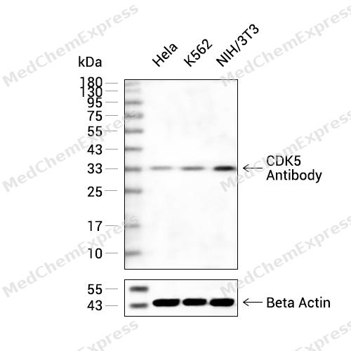 Western blot analysis of extracts from Hela(lane 2(20ug) , K562(lane 3(20ug) and NIH/3T3(lane 4(20ug) using CDK5 Antibody (HY-P80613) Rabbit mAb. Proteins were transferred to a PVDF membrane and blocked with 5% non-fat milk in TBST for 2 hour at room temperature. The primary antibody (1/1000) and Loading control antibody (Beta Actin, HY-P80438, 1/10000) was used in 5% non-fat milk in TBST at 4°C overnight. Goat Anti-Mouse/Rabbit IgG-HRP Secondary Antibody (1/10000) was used for 1 hour at room temperature.
 -
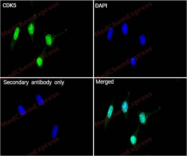 Immunocytochemistry analysis of SH-SY5Y cells labeling CDK5 with CDK5 Antibody (HY-P80613) at 1/100 dilution. Cells were fixed in 4% paraformaldehyde for 15 minutes at room temperature, permeabilized with 0.1% Triton X-100 in PBS for 15 minutes at room temperature, then blocked with quick block buffer for 10 minutes at room temperature. Cells were then incubated with CDK5 Antibody (HY-P80613) at 1/100 dilution in quick block buffer overnight at 4 ℃. AF488-conjugated Goat Anti-Mouse IgG H&L(HY-P8005, Green) was used as the secondary antibody at 1/1,000 dilution. PBS instead of the primary antibody was used as the secondary antibody only control. The Nuclear counterstain was DAPI (Blue).
 -
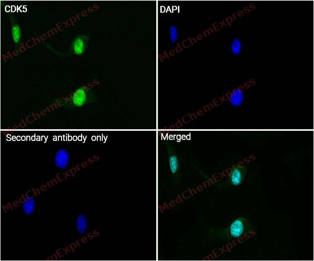 Immunocytochemistry analysis of SH-SY5Y cells labeling CDK5 with CDK5 Antibody (HY-P80613) at 1/200 dilution. Cells were fixed in 4% paraformaldehyde for 15 minutes at room temperature, permeabilized with 0.1% Triton X-100 in PBS for 15 minutes at room temperature, then blocked with quick block buffer for 10 minutes at room temperature. Cells were then incubated with CDK5 Antibody (HY-P80613) at 1/200 dilution in quick block buffer overnight at 4 ℃. AF488-conjugated Goat Anti-Mouse IgG H&L(HY-P8005, Green) was used as the secondary antibody at 1/1,000 dilution. PBS instead of the primary antibody was used as the secondary antibody only control. The Nuclear counterstain was DAPI (Blue).


Background
-
Function
CDK5 is a proline-directed serine/threonine kinase that controls neuronal development, postsynaptic signal integration, neurotransmitter release, and neurite outgrowth[1][2]. Mechanistically, CDK5 activity depends on neural activators such as p35, and p35-deficient mice show cortical lamination defects, seizures, and adult lethality[3][4]. In disease models, calpain-mediated conversion of p35 to p25 deregulates CDK5 activity and promotes neurodegeneration, linking the CDK5-p25 axis to tau phosphorylation research[5]. Compared with proliferative CDKs, CDK5 differs because p35 or p39 binding activates it, whereas Tyr-15 phosphorylation does not activate the CDK5-p35 complex[6]. For experimental applications, roscovitine inhibits cdc2, CDK2, and CDK5, while p35-derived peptides specifically inhibit deregulated CDK5 activity[7][8].
-
Subcellular Localization
Cytoplasm; Nucleus; Cell membrane; Peripheral membrane protein; Perikaryon; Cell projection, lamellipodium; Cell projection, growth cone; Postsynaptic density; Synapse; Nucleus
-
Expression
Tissue_specificity:Widely expressed (PubMed:17009320, PubMed:19693690) . Accumulates in cortical neurons (protein level) (PubMed:17009320) ; expressed in the testes, skeletal muscle, colon, bone marrow, and ovaries. -
Isoforms & Post-Translational Modification
Q00535 has 2 isomers: Q00535-1: 33304 Da (predicted); Q00535-2: 29544 Da (predicted).
Phosphorylation on Tyr-15 by ABL1 and FYN, and on Ser-159 by casein kinase 1 promotes kinase activity. By contrast, phosphorylation at Thr-14 inhibits activity;Phosphorylation at Ser-159 is essential for maximal catalytic activity -
Subunit
Heterodimer composed of a catalytic subunit CDK5 and a regulatory subunit CDK5R1 (p25) and macromolecular complex composed of at least CDK5, CDK5R1 (p35) and CDK5RAP1 or CDK5RAP2 or CDK5RAP3. Only the heterodimer shows kinase activity. Under neurotoxic stress and neuronal injury conditions, p35 is cleaved by calpain to generate p25 that hyperactivates CDK5, that becomes functionally disabled and often toxic. Found in a trimolecular complex with CABLES1 and ABL1. Interacts with CABLES1 and CABLES2 (By similarity). Interacts with AATK and GSTP1. Binds to HDAC1 when in complex with p25. Interaction with myristoylation p35 promotes CDK5 association with membranes. Both isoforms 1 and 2 interacts with beta-catenin/CTNNB1. Interacts with delta-catenin/CTNND2 and APEX1. Interacts with P53/TP53 in neurons. Interacts with EPHA4; may mediate the activation of NGEF by EPHA4. Interacts with PTK2/FAK1 (By similarity). The complex p35/CDK5 interacts with CLOCK. Interacts with HTR6 (By similarity)
-
SwissProt ID
-
Synonyms
Cdk 5; Cdk5; CDK5_HUMAN; Cell division protein kinase 5; Crk6; Cyclin dependent kinase 5; Cyclin-dependent kinase 5; Protein kinase CDK5 splicing; PSSALRE; Serine threonine protein kinase PSSALRE; Serine/threonine-protein kinase PSSALRE; Tau protein kinas
-
Research Field
Cell Biology


Documentation
-
Fiche technique (262 KB)
-
SDS (252 KB)
- English - EN (252 KB)
- Français - FR (252 KB)
- Deutsch - DE (252 KB)
- Norwegian - NO (252 KB)
- Español - ES (252 KB)
- Swedish - SV (252 KB)
- Italian - IT (252 KB)
- Korean - KR (252 KB)
- Portuguese - PT (252 KB)
-
User Guide for Antibodies (1077 KB)
References
[1]. Kim SH, et al. CDK5 serves as a major control point in neurotransmitter release. Neuron. 2010 Sep 9;67(5):797-809. [Content Brief]
[2]. Nikolic M, et al. The cdk5/p35 kinase is essential for neurite outgrowth during neuronal differentiation. Genes Dev. 1996 Apr 1;10(7):816-25. [Content Brief]
[3]. Tsai LH, et al. p35 is a neural-specific regulatory subunit of cyclin-dependent kinase 5. Nature. 1994 Sep 29;371(6496):419-23. [Content Brief]
[4]. Chae T, et al. Mice lacking p35, a neuronal specific activator of Cdk5, display cortical lamination defects, seizures, and adult lethality. Neuron. 1997 Jan;18(1):29-42. [Content Brief]
[5]. Patrick GN, et al. Conversion of p35 to p25 deregulates Cdk5 activity and promotes neurodegeneration. Nature. 1999 Dec 9;402(6762):615-22. [Content Brief]
[6]. Kobayashi H, et al. Phosphorylation of cyclin-dependent kinase 5 (Cdk5) at Tyr-15 is inhibited by Cdk5 activators and does not contribute to the activation of Cdk5. J Biol Chem. 2014 Jul 11;289(28):19627-36. [Content Brief]
[7]. Meijer L, et al. Biochemical and cellular effects of roscovitine, a potent and selective inhibitor of the cyclin-dependent kinases cdc2, cdk2 and cdk5. Eur J Biochem. 1997 Jan 15;243(1-2):527-36. [Content Brief]
[8]. Kesavapany S, et al. Peptides derived from Cdk5 activator p35, specifically inhibit deregulated activity of Cdk5. Biotechnol J. 2007 Aug;2(8):978-87. [Content Brief]

 Powered by Bioz
