



DGAT1 Antibody (YA3508)
(Synonyms: ACAT related gene product 1; ACAT-related gene product 1; Acyl coenzyme A:cholesterol acyltransferase related gene 1; Acyl-CoA retinol O-fatty-acyltransferase; Acyl-CoA:diacylglycerol acyltransferase; ARAT; ARGP1; C75990; D15Ertd23e; Dgat; DGAT1; DGAT1_HUMAN; Diacylglycerol O acyltransferase 1; Diacylglycerol O-acyltransferase 1; Diglyceride acyltransferase; EC 2.3.1.20; hCG_24006; MGC139064; Retinol O fatty acyltransferase. )
Based on 1 Customer Validation
DGAT1 Antibody (YA3508) is a Rabbit-derived and non-conjugated IgG monoclonal antibody, targeting to DGAT1.
-
Host:
Rabbit
-
Isotype:
IgG
-
Application:
WB, IHC-P, IHC-F, ICC/IF, ELISA
-
Reactivity :
Human
-
Formulation:
Supplied in 50mM Tris-Glycine(pH 7.4), 0.15M NaCl, 40% Glycerol, 0.01% Sodium azide and 0.05% BSA.
-
Conjugation:
Non-conjugated


To place orders, for customer services and technical support, please contact: MedChemExpress USA
Tel: 609-228-6898 E-mail: [email protected] [email protected]





Applications
| Application |
WB

WB: Western Blot
|
ICC/IF
ICC/IF: Immunocytochemistry/
Immunofluorescence |
IHC-P
IHC-P: Immunohistochemistry-Paraffin
|
IHC-F
IHC-F: Immunohistochemistry-Frozen
|
IP
IP: Immunoprecipitation
|
|---|---|---|---|---|---|
| Dilution Ratio | 1:500-1:1000 | l1:50-1:100 | 1:50-1:100 | 1:50-1:100 | 1:20 |


Product Details
DGAT1 Antibody (YA3508) is a Rabbit-derived and non-conjugated IgG monoclonal antibody, targeting to DGAT1.
-
Host Rabbit

-
Clonality Monoclonal,Recombinant

-
Species ReactivityHuman
-
Observed Molecular WeightObserved band size: 55 kDa
Note: Due to possible protein modifications or aggregation, the molecular weight should be confirmed by actual measurement, and the predicted value is for reference only.

-
Calculated Molecular Weight Predicted band size: 55 kDa

Recombinant protein of human DGAT1.
Endogenous
Affinity Purified
Non-conjugated
Unmodified
IgG


Product Properties
-
Appearance
Solution
-
Formulation
Supplied in 50mM Tris-Glycine(pH 7.4), 0.15M NaCl, 40% Glycerol, 0.01% Sodium azide and 0.05% BSA.
-
Concentration
Batch-dependent, Please check the COA for the concentration of each lot. Check Lot Concentration
-
Storage & Stability
Stored at -20°C for 1 year. Avoid repeated freeze / thaw cycles.
-
Shipping
Shipping with blue ice.


Verification Images
-
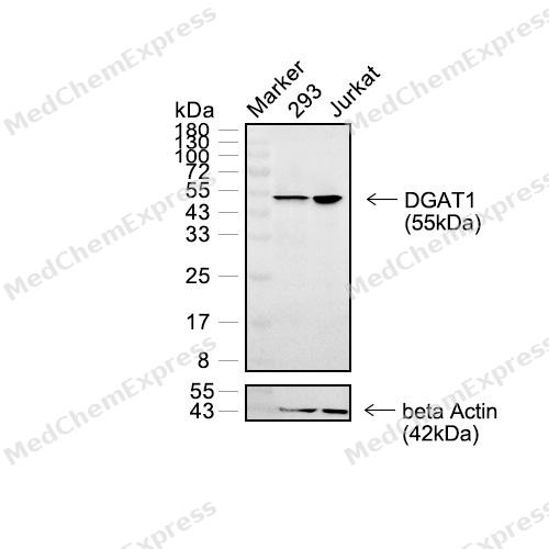 Western blot analysis of extracts from 293 (lane 2(20μg)) and Jurkat (lane 3(20μg)) using DGAT1 Rabbit mAb. Proteins were transferred to a PVDF membrane and blocked with 5% non-fat milk in TBST for 2 hour at room temperature. The primary antibody (1/1000) and Loading control antibody (beta Actin, HY-P83730, 1/5000) was used in 5% non-fat milk in TBST at 4°C overnight. Goat Anti-Rabbit/Mouse IgG-HRP Secondary Antibody (1/10000) was used for 1 hour at room temperature.
 -
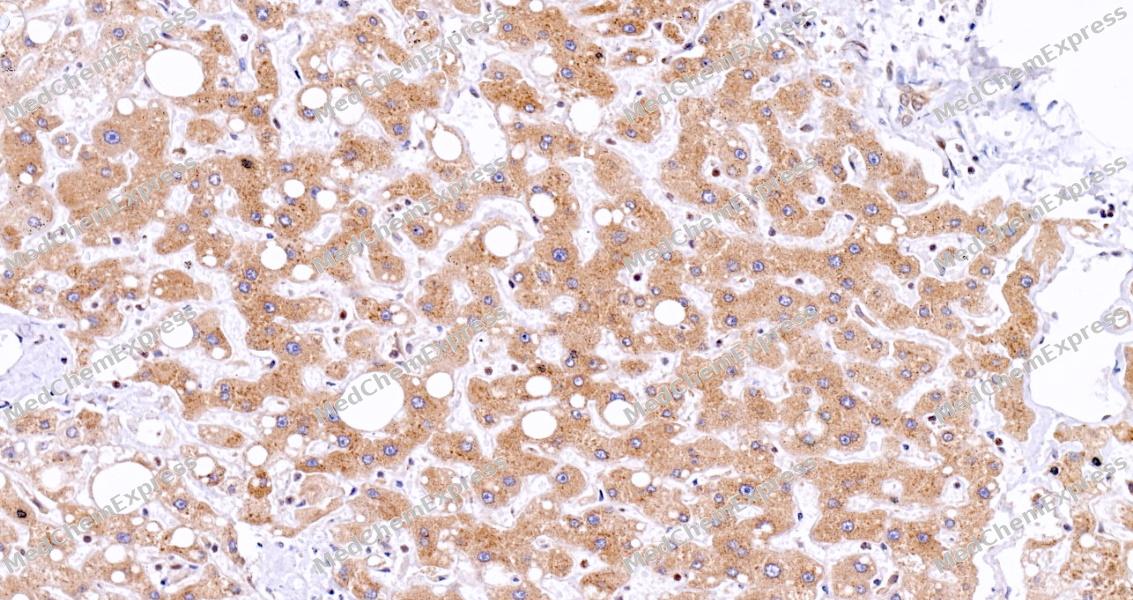 Immunohistochemical analysis of paraffin-embedded human liver tissue using DGAT1 Antibody (HY-P81246, 1/100). The section was pretreated using heat mediated antigen retrieval with sodium citrate buffer (pH 6.0) for 8 minutes. The tissues were blocked with quick block buffer for 0.5 hours at room temperature, washed with PBS and PBST, and then incubated with the primary antibody overnight at 4℃. The detection was performed using an HRP conjugated compact polymer system. DAB was used as the chromogen. Tissues were counterstained with hematoxylin and mounted with DPX.
 -
 Immunohistochemical analysis of paraffin-embedded human salivary gland tissue using DGAT1 Antibody (HY-P81246, 1/100). The section was pretreated using heat mediated antigen retrieval with sodium citrate buffer (pH 6.0) for 8 minutes. The tissues were blocked with quick block buffer for 0.5 hours at room temperature, washed with PBS and PBST, and then incubated with the primary antibody overnight at 4℃. The detection was performed using an HRP conjugated compact polymer system. DAB was used as the chromogen. Tissues were counterstained with hematoxylin and mounted with DPX.
 -
 Immunohistochemical analysis of paraffin-embedded human kidney tissue using DGAT1 Antibody (HY-P81246, 1/100). The section was pretreated using heat mediated antigen retrieval with sodium citrate buffer (pH 6.0) for 8 minutes. The tissues were blocked with quick block buffer for 0.5 hours at room temperature, washed with PBS and PBST, and then incubated with the primary antibody overnight at 4℃. The detection was performed using an HRP conjugated compact polymer system. DAB was used as the chromogen. Tissues were counterstained with hematoxylin and mounted with DPX.
 -
 Immunohistochemical analysis of paraffin-embedded human testis tissue using DGAT1 Antibody (HY-P81246, 1/100). The section was pretreated using heat mediated antigen retrieval with sodium citrate buffer (pH 6.0) for 8 minutes. The tissues were blocked with quick block buffer for 0.5 hours at room temperature, washed with PBS and PBST, and then incubated with the primary antibody overnight at 4℃. The detection was performed using an HRP conjugated compact polymer system. DAB was used as the chromogen. Tissues were counterstained with hematoxylin and mounted with DPX.
 -
 Immunohistochemical analysis of paraffin-embedded human pancreas using DGAT1 Antibody (HY-P81246, 1/100). The section was pretreated using heat mediated antigen retrieval with sodium citrate buffer (pH 6.0) for 8 minutes. The tissues were blocked with quick block buffer for 0.5 hours at room temperature, washed with PBS and PBST, and then incubated with the primary antibody overnight at 4℃. The detection was performed using an HRP conjugated compact polymer system. DAB was used as the chromogen. Tissues were counterstained with hematoxylin and mounted with DPX.
 -
 Immunohistochemical analysis of paraffin-embedded human gallbladder tissue using DGAT1 Antibody (HY-P81246, 1/100). The section was pretreated using heat mediated antigen retrieval with sodium citrate buffer (pH 6.0) for 8 minutes. The tissues were blocked with quick block buffer for 0.5 hours at room temperature, washed with PBS and PBST, and then incubated with the primary antibody overnight at 4℃. The detection was performed using an HRP conjugated compact polymer system. DAB was used as the chromogen. Tissues were counterstained with hematoxylin and mounted with DPX.
 -
 Immunohistochemical analysis of paraffin-embedded human liver tissue using DGAT1 Antibody (HY-P81246, 1/100). The section was pretreated using heat mediated antigen retrieval with sodium citrate buffer (pH 6.0) for 8 minutes. The tissues were blocked with quick block buffer for 0.5 hours at room temperature, washed with PBS and PBST, and then incubated with the primary antibody overnight at 4℃. The detection was performed using an HRP conjugated compact polymer system. DAB was used as the chromogen. Tissues were counterstained with hematoxylin and mounted with DPX.
 -
 Immunohistochemical analysis of paraffin-embedded human adrenal gland tissue using DGAT1 Antibody (HY-P81246, 1/100). The section was pretreated using heat mediated antigen retrieval with sodium citrate buffer (pH 6.0) for 8 minutes. The tissues were blocked with quick block buffer for 0.5 hours at room temperature, washed with PBS and PBST, and then incubated with the primary antibody overnight at 4℃. The detection was performed using an HRP conjugated compact polymer system. DAB was used as the chromogen. Tissues were counterstained with hematoxylin and mounted with DPX.
 -
 Immunohistochemical analysis of paraffin-embedded human thyroid gland tissue using DGAT1 Antibody (HY-P81246, 1/100). The section was pretreated using heat mediated antigen retrieval with sodium citrate buffer (pH 6.0) for 8 minutes. The tissues were blocked with quick block buffer for 0.5 hours at room temperature, washed with PBS and PBST, and then incubated with the primary antibody overnight at 4℃. The detection was performed using an HRP conjugated compact polymer system. DAB was used as the chromogen. Tissues were counterstained with hematoxylin and mounted with DPX.
 -
 Immunohistochemical analysis of paraffin-embedded human cerebral cortex tissue using DGAT1 Antibody (HY-P81246, 1/100). The section was pretreated using heat mediated antigen retrieval with sodium citrate buffer (pH 6.0) for 8 minutes. The tissues were blocked with quick block buffer for 0.5 hours at room temperature, washed with PBS and PBST, and then incubated with the primary antibody overnight at 4℃. The detection was performed using an HRP conjugated compact polymer system. DAB was used as the chromogen. Tissues were counterstained with hematoxylin and mounted with DPX.
 -
 Immunohistochemical analysis of paraffin-embedded human heart muscle tissue using DGAT1 Antibody (HY-P81246, 1/100). The section was pretreated using heat mediated antigen retrieval with sodium citrate buffer (pH 6.0) for 8 minutes. The tissues were blocked with quick block buffer for 0.5 hours at room temperature, washed with PBS and PBST, and then incubated with the primary antibody overnight at 4℃. The detection was performed using an HRP conjugated compact polymer system. DAB was used as the chromogen. Tissues were counterstained with hematoxylin and mounted with DPX.
 -
 Immunocytochemistry analysis of Hela cells labeling DGAT1 with DGAT1 Antibody (HY-P81246) at 1/50 dilution. Cells were fixed in 4% paraformaldehyde for 15 minutes at room temperature, permeabilized with 0.1% Triton X-100 in PBS for 15 minutes at room temperature, then blocked with quick block buffer for 10 minutes at room temperature. Cells were then incubated with DGAT1 Antibody (HY-P81246) at 1/50 dilution in quick block buffer overnight at 4 ℃. AF488-conjugated Goat Anti-Rabbit IgG H&L (HY-P8002, Green) was used as the secondary antibody at 1/1,000 dilution. PBS instead of the primary antibody was used as the secondary antibody only control. The Nuclear counterstain was DAPI (Blue).
 -
 Immunocytochemistry analysis of Hela cells labeling DGAT1 with DGAT1 Antibody (HY-P81246) at 1/100 dilution. Cells were fixed in 4% paraformaldehyde for 15 minutes at room temperature, permeabilized with 0.1% Triton X-100 in PBS for 15 minutes at room temperature, then blocked with quick block buffer for 10 minutes at room temperature. Cells were then incubated with DGAT1 Antibody (HY-P81246) at 1/100 dilution in quick block buffer overnight at 4 ℃. AF488-conjugated Goat Anti-Rabbit IgG H&L (HY-P8002, Green) was used as the secondary antibody at 1/1,000 dilution. PBS instead of the primary antibody was used as the secondary antibody only control. The Nuclear counterstain was DAPI (Blue).


Background
-
Function
DGAT1 (diacylglycerol O-acyltransferase 1) catalyzes the terminal step of triacylglycerol (TAG) synthesis by esterifying diacylglycerol with fatty acyl-CoA at the endoplasmic reticulum, making it a central regulator of cellular lipid storage and lipid droplet formation[1]. Mechanistically, DGAT1 functions within the Kennedy pathway and contributes substantially to triglyceride turnover, fatty acid utilization, and metabolic homeostasis in multiple tissues, including intestine, adipose tissue, and heart[1][2]. In disease-related models, genetic or pharmacological modulation of DGAT activity alters triglyceride accumulation, fatty acid oxidation, endoplasmic reticulum stress responses, and susceptibility to diet-induced metabolic dysfunction, supporting its relevance to obesity, hepatic steatosis, and cardiometabolic disorders[2][3][4]. Compared with the closely related isoform DGAT2, DGAT1 and DGAT2 catalyze the same biochemical reaction but exhibit distinct physiological functions, tissue distributions, structural origins, and compensatory capacities despite jointly accounting for most mammalian TAG synthesis[2][5]. Experimental studies further demonstrate that DGAT1 uniquely contributes to protection against lipid-induced endoplasmic reticulum stress under high-fat dietary conditions, whereas DGAT2 is not essential for adipose triglyceride storage in several mouse models[3]. For research applications, selective DGAT inhibitors have been widely used to interrogate triglyceride synthesis pathways and evaluate therapeutic strategies targeting metabolic disease, although gastrointestinal tolerability has limited clinical development of several DGAT1-directed compounds[4].
-
Subcellular Localization
Endoplasmic reticulum membrane; Multi-pass membrane protein
-
Subunit
Homodimer or homotetramer; both forms have similar enzymatic activities
-
SwissProt ID
-
Synonyms
ACAT related gene product 1; ACAT-related gene product 1; Acyl coenzyme A:cholesterol acyltransferase related gene 1; Acyl-CoA retinol O-fatty-acyltransferase; Acyl-CoA:diacylglycerol acyltransferase; ARAT; ARGP1; C75990; D15Ertd23e; Dgat; DGAT1; DGAT1_HUMAN; Diacylglycerol O acyltransferase 1; Diacylglycerol O-acyltransferase 1; Diglyceride acyltransferase; EC 2.3.1.20; hCG_24006; MGC139064; Retinol O fatty acyltransferase.


Documentation
-
Data Sheet (261 KB)
-
SDS (254 KB)
- English - EN (254 KB)
- Français - FR (254 KB)
- Deutsch - DE (254 KB)
- Norwegian - NO (254 KB)
- Español - ES (254 KB)
- Swedish - SV (254 KB)
- Italian - IT (254 KB)
- Korean - KR (254 KB)
- Portuguese - PT (254 KB)
-
User Guide for Antibodies (1077 KB)
References
[1]. Yen CL, et al. Thematic review series: glycerolipids. DGAT enzymes and triacylglycerol biosynthesis. J Lipid Res. 2008 Nov;49(11):2283-301. [Content Brief]
[2]. Roe ND, et al. The Role of Diacylglycerol Acyltransferase (DGAT) 1 and 2 in Cardiac Metabolism and Function. Sci Rep. 2018 Mar 21;8(1):4983. [Content Brief]
[3]. Liu X, et al. Physical layer encryption scheme based on biological genetic variation mechanisms in CO-OFDM system. Opt Express. 2025 Oct 6;33(20):42528-42541. [Content Brief]
[4]. Devasthale P, et al. Monoacylglycerol Acyltransferase 2 (MGAT2) Inhibitors for the Treatment of Metabolic Diseases and Nonalcoholic Steatohepatitis (NASH). J Med Chem. 2018 Nov 21;61(22):9879-9888. [Content Brief]
[5]. Kiros M, et al. Trends in HIV-1 pretreatment drug resistance and HIV-1 variant dynamics among antiretroviral therapy-naive Ethiopians from 2003 to 2018: a pooled sequence analysis. Virol J. 2023 Oct 25;20(1):243. [Content Brief]

 Powered by Bioz
