



Glutaminase Antibody (YA406)
(Synonyms: Glutaminase kidney isoform; GLS; GLS1; KGA; K-glutaminase; GAM; GAC; Glutaminase C; L-glutamine amidohydrolase)
Based on 1 Customer Validation
Glutaminase Antibody (YA406) is a Rabbit-derived and non-conjugated IgG monoclonal antibody, targeting to Glutaminase.
-
Host:
Rabbit
-
Isotype:
IgG
-
Application:
WB, ICC/IF, IP, FC
-
Reactivity :
Human, Mouse, Rat
-
Formulation:
Supplied in 50 mM Tris-Glycine (pH 7.4), 0.15 M NaCl, 40% Glycerol and 0.05% BSA. Preservative: 0.01% Sodium azide
-
Conjugation:
Non-conjugated


To place orders, for customer services and technical support, please contact: MedChemExpress USA
Tel: 609-228-6898 E-mail: [email protected] [email protected]





Applications
| Application |
WB

WB: Western Blot
|
ICC/IF
ICC/IF: Immunocytochemistry/
Immunofluorescence |
FC
FC: Flow Cytometry
|
IP
IP: Immunoprecipitation
|
|---|---|---|---|---|
| Dilution Ratio | 1:500-1000 | 1:200-500 | 1:200-500 | 1:20-50 |


Product Details
Glutaminase Antibody (YA406) is a Rabbit-derived and non-conjugated IgG monoclonal antibody, targeting to Glutaminase.
-
Host Rabbit

-
Clonality Recombinant,Monoclonal

-
Species ReactivityHuman, Mouse, Rat
-
Calculated Molecular Weight Predicted band size: 73 kDa;Observed band size: 68 kDa

Entrez Gene: 2744 Human ; 14660 Mouse ; 24398 Rat
SwissProt: O94925 Human ; D3Z7P3 Mouse ; P13264 Rat
OMIM: 618328 Human
Synthetic peptide corresponding to Human Glutaminase C.The exact sequence is proprietary to MCE.
Endogenous
affinity purified
Non-conjugated
Unmodified
IgG


Product Properties
-
Appearance
Solution
-
Formulation
Supplied in 50 mM Tris-Glycine (pH 7.4), 0.15 M NaCl, 40% Glycerol and 0.05% BSA. Preservative: 0.01% Sodium azide
-
Concentration
Batch-dependent, Please check the COA for the concentration of each lot. Check Lot Concentration
-
Storage & Stability
Stored at -20°C for 1 year. Avoid repeated freeze / thaw cycles.
-
Shipping
Shipping with blue ice.


Verification Images
-
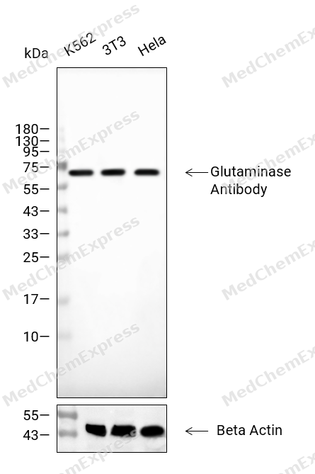 Western blot analysis of extracts from K562 (lane 2(20μg), 3T3 (lane 3(20μg), Hela (lane 4(20μg) using Glutaminase Antibody. Proteins were transferred to a PVDF membrane and blocked with 5% BSA in TBST for 2 hour at room temperature. The primary antibody and Loading control antibody (Beta Actin, HY-P80438, 1/3000) was used in 5% BSA in TBST at 4°C overnight. Goat Anti-Mouse/Rabbit IgG-HRP Secondary Antibody (HY-P8004/HY-P8001, 1/10,000) was used for 1 hour at room temperature.
 -
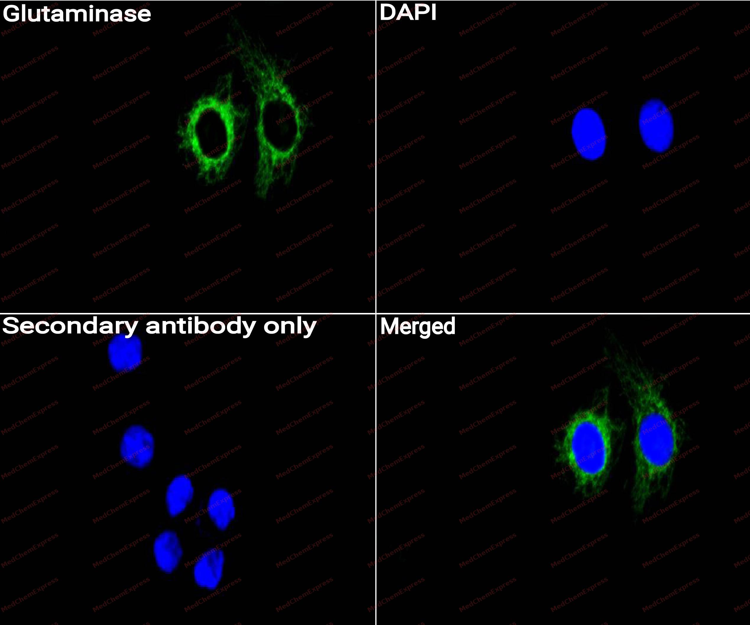 Immunocytochemistry analysis of Siha cells labeling Glutaminase with Glutaminase Antibody (HY-P80690) at 1/50 dilution. Cells were fixed in 4% paraformaldehyde for 15 minutes at room temperature, permeabilized with 0.1% Triton X-100 for 10 minutes at room temperature, then blocked with QuickBlock™ Blocking Buffer for Immunol Staining for 10 min at room temperature. Cells were then incubated with Glutaminase Antibody (HY-P80690) at 1/50 dilution in QuickBlock™ Blocking Buffer for Immunol Staining at 4 ℃. Alexa Fluor® 488-conjugated AffiniPure Goat Anti-Rabbit IgG H&L(HY-P8002, Green) was used as the secondary antibody at 1/1,000 dilution. PBS instead of the primary antibody was used as the secondary antibody only control. The Nuclear counterstain was DAPI (Blue).
 -
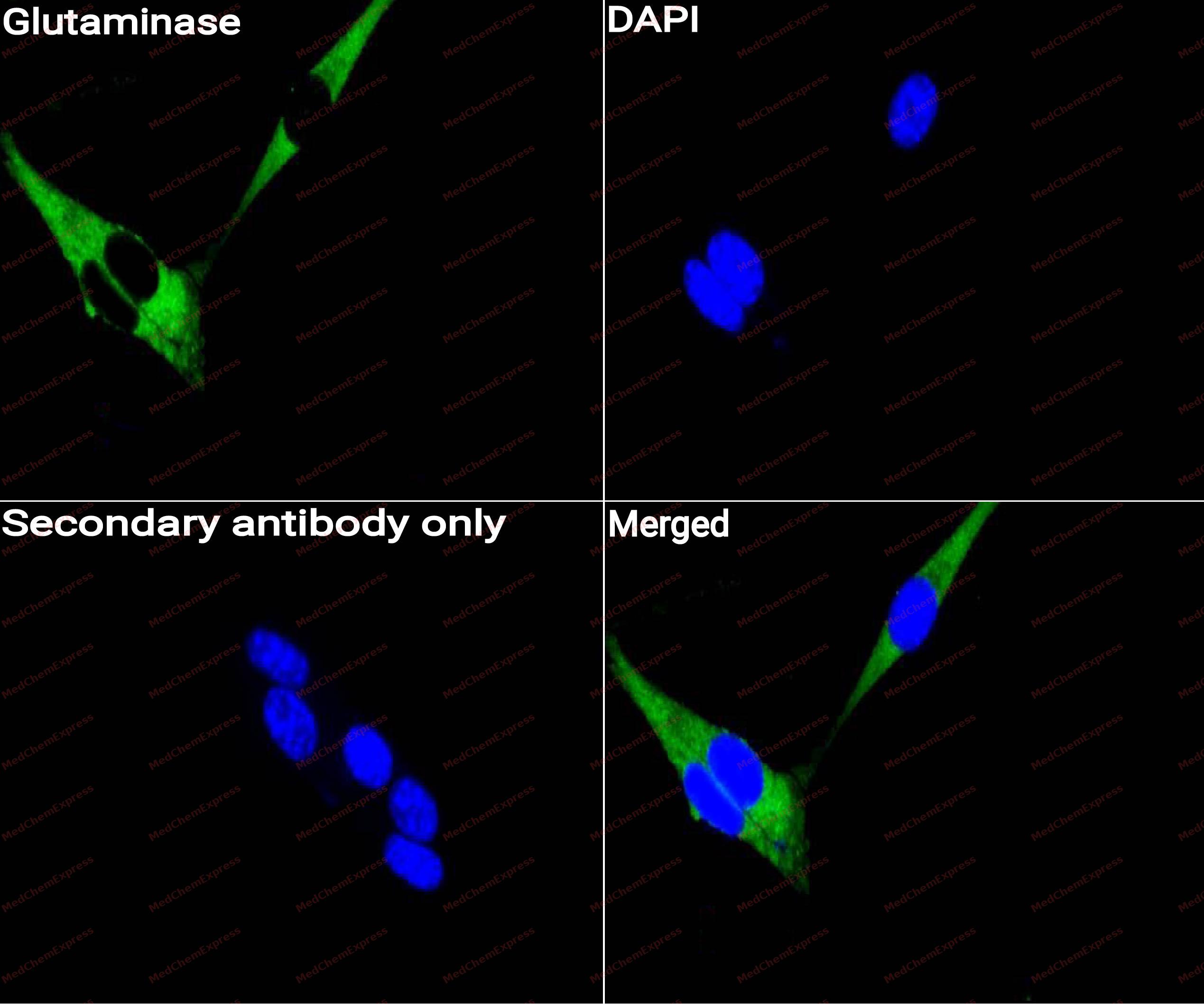 Immunocytochemistry analysis of NIH3T3 cells labeling Glutaminase with Glutaminase Antibody (HY-P80690) at 1/50 dilution. Cells were fixed in 4% paraformaldehyde for 15 minutes at room temperature, permeabilized with 0.1% Triton X-100 for 10 minutes at room temperature, then blocked with QuickBlock™ Blocking Buffer for Immunol Staining for 10 min at room temperature. Cells were then incubated with Glutaminase Antibody (HY-P80690)at 1/50 dilution in QuickBlock™ Blocking Buffer for Immunol Staining at 4 ℃. Alexa Fluor® 488-conjugated AffiniPure Goat Anti-Rabbit IgG H&L(HY-P8002,Green) was used as the secondary antibody at 1/1,000 dilution. PBS instead of the primary antibody was used as the secondary antibody only control. The Nuclear counterstain was DAPI (Blue).


Background
-
Function
Glutaminase is the initial enzyme in glutamine metabolism, which catalyzes the hydrolysis of glutamine to glutamate in cells. Glutaminase plays a key role in cancer cell metabolism, growth, and proliferation. In mammalian cells, there are two paralogous GLS genes, GLS1 (or GLS) and GLS2. GLS1 encodes two alternatively spliced isozymes: kidney glutaminase (KGA) and glutaminase C (GAC) . GLS2 also encodes two isozymes: liver glutaminase (LGA) and glutaminase B.
GLS1 is ubiquitously expressed in various tissues, and its expression can be induced by the oncogene MYC. GLS1 is frequently activated and/or overexpressed in various types of cancer, including hepatocellular carcinoma (HCC) . GLS1 has been reported to promote tumorigenesis in different types of cancer, including HCC, which is mainly attributable to its glutaminase activity and role in promoting glutamine metabolism. GLS has emerged as a critical enzyme in a number of cancer types. Elevated GLS2 enzymatic activity has also been correlated with tumor cell growth in vitro and in vivo. N-Myc activates GLS2 to promote conversion of glutamine to glutamate in MYCN-amplified neuroblastoma cells. Abrogation of GLS2 function profoundly inhibits glutaminolysis and dramatically decreases cell proliferation and survival in vitro and in vivo. However, there is controversy over the role of GLS2 as a tumor suppressor. Enzymatic activity independent of GLS2 is up-regulated via p53 or p63 and plays a role of tumor suppressor.
-
Subcellular Localization
Mitochondrion; Cytoplasm, cytosol; Mitochondrion; Mitochondrion matrix; Mitochondrion matrix
-
Expression
Tissue_specificity:Isomers 1 and 3 were detected in the cerebral cortex. Isomer 3 was highly expressed in astrocytomas, gangliogliomas, and ependymomas. Isomer 1 was highly expressed in the brain and kidneys, but not detected in the liver. Isomer 3 was highly expressed in the heart and pancreas, and expressed at lower levels in the placenta, lungs, pancreas, and kidneys, but not detected in the liver. Isomer 2 was expressed in cardiac and skeletal muscle. -
Isoforms & Post-Translational Modification
O94925 has 3 isomers: O94925-1: 73461 Da (predicted); O94925-2: 17738 Da (predicted); O94925-3: 65460 Da (predicted).
Synthesized as a 74-kDa cytosolic precursor which is proteolytically processed by the mitochondrial-processing peptidase (MPP) via a 72-kDa intermediate to yield the mature mitochondrial 68- and 65-kDa subunits -
Subunit
Homotetramer, dimer of dimers (PubMed:22538822, PubMed:26988803, PubMed:28526749, PubMed:29317493). The tetramers can assemble into rod-like oligomers (in vitro), but the physiological significance of this is not clear (By similarity). Interacts with RAF1 and MAP2K2 (PubMed:22538822). Interacts with ATCAY; the interaction is direct and may control GLS localization, negatively regulating its activity
-
SwissProt ID
-
Synonyms
Glutaminase kidney isoform; GLS; GLS1; KGA; K-glutaminase; GAM; GAC; Glutaminase C; L-glutamine amidohydrolase
-
Research Field
Neuroscience


Documentation
-
Data Sheet (234 KB)
-
SDS (313 KB)
- English - EN (313 KB)
- Français - FR (313 KB)
- Deutsch - DE (313 KB)
- Norwegian - NO (313 KB)
- Español - ES (313 KB)
- Swedish - SV (313 KB)
- Italian - IT (313 KB)
- Korean - KR (313 KB)
- Portuguese - PT (313 KB)
-
User Guide for Antibodies (1077 KB)

 Powered by Bioz
