



HDAC4 Antibody (YA391)
(Synonyms: KIAA0288, HDAC4, Histone deacetylase 4, HD4)
Based on 1 Customer Validation
HDAC4 Antibody (YA391) is a Rabbit-derived and non-conjugated IgG monoclonal antibody, targeting to HDAC4.
-
Host:
Rabbit
-
Isotype:
IgG
-
Application:
WB, ICC/IF
-
Reactivity :
Human
-
Formulation:
Supplied in 1*TBS (pH7.4), 0.05% BSA and 40% Glycerol. Preservative: 0.05% Sodium Azide.
-
Conjugation:
Non-conjugated


To place orders, for customer services and technical support, please contact: MedChemExpress USA
Tel: 609-228-6898 E-mail: [email protected] [email protected]





Applications
| Application |
WB

WB: Western Blot
|
ICC/IF
ICC/IF: Immunocytochemistry/
Immunofluorescence |
|---|---|---|
| Dilution Ratio | 1:1000-1:2000 | 1:100-1:500 |


Product Details
HDAC4 Antibody (YA391) is a Rabbit-derived and non-conjugated IgG monoclonal antibody, targeting to HDAC4.
-
Host Rabbit

-
Clonality Recombinant,Monoclonal

-
Species ReactivityHuman
-
Observed Molecular WeightObserved band size: 150 kDa
Note: Due to possible protein modifications or aggregation, the molecular weight should be confirmed by actual measurement, and the predicted value is for reference only.

-
Calculated Molecular Weight Predicted band size: 119 kDa

Synthetic peptide corresponding to Human HDAC4.AA range:11-125.
Endogenous
Protein A affinity purified.
Non-conjugated
Unmodified
IgG


Product Properties
-
Appearance
Solution
-
Formulation
Supplied in 1*TBS (pH7.4), 0.05% BSA and 40% Glycerol. Preservative: 0.05% Sodium Azide.
-
Concentration
Batch-dependent, Please check the COA for the concentration of each lot. Check Lot Concentration
-
Storage & Stability
Stored at -20°C for 1 year. Avoid repeated freeze / thaw cycles.
-
Livraison
Shipping with blue ice.


Verification Images
-
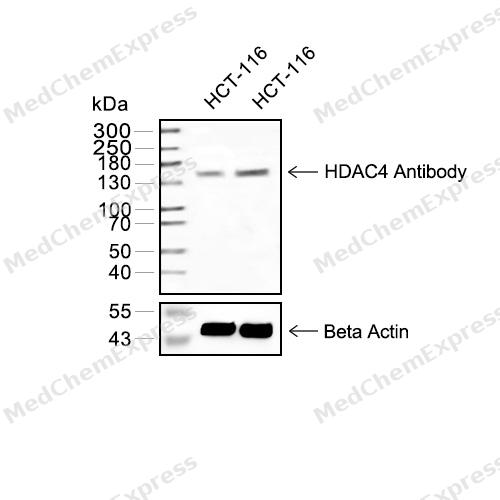 Western blot analysis of extracts from HCT116(lane 2(20μg) and HCT116(lane 3(40μg) using HDAC4(HY-P80153 Rabbit mAb. Proteins were transferred to a PVDF membrane and blocked with 5% non-fat milk in TBST for 2 hour at room temperature. The primary antibody (1/1000) and Loading control antibody (Beta Actin, HY-P80438, 1/10000) was used in 5% non-fat milk in TBST at 4°C overnight. Goat Anti-Mouse/Rabbit IgG-HRP Secondary Antibody (1/10000) was used for 1 hour at room temperature.
 -
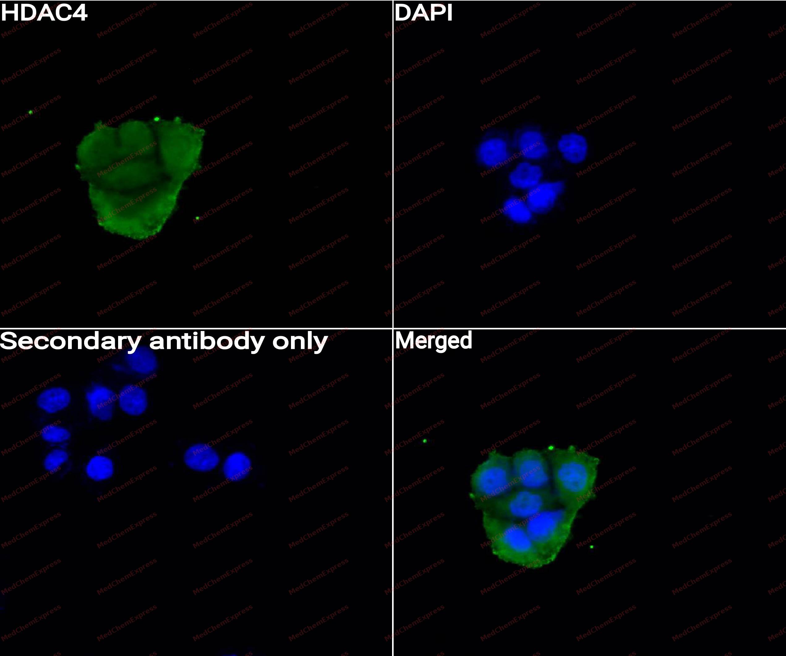 Immunocytochemistry analysis of Hela cells labeling HDAC4 with HDAC4 Antibody (HY-P80153) at 1/50 dilution. Cells were fixed in 4% paraformaldehyde for 15 minutes at room temperature, permeabilized with 0.1% Triton X-100 for 10 minutes at room temperature, then blocked with QuickBlock™ Blocking Buffer for Immunol Staining for 10 min at room temperature. Cells were then incubated with HDAC4 Antibody (HY-P80153)at 1/50 dilution in QuickBlock™ Blocking Buffer for Immunol Staining at 4 ℃. Alexa Fluor® 488-conjugated AffiniPure Goat Anti-Rabbit IgG H&L(HY-P8002,Green) was used as the secondary antibody at 1/1,000 dilution. PBS instead of the primary antibody was used as the secondary antibody only control. The Nuclear counterstain was DAPI (Blue).
 -
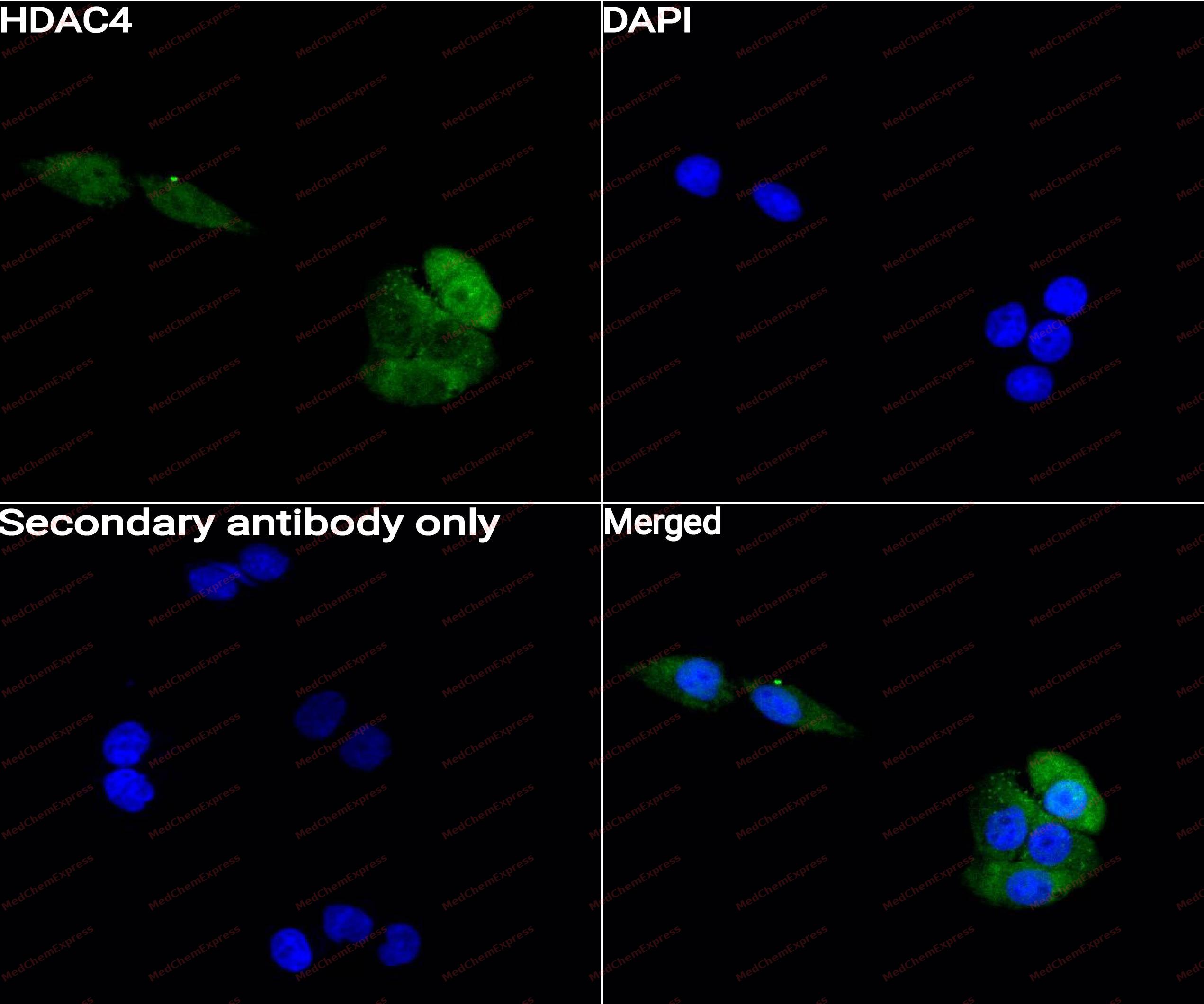 Immunocytochemistry analysis of HepG2 cells labeling HDAC4 with HDAC4 Antibody (HY-P80153) at 1/50 dilution. Cells were fixed in 4% paraformaldehyde for 15 minutes at room temperature, permeabilized with 0.1% Triton X-100 for 10 minutes at room temperature, then blocked with QuickBlock™ Blocking Buffer for Immunol Staining for 10 min at room temperature. Cells were then incubated with HDAC4 Antibody (HY-P80153) at 1/50 dilution in QuickBlock™ Blocking Buffer for Immunol Staining at 4 ℃. Alexa Fluor® 488-conjugated AffiniPure Goat Anti-Rabbit IgG H&L(HY-P8002, Green) was used as the secondary antibody at 1/1,000 dilution. PBS instead of the primary antibody was used as the secondary antibody only control. The Nuclear counterstain was DAPI (Blue).


Background
-
Function
HDAC4 is a class IIa histone deacetylase that acts as a signal-dependent transcriptional regulator linking extracellular cues to chromatin remodeling, gene expression, and cell differentiation[1][2]. Mechanistically, class IIa HDACs, including HDAC4, shuttle between nuclear and cytoplasmic compartments through post-translational modification, and this localization controls their transcriptional functions[3]. In liver, HDAC4, HDAC5, and HDAC7 respond to glucagon by entering the nucleus, associating with gluconeogenic promoters, recruiting HDAC3, activating FOXO transcription factors, and increasing blood-glucose-related gene expression[4]. In skeletal muscle, class II HDAC proteins repress MEF2 activity and suppress slow-twitch oxidative myofiber formation, while MEF2 activation promotes endurance-associated fiber programs[5]. In pancreatic endocrine models, HDAC4, HDAC5, and HDAC9 regulate β-cell and δ-cell lineage control, and class IIa HDAC inhibition with MC1568 amplifies endocrine β- and δ-cells[6]. Compared with related isoforms, HDAC4 belongs to the HDAC4/5/7/9 class IIa subgroup, which shares MEF2-related transcriptional partners but shows distinct disease-linked regulatory roles[7]. For experimental applications, LBH589 confined HDAC4 to the cytoplasm, prolonged γ-H2AX foci after irradiation, and sensitized non-small cell lung cancer models to radiation-induced DNA double-strand breaks[8]. Selective inhibitor design also identified LMK235 as showing nanomolar inhibition of HDAC4 and HDAC5, unlike vorinostat and TSA, which inhibited these isoforms in the higher micromolar range[9].
-
Subcellular Localization
Nucleus; Cytoplasm
-
Expression
Tissue_specificity:general expression -
Isoforms & Post-Translational Modification
P56524 has 2 isomers: P56524-1: 119040 Da (predicted); P56524-2: 106367 Da (predicted).
Phosphorylated by CaMK4 at Ser-246, Ser-467 and Ser-632. Phosphorylation at other residues by CaMK2D is required for the interaction with 14-3-3. Phosphorylation at Ser-350, within the PxLPxI/L motif, impairs the binding of ANKRA2 but generates a high-affinity docking site for 14-3-3;Sumoylation on Lys-559 is promoted by the E3 SUMO-protein ligase RANBP2, and prevented by phosphorylation by CaMK4 -
Subunit
Homodimer. Homodimerization via its N-terminal domain (PubMed:12032081). Interacts with MEF2A (PubMed:10487761). Interacts with MEF2C and MEF2D (PubMed:10523670). Interacts with AHRR (By similarity). Interacts with NR2C1 (PubMed:11463856). Interacts with HDAC7 (By similarity). Interacts with a 14-3-3 chaperone proteins in a phosphorylation dependent manner (PubMed:10958686). Interacts with 14-3-3 protein YWHAB (PubMed:33537682). Interacts with BTBD14B (By similarity). Interacts with KDM5B (PubMed:17373667). Interacts with MYOCD (By similarity). Interacts with MORC2 (PubMed:20110259). Interacts (via PxLPxI/L motif) with ANKRA2 (via ankyrin repeats). Interacts with CUL7 (as part of the 3M complex); negatively regulated by ANKRA2 (PubMed:25752541). Interacts with EP300 in the presence of TFAP2C (PubMed:24413532). Interacts with HSPA1A and HSPA1B leading to their deacetylation at 'Lys-77' (PubMed:27708256). Interacts with ZBTB7B; the interaction allows the recruitment of HDAC4 on CD8 loci for deacetylation and possible inhibition of CD8 genes expression (By similarity). Interacts with DHX36 (By similarity). Interacts with SIK3; this interaction leads to HDAC4 retention in the cytoplasm (By similarity). Interacts with ZNF638 (PubMed:30487602)
-
SwissProt ID
-
Synonyms
KIAA0288, HDAC4, Histone deacetylase 4, HD4
-
Research Field
Epigenetics and Nuclear Signaling


Documentation
-
Fiche technique (262 KB)
-
SDS (251 KB)
- English - EN (251 KB)
- Français - FR (251 KB)
- Deutsch - DE (251 KB)
- Norwegian - NO (251 KB)
- Español - ES (251 KB)
- Swedish - SV (251 KB)
- Italian - IT (251 KB)
- Korean - KR (251 KB)
- Portuguese - PT (251 KB)
-
User Guide for Antibodies (1077 KB)
References
[1]. Verdin E, et al. Class II histone deacetylases: versatile regulators. Trends Genet. 2003 May;19(5):286-93. [Content Brief]
[2]. Yang XJ, et al. Class II histone deacetylases: from sequence to function, regulation, and clinical implication. Mol Cell Biol. 2005 Apr;25(8):2873-84. [Content Brief]
[3]. Mathias RA, et al. Post-translational modifications regulate class IIa histone deacetylase (HDAC) function in health and disease. Mol Cell Proteomics. 2015 Mar;14(3):456-70. [Content Brief]
[4]. Mihaylova MM, et al. Class IIa histone deacetylases are hormone-activated regulators of FOXO and mammalian glucose homeostasis. Cell. 2011 May 13;145(4):607-21. [Content Brief]
[5]. Potthoff MJ, et al. Histone deacetylase degradation and MEF2 activation promote the formation of slow-twitch myofibers. J Clin Invest. 2007 Sep;117(9):2459-67. [Content Brief]
[6]. Lenoir O, et al. Specific control of pancreatic endocrine β- and δ-cell mass by class IIa histone deacetylases HDAC4, HDAC5, and HDAC9. Diabetes. 2011 Nov;60(11):2861-71. [Content Brief]
[7]. Clocchiatti A, et al. Class IIa HDACs: from important roles in differentiation to possible implications in tumourigenesis. J Cell Mol Med. 2011 Sep;15(9):1833-46. [Content Brief]
[8]. Geng L, et al. Histone deacetylase (HDAC) inhibitor LBH589 increases duration of gamma-H2AX foci and confines HDAC4 to the cytoplasm in irradiated non-small cell lung cancer. Cancer Res. 2006 Dec 1;66(23):11298-304. [Content Brief]
[9]. Marek L, et al. Histone deacetylase (HDAC) inhibitors with a novel connecting unit linker region reveal a selectivity profile for HDAC4 and HDAC5 with improved activity against chemoresistant cancer cells. J Med Chem. 2013 Jan 24;56(2):427-36. [Content Brief]

 Powered by Bioz
