



JNK1 Antibody (YA720)
(Synonyms: AI849689 antibody; c Jun N terminal kinase 1 antibody; C-JUN kinase 1 antibody; c-Jun N-terminal kinase 1 antibody; EC 2.7.11.24 antibody; JNK 1 antibody; JNK antibody; JNK-46 antibody; JNK1A2 antibody; JNK21B1/2 antibody; MAP kinase 8 antibody; MAPK 8 antibody; mapk8 antibody; Mitogen activated protein kinase 8 antibody; MK08_HUMAN antibody; p54 gamma antibody; Prkm8 antibody; Protein kinase JNK1 antibody; Protein kinase, mitogen-activated, 8 antibody; SAPK 1 antibody; SAPK gamma antibody; SAPK1 antibody; Stress-activated protein kinase 1 antibody)
Based on 1 Customer Validation
JNK1 Antibody (YA720) is a Mouse-derived and non-conjugated IgG2a monoclonal antibody, targeting to JNK1.
-
Host:
Mouse
-
Isotype:
IgG
-
Application:
WB, ICC/IF
-
Reactivity :
Human, Mouse, Rat
-
Formulation:
Supplied in 1*PBS (pH 7.3), 50% glycerol and 0.5% BSA. Preservative: 0.02% sodium azide.
-
Conjugation:
Non-conjugated


To place orders, for customer services and technical support, please contact: MedChemExpress USA
Tel: 609-228-6898 E-mail: [email protected] [email protected]





Applications
| Application |
WB

WB: Western Blot
|
ICC/IF
ICC/IF: Immunocytochemistry/
Immunofluorescence |
|---|---|---|
| Dilution Ratio | 1:500-1:1000 | 1:50-1:200 |


Product Details
JNK1 Antibody (YA720) is a Mouse-derived and non-conjugated IgG2a monoclonal antibody, targeting to JNK1.
-
Host Mouse

-
Clonality Monoclonal

-
Species ReactivityHuman, Mouse, Rat
-
Observed Molecular WeightObserved band size: 46/54 kDa
Note: Due to possible protein modifications or aggregation, the molecular weight should be confirmed by actual measurement, and the predicted value is for reference only.

-
Calculated Molecular Weight Predicted band size: 48 kDa

Entrez Gene: 5599 Human ; 26419 Mouse ; 116554 Rat
SwissProt: P45983 Human ; Q91Y86 Mouse ; P49185 Rat
OMIM: 601158 Human
Synthetic peptide corresponding to Human JNK1.The exact sequence is proprietary to MCE.
Endogenous
affinity purified
Non-conjugated
Unmodified
IgG


Product Properties
-
Appearance
Solution
-
Formulation
Supplied in 1*PBS (pH 7.3), 50% glycerol and 0.5% BSA. Preservative: 0.02% sodium azide.
-
Concentration
Batch-dependent, Please check the COA for the concentration of each lot. Check Lot Concentration
-
Storage & Stability
Stored at -20°C for 1 year. Avoid repeated freeze / thaw cycles.
-
Shipping
Shipping with blue ice.


Verification Images
-
 Western blot analysis of extracts from HeLa (lane 1(20μg)) 、HEK293 (lane 2(20μg)) 、Neuro-2a (lane 3(20μg)) 、NIH-3T3 (lane 4(20μg)) and C6 (lane 5(20μg) using JNK1 Antibody (HY-P80729) . Proteins were transferred to a PVDF membrane and blocked with 5% nonfat dry milk in TBST for 1.5 hour at room temperature. The primary antibody (1/1000) and Loading control antibody (β Actin, 1/10000) was used in 5% nonfat dry milk in TBST at 4℃ overnight. Goat Anti-Mouse IgG-HRP Secondary Antibody (1/10,000) was used for 1 hour at room temperature.
 -
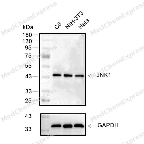 Western blot analysis of extracts from C6(lane 2(20μg))、NIH-3T3 (lane 3(20μg)) and Hela(lane 4(20μg)), using JNK1 (HY-P80729) Mouse mAb. Proteins were transferred to a PVDF membrane and blocked with 5% BSA in TBST for 2 hour at room temperature. The primary antibody (HY-P80729, 1/1000) and Loading control antibody (GAPDH, HY-P80954, 1/3000) was used in 5% BSA in TBST at 4°C overnight. Goat Anti-Mouse/Rabbit IgG-HRP Secondary Antibody (HY-P8004/HY-P8001, 1/10,000) was used for 1 hour at room temperature.
 -
 Immunocytochemistry analysis of HepG2 cells labeling JNK1 with JNK1 Antibody (HY-P80729) at 1/100 dilution. Cells were fixed in 4% paraformaldehyde for 15 minutes at room temperature, permeabilized with 0.1% Triton X-100 in PBS for 15 minutes at room temperature, then blocked with quick block buffer for 10 minutes at room temperature. Cells were then incubated with JNK1 Antibody (HY-P80729) at 1/100 dilution in quick block buffer overnight at 4 ℃.AF488-conjugated Goat Anti-Mouse IgG H&L (HY-P8005, Green) was used as the secondary antibody at 1/1,000 dilution. PBS instead of the primary antibody was used as the secondary antibody only control. The Nuclear counterstain was DAPI (Blue).
 -
 Immunocytochemistry analysis of HepG2 cells labeling JNK1 with JNK1 Antibody (HY-P80729) at 1/200 dilution. Cells were fixed in 4% paraformaldehyde for 15 minutes at room temperature, permeabilized with 0.1% Triton X-100 in PBS for 15 minutes at room temperature, then blocked with quick block buffer for 10 minutes at room temperature. Cells were then incubated with JNK1 Antibody (HY-P80729) at 1/200 dilution in quick block buffer overnight at 4 ℃.AF488-conjugated Goat Anti-Mouse IgG H&L (HY-P8005, Green) was used as the secondary antibody at 1/1,000 dilution. PBS instead of the primary antibody was used as the secondary antibody only control. The Nuclear counterstain was DAPI (Blue).


Background
-
Function
c-Jun N-terminal kinase 1 (JNK1) is a mitogen-activated protein kinase (MAPK) that regulates autophagy, apoptosis, and cell proliferation across diverse tissues[1][2]. Mechanistically, JNK1 integrates stress signals from cytokines, reactive oxygen species, and metabolic cues to modulate downstream transcription factors and cytoplasmic substrates, influencing cell survival and differentiation[3][4]. In oligodendrocyte progenitor cells, JNK1 controls branching architecture, proliferation, and myelination, indicating a critical role in central nervous system development[5]. Compared with JNK2 and JNK3, JNK1 uniquely mediates inflammatory responses in arthritis and contributes to hepatic steatosis under high-fat diet conditions[6][7]. In disease models, JNK1 inhibition reduces hypoxia-induced autophagy and sensitizes cancer cells to chemotherapeutic agents, demonstrating isoform-specific therapeutic potential[8]. The development of JNK1-selective inhibitors remains challenging due to conserved ATP-binding sites among JNK isoforms; however, computational approaches using molecular descriptors have been applied to predict candidate inhibitors targeting autophagy regulation in cancer[1]. Collectively, JNK1 functions as a central regulator of stress signaling, with distinct contributions to metabolic, inflammatory, and neurological disease models, setting it apart from its isoform counterparts and informing experimental design for isoform-specific modulation[1][2][6][7][8].
-
Subcellular Localization
Cytoplasm; Nucleus; Synapse
-
Isoforms & Post-Translational Modification
P45983 has 5 isomers: P45983-1: 48296 Da (predicted); P45983-2: 44229 Da (predicted); P45983-3: 44022 Da (predicted); P45983-4: 48088 Da (predicted); P45983-5: 35333 Da (predicted).
Dually phosphorylated on Thr-183 and Tyr-185 by MAP2K7 and MAP2K4, which activates the enzyme (PubMed:11062067). Phosphorylated by TAOK2 (PubMed:17158878). May be phosphorylated at Thr-183 and Tyr-185 by MAP3K1/MEKK1 (PubMed:17761173). Phosphorylated form is more concentrated at synapses than none-phosphorylated (By similarity) -
Subunit
Forms a complex with MAPK8IP1 and ARHGEF28 (By similarity). Found in a complex with SH3RF1, RAC1, MAP3K11/MLK3, MAP2K7/MKK7 and MAPK8IP1/JIP1.
-
SwissProt ID
-
Synonyms
AI849689 antibody; c Jun N terminal kinase 1 antibody; C-JUN kinase 1 antibody; c-Jun N-terminal kinase 1 antibody; EC 2.7.11.24 antibody; JNK 1 antibody; JNK antibody; JNK-46 antibody; JNK1A2 antibody; JNK21B1/2 antibody; MAP kinase 8 antibody; MAPK 8 antibody; mapk8 antibody; Mitogen activated protein kinase 8 antibody; MK08_HUMAN antibody; p54 gamma antibody; Prkm8 antibody; Protein kinase JNK1 antibody; Protein kinase, mitogen-activated, 8 antibody; SAPK 1 antibody; SAPK gamma antibody; SAPK1 antibody; Stress-activated protein kinase 1 antibody
-
Research Field
Signal Transduction


Documentation
-
Data Sheet (262 KB)
-
SDS (254 KB)
- English - EN (254 KB)
- Français - FR (254 KB)
- Deutsch - DE (254 KB)
- Norwegian - NO (254 KB)
- Español - ES (254 KB)
- Swedish - SV (254 KB)
- Italian - IT (254 KB)
- Korean - KR (254 KB)
- Portuguese - PT (254 KB)
-
User Guide for Antibodies (1077 KB)
References
[2]. Kumar A, et al. JNK pathway signaling: a novel and smarter therapeutic targets for various biological diseases. Future Med Chem. 2015;7(15):2065-86. [Content Brief]
[3]. Seki E, et al. A liver full of JNK: signaling in regulation of cell function and disease pathogenesis, and clinical approaches. Gastroenterology. 2012 Aug;143(2):307-20. [Content Brief]
[4]. Shashikanth N, et al. Role of C-Jun N-Terminal Kinases on a Stressed Epithelium: Time for Testing Isoform Specificity. Biology (Basel). 2025 Jun 3;14(6):649. [Content Brief]
[5]. Lorenzati M, et al. c-Jun N-terminal kinase 1 (JNK1) modulates oligodendrocyte progenitor cell architecture, proliferation and myelination. Sci Rep. 2021 Mar 31;11(1):7264. [Content Brief]
[6]. Singh R, et al. Differential effects of JNK1 and JNK2 inhibition on murine steatohepatitis and insulin resistance. Hepatology. 2009 Jan;49(1):87-96. [Content Brief]
[7]. Denninger K, et al. JNK1, but not JNK2, is required in two mechanistically distinct models of inflammatory arthritis. Am J Pathol. 2011 Oct;179(4):1884-93. [Content Brief]
[8]. Vasilevskaya IA, et al. JNK1 Inhibition Attenuates Hypoxia-Induced Autophagy and Sensitizes to Chemotherapy. Mol Cancer Res. 2016 Aug;14(8):753-63. [Content Brief]

 Powered by Bioz
