



Cytokeratin 2e Antibody (YA3977)
(Synonyms: K2e; KRTE; CK-2e; KRT2A; KRT2E)
Based on 1 Customer Validation
Cytokeratin 2e Antibody (YA3977) is a Mouse-derived and non-conjugated IgG2a monoclonal antibody, targeting to Cytokeratin 2e.
-
Host:
Mouse
-
Isotype:
IgG
-
Application:
WB, IHC-P, ICC/IF, FC, ELISA
-
Reactivity :
Human
-
Formulation:
Supplied in PBS with 0.05% sodium azide
-
Conjugation:
Non-conjugated


To place orders, for customer services and technical support, please contact: MedChemExpress USA
Tel: 609-228-6898 E-mail: [email protected] [email protected]





Applications
| Application |
WB

WB: Western Blot
|
IHC-P
IHC-P: Immunohistochemistry-Paraffin
|
ICC/IF
ICC/IF: Immunocytochemistry/
Immunofluorescence |
FC
FC: Flow Cytometry
|
ELISA
ELISA: Enzyme Linked Immunosorbent Assay
|
|---|---|---|---|---|---|
| Dilution Ratio | 1:500-1:2000 | 1:200-1:1000 | 1:200-1:1000 | 1:200-1:400 | 1:10000 |


Product Details
Cytokeratin 2e Antibody (YA3977) is a Mouse-derived and non-conjugated IgG2a monoclonal antibody, targeting to Cytokeratin 2e.
-
Host Mouse

-
Clonality Monoclonal

-
Species ReactivityHuman
-
Observed Molecular WeightObserved band size: 65 kDa
Note: Due to possible protein modifications or aggregation, the molecular weight should be confirmed by actual measurement, and the predicted value is for reference only.

-
Calculated Molecular Weight Predicted band size: 65 kDa

Purified recombinant fragment of human KRT2 (AA: 177-490) expressed in E. Coli.
affinity purified.
Non-conjugated
Unmodified
IgG


Product Properties
-
Appearance
Solution
-
Formulation
Supplied in PBS with 0.05% sodium azide
-
Concentration
Batch-dependent, Please check the COA for the concentration of each lot. Check Lot Concentration
-
Storage & Stability
Stored at -20°C for 1 year. Avoid repeated freeze / thaw cycles.
-
Shipping
Shipping with blue ice.


Verification Images
-
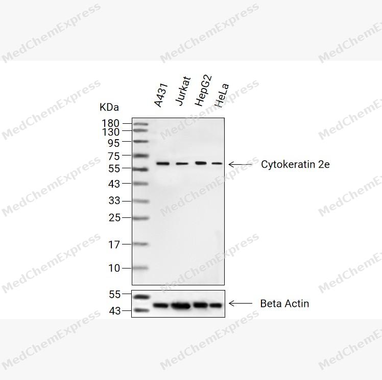 Western blot analysis of extracts from A431(lane 2(20μg) , Jurkat(lane 3(20μg) ,HepG2(lane 4(20μg)and HeLa(lane 5(20μg) using Cytokeratin 2e Antibody(HY-P84280).Proteins were transferred to a PVDF membrane and blocked with 5% non-fat milk in TBST for 2 hour at room temperature. The primary antibody ( 1/1000) and Loading control antibody (Beta Actin, HY-P80993,1/10000) was used in 5% non-fat milk in TBST at 4°C overnight. Goat Anti-Mouse IgG-HRP Secondary Antibody (HY-P8004,1/10,000) was used for 1 hour at room temperature.
 -
 Immunohistochemical analysis of paraffin-embedded human skin tissue using Cytokeratin 2e Antibody (HY-P84280, 1/200). The section was pretreated using heat mediated antigen retrieval with Tris-EDTA buffer (pH 8.0) for 10 minutes. The tissues were blocked with quick block buffer for 0.5 hours at room temperature, washed with PBS and PBST, and then incubated with the primary antibody overnight at 4℃. The detection was performed using an HRP conjugated compact polymer system. DAB was used as the chromogen. Tissues were counterstained with hematoxylin and mounted with DPX.
 -
 Immunohistochemical analysis of paraffin-embedded human tonsil tissue using Cytokeratin 2e Antibody (HY-P84280, 1/200). The section was pretreated using heat mediated antigen retrieval with Tris-EDTA buffer (pH 8.0) for 10 minutes. The tissues were blocked with quick block buffer for 0.5 hours at room temperature, washed with PBS and PBST, and then incubated with the primary antibody overnight at 4℃. The detection was performed using an HRP conjugated compact polymer system. DAB was used as the chromogen. Tissues were counterstained with hematoxylin and mounted with DPX.
 -
 Immunohistochemical analysis of paraffin-embedded human vagina tissue using Cytokeratin 2e Antibody (HY-P84280, 1/200). The section was pretreated using heat mediated antigen retrieval with Tris-EDTA buffer (pH 8.0) for 10 minutes. The tissues were blocked with quick block buffer for 0.5 hours at room temperature, washed with PBS and PBST, and then incubated with the primary antibody overnight at 4℃. The detection was performed using an HRP conjugated compact polymer system. DAB was used as the chromogen. Tissues were counterstained with hematoxylin and mounted with DPX.
 -
 Immunohistochemical analysis of paraffin-embedded human cervix tissue using Cytokeratin 2e Antibody (HY-P84280, 1/200). The section was pretreated using heat mediated antigen retrieval with Tris-EDTA buffer (pH 8.0) for 10 minutes. The tissues were blocked with quick block buffer for 0.5 hours at room temperature, washed with PBS and PBST, and then incubated with the primary antibody overnight at 4℃. The detection was performed using an HRP conjugated compact polymer system. DAB was used as the chromogen. Tissues were counterstained with hematoxylin and mounted with DPX.


Background
-
Function
Cytokeratin 2e is a Probably contributes to terminal cornification. Associated with keratinocyte activation, proliferation and keratinization. Required for maintenance of corneocytes and keratin filaments in suprabasal keratinocytes in the epidermis of the ear, potentially via moderation of expression and localization of keratins and their partner proteins. Plays a role in the establishment of the epidermal barrier on plantar skin[1][2].
-
Subcellular Localization
Cytoplasm
-
Expression
Tissue_specificity:It is expressed in the upper spinous and granular layers of normal adult epidermal tissue in most body parts, including the thighs, nipples, soles of the feet, penile shaft, and armpits. It is not expressed in the foreskin, squamous metaplasia, or carcinoma. In hypertrophic scars and keloids, expression begins in the deepest upper layers. Expression is weak in normal gingiva and tongue, but is enhanced in benign keratosis of the lingual mucosa and mild to moderate oral dysplasia with orthokeratosis.
Induction:Induced in keratinocytes by all-trans retinoic acid (ATRA) , via increase in mRNA stability -
Subunit
Heterotetramer of two type I and two type II keratins. Associates with KRT10
-
SwissProt ID
-
Synonyms
K2e; KRTE; CK-2e; KRT2A; KRT2E


Documentation
-
Data Sheet (260 KB)
-
SDS (251 KB)
- English - EN (251 KB)
- Français - FR (251 KB)
- Deutsch - DE (251 KB)
- Norwegian - NO (251 KB)
- Español - ES (251 KB)
- Swedish - SV (251 KB)
- Italian - IT (251 KB)
- Korean - KR (251 KB)
- Portuguese - PT (251 KB)
-
User Guide for Antibodies (1077 KB)
References
[1]. Collin C, et al. Characterization of human cytokeratin 2, an epidermal cytoskeletal protein synthesized late during differentiation. Exp Cell Res. 1992 Sep;202(1):132-41. [Content Brief]
[2]. Bloor BK, et al. Expression of keratin K2e in cutaneous and oral lesions: association with keratinocyte activation, proliferation, and keratinization. Am J Pathol. 2003 Mar;162(3):963-75. [Content Brief]

 Powered by Bioz
