



PD-L2 Antibody (YA3391)
(Synonyms: PD-1 ligand 2; PD-L2; PDCD1 ligand 2; B7-DC; CD273; PD L2; PDL2)Based on 1 Customer Validation
PD-L2 Antibody (YA3391) is a Mouse-derived and non-conjugated IgG2b monoclonal antibody, targeting to PD-L2.
-
Host:
Mouse
-
Isotype:
IgG2b
-
Application:
WB, ICC/IF, FC, ELISA
-
Reactivity :
Human, Mouse, Rat
-
Formulation:
Supplied in PBS with 0.05% sodium azide.
-
Conjugation:
Non-conjugated


To place orders, for customer services and technical support, please contact: MedChemExpress USA
Tel: 609-228-6898 E-mail: [email protected] [email protected]





Applications
| Application |
WB

WB: Western Blot
|
ICC/IF
ICC/IF: Immunocytochemistry/
Immunofluorescence |
FC
FC: Flow Cytometry
|
ELISA
ELISA: Enzyme Linked Immunosorbent Assay
|
|---|---|---|---|---|
| Dilution Ratio | 1:500-1:1000 | 1:50-1:200 | 1:50-1:100 | 1:10000 |


Product Details
PD-L2 Antibody (YA3391) is a Mouse-derived and non-conjugated IgG2b monoclonal antibody, targeting to PD-L2.
-
Host Mouse

-
Clonality Monoclonal

-
Species ReactivityHuman, Mouse, Rat
-
Observed Molecular WeightObserved band size: 35-70kDa
Note: Due to possible protein modifications or aggregation, the molecular weight should be confirmed by actual measurement, and the predicted value is for reference only.

-
Calculated Molecular Weight Predicted band size: 31 kDa

Entrez Gene: 80380 Human ; 58205 Mouse ; 309304 Rat
SwissProt: Q9BQ51 Human ; Q9WUL5 Mouse ;
OMIM: 605723 Human
Purified recombinant fragment of human CD273 aa 20-220.
Endogenous
Affinity Purified
Non-conjugated
Unmodified
IgG2b


Product Properties
-
Appearance
Liquid
-
Formulation
Supplied in PBS with 0.05% sodium azide.
-
Concentration
Batch-dependent, Please check the COA for the concentration of each lot. Check Lot Concentration
-
Storage & Stability
Stored at -20°C for 1 year. Avoid repeated freeze / thaw cycles.
-
Shipping
Shipping with blue ice.


Verification Images
-
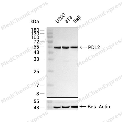 Western blot analysis was performed on extracts from U2OS (lane 1, 20 μg), 3T3 (lane 2, 20 μg), and Raji (lane 3, 20 μg) using PDL2 Mouse mAb.Proteins were transferred to a PVDF membrane and blocked with 5% non - fat milk in TBST at 4°C overnight.The primary antibody (1:1000 dilution) and the loading control antibody (beta-Actin,HY-P83730, 1:5000 dilution) were incubated in 5% non-fat milk in TBST for 1 hour at 37°C.Goat Anti - Mouse/Rabbit IgG - HRP Secondary Antibody (1:20000 dilution) was then applied for 40 minutes at 37°C.


Background
-
Function
PD-L2 is involved in the costimulatory signal, essential for T-cell proliferation and IFNG production in a PDCD1-independent manner. Interaction with PDCD1 inhibits T-cell proliferation by blocking cell cycle progression and cytokine production
-
Subcellular Localization
Secreted; Endomembrane system; Single-pass type I membrane protein; Cell membrane; Single-pass type I membrane protein
-
Expression
Tissue_specificity:It is highly expressed in the heart, placenta, pancreas, lungs, and liver, and lowly expressed in the spleen, lymph nodes, and thymus.
Induction:Up-regulated by IFNG/IFN-gamma stimulation in monocytes and induced on dendritic cells grown from peripheral blood mononuclear cells with CSF2 and IL4/interleukin-4 -
Isoforms & Post-Translational Modification
Q9BQ51 has 3 isomers: Q9BQ51-1: 30957 Da (predicted); Q9BQ51-2: 20867 Da (predicted); Q9BQ51-3: 20745 Da (predicted).
-
Subunit
Interacts with PDCD1
-
SwissProt ID
-
Synonyms
PD-1 ligand 2; PD-L2; PDCD1 ligand 2; B7-DC; CD273; PD L2; PDL2
-
Research Field
Immunology


Documentation
-
Data Sheet (234 KB)
-
SDS (251 KB)
- English - EN (251 KB)
- Français - FR (251 KB)
- Deutsch - DE (251 KB)
- Norwegian - NO (251 KB)
- Español - ES (251 KB)
- Swedish - SV (251 KB)
- Italian - IT (251 KB)
- Korean - KR (251 KB)
- Portuguese - PT (251 KB)
-
User Guide for Antibodies (1077 KB)

 Powered by Bioz
