



PI3 Kinase p85 alpha Antibody (YA5432)
(Synonyms: PIK3R1; GRB1; Phosphatidylinositol 3-kinase regulatory subunit alpha; PI3-kinase regulatory subunit alpha; PI3K regulatory subunit alpha; PtdIns-3-kinase regulatory subunit alpha; Phosphatidylinositol 3-kinase 85 kDa regulatory subunit alpha; PI3-kinase subunit p85-alpha; PtdIns-3-kinase regulatory subunit p85-alpha)
Based on 1 Customer Validation
PI3 Kinase p85 alpha Antibody (YA5432) is a Mouse-derived and non-conjugated monoclonal antibody, targeting to PI3 Kinase p85 alpha.
-
Host:
Mouse
-
Application:
WB, IHC-P, ICC/IF
-
Reactivity :
Rat, Mouse
-
Formulation:
Supplied in PBS containing 50% glycerol, 0.5% BSA and 0.02% sodium azide.
-
Conjugation:
Non-conjugated


To place orders, for customer services and technical support, please contact: MedChemExpress USA
Tel: 609-228-6898 E-mail: [email protected] [email protected]





Applications
| Application |
WB

WB: Western Blot
|
IHC-P
IHC-P: Immunohistochemistry-Paraffin
|
ICC/IF
ICC/IF: Immunocytochemistry/
Immunofluorescence |
|---|---|---|---|
| Dilution Ratio | 1:1000-2000 | 1:100-200 | 1:50-200 |


Product Details
PI3 Kinase p85 alpha Antibody (YA5432) is a Mouse-derived and non-conjugated monoclonal antibody, targeting to PI3 Kinase p85 alpha.
-
Host Mouse

-
Clonality Monoclonal

-
Species ReactivityRat, Mouse
-
Observed Molecular WeightObserved band size: 85 kDa
Note: Due to possible protein modifications or aggregation, the molecular weight should be confirmed by actual measurement, and the predicted value is for reference only.

Recombinant Protein of PI3 Kinase P85α
affinity chromatography.
Non-conjugated
Unmodified


Product Properties
-
Appearance
Liquid
-
Formulation
Supplied in PBS containing 50% glycerol, 0.5% BSA and 0.02% sodium azide.
-
Concentration
Batch-dependent, Please check the COA for the concentration of each lot. Check Lot Concentration
-
Storage & Stability
Stored at -20°C for 1 year. Avoid repeated freeze / thaw cycles.
-
Shipping
Shipping with blue ice.


Verification Images
-
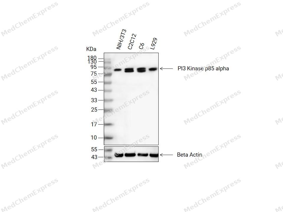 Western blot analysis of extracts from NIH/3T3(lane2(20μg), C2C12(lane3(20μg), C6(lane4(20μg) and L929(lane5(20μg) using PI3 Kinase p85 alpha Antibody (YA5432)(HY-P85740). Proteins were transferred to a PVDF membrane and blocked with 5% non-fat milk in TBST at 4°C overnight. The primary antibody (1/1000) and Loading control antibody (Beta Actin, HY-P80993, 1/10,000) was used in 5% non-fat milk in TBST for 2 hour at room temperature. Goat Anti-Mouse IgG-HRP Secondary Antibody (HY-P8004, 1/10,000) was used for 1 hour at room temperature.
 -
 Immunohistochemical analysis of paraffin-embedded Rat spleen tissue using PI3 Kinase p85 alpha Antibody (YA5432). The section was pre-treated using heat mediated antigen retrieval with Tris/EDTA buffer (pH 9.0) for 20 minutes. The tissues were probed with the primary antibody (HY-P85740, 1/100) overnight at 4℃. The detection was performed using an HRP conjugated compact polymer system. DAB was used as the chromogen. Tissues were counterstained with hematoxylin and mounted with neutral balsam.
 -
 Immunohistochemical analysis of paraffin-embedded Rat spleen tissue using PI3 Kinase p85 alpha Antibody (YA5432). The section was pre-treated using heat mediated antigen retrieval with Tris/EDTA buffer (pH 9.0) for 20 minutes. The tissues were probed with the primary antibody (HY-P85740, 1/100) overnight at 4℃. The detection was performed using an HRP conjugated compact polymer system. DAB was used as the chromogen. Tissues were counterstained with hematoxylin and mounted with neutral balsam.
 -
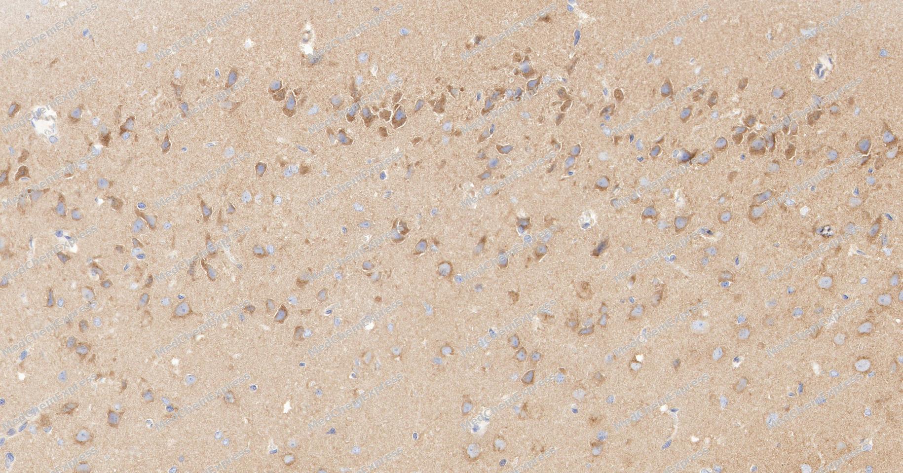 Immunohistochemical analysis of paraffin-embedded Rat brain tissue using PI3 Kinase p85 alpha Antibody (YA5432). The section was pre-treated using heat mediated antigen retrieval with Tris/EDTA buffer (pH 9.0) for 20 minutes. The tissues were probed with the primary antibody (HY-P85740, 1/100) overnight at 4℃. The detection was performed using an HRP conjugated compact polymer system. DAB was used as the chromogen. Tissues were counterstained with hematoxylin and mounted with neutral balsam.
 -
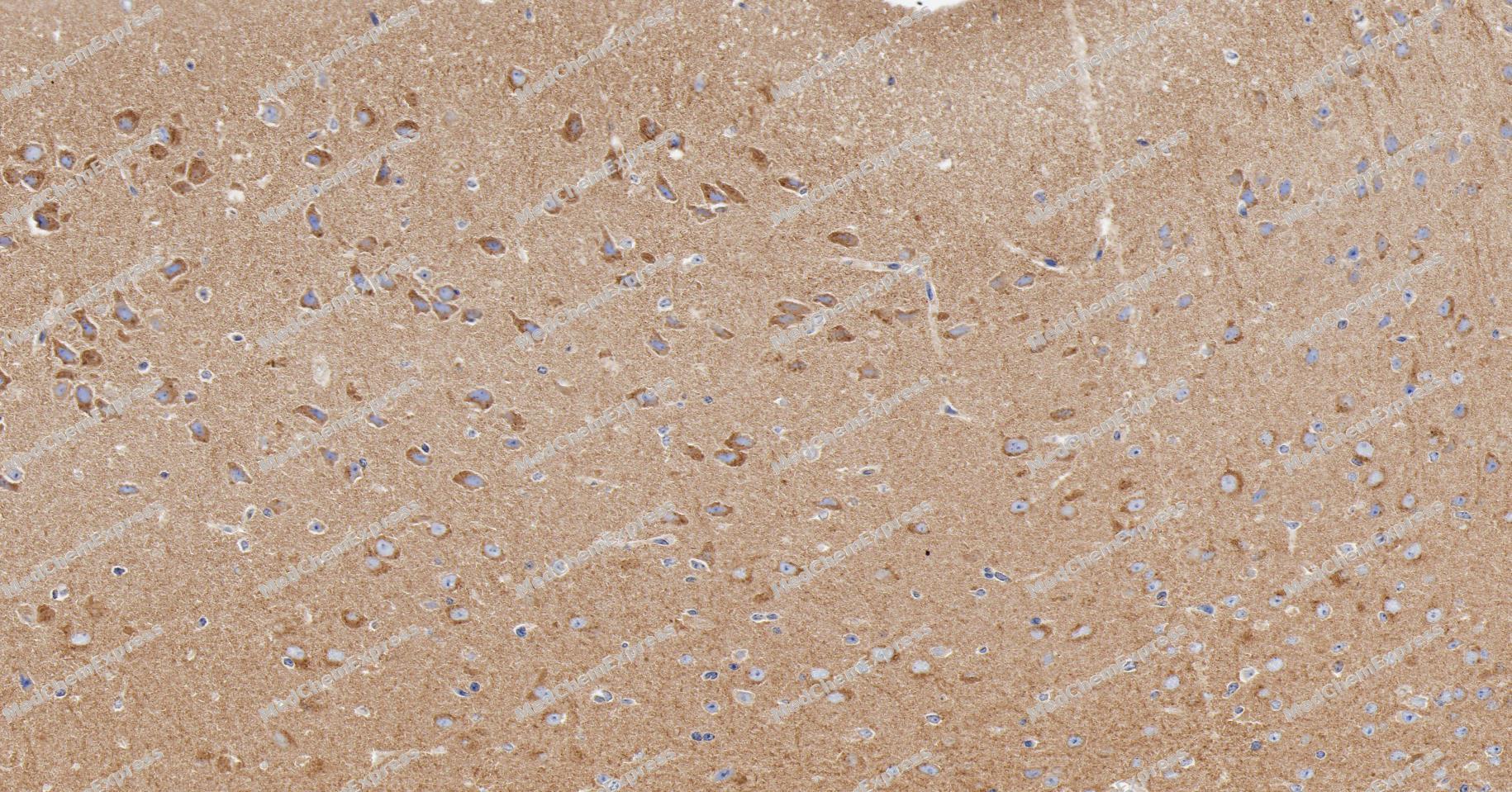 Immunohistochemical analysis of paraffin-embedded mouse brain tissue using PI3 Kinase p85 alpha Antibody (YA5432). The section was pre-treated using heat mediated antigen retrieval with Tris/EDTA buffer (pH 9.0) for 20 minutes. The tissues were probed with the primary antibody (HY-P85740, 1/100) overnight at 4℃. The detection was performed using an HRP conjugated compact polymer system. DAB was used as the chromogen. Tissues were counterstained with hematoxylin and mounted with neutral balsam.
 -
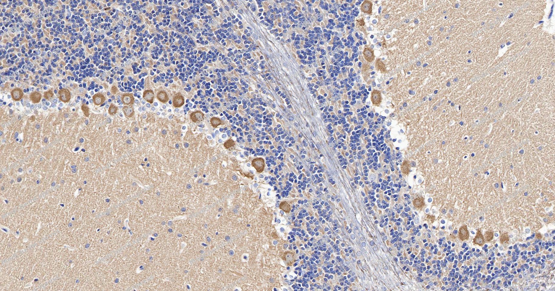 Immunohistochemical analysis of paraffin-embedded Rat brain tissue using PI3 Kinase p85 alpha Antibody (YA5432). The section was pre-treated using heat mediated antigen retrieval with Tris/EDTA buffer (pH 9.0) for 20 minutes. The tissues were probed with the primary antibody (HY-P85740, 1/100) overnight at 4℃. The detection was performed using an HRP conjugated compact polymer system. DAB was used as the chromogen. Tissues were counterstained with hematoxylin and mounted with neutral balsam.
 -
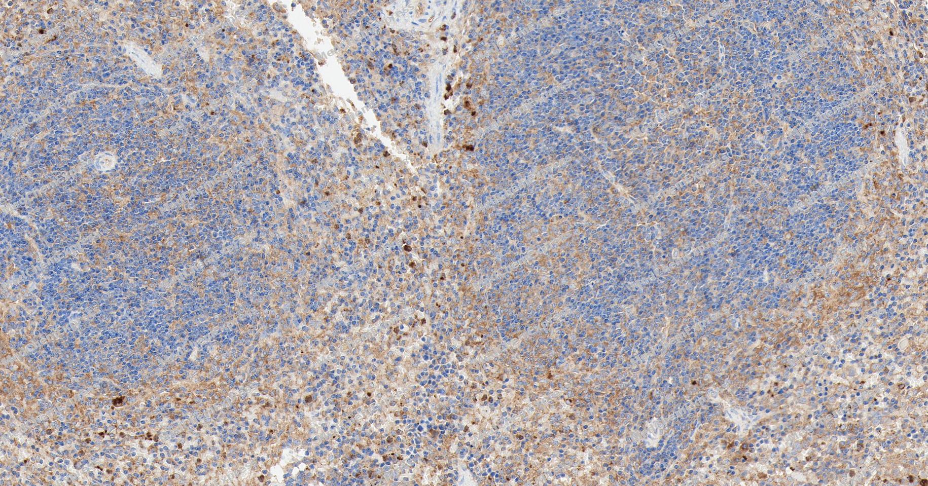 Immunohistochemical analysis of paraffin-embedded mouse spleen tissue using PI3 Kinase p85 alpha Antibody (YA5432). The section was pre-treated using heat mediated antigen retrieval with Tris/EDTA buffer (pH 9.0) for 20 minutes. The tissues were probed with the primary antibody (HY-P85740, 1/100) overnight at 4℃. The detection was performed using an HRP conjugated compact polymer system. DAB was used as the chromogen. Tissues were counterstained with hematoxylin and mounted with neutral balsam.
 -
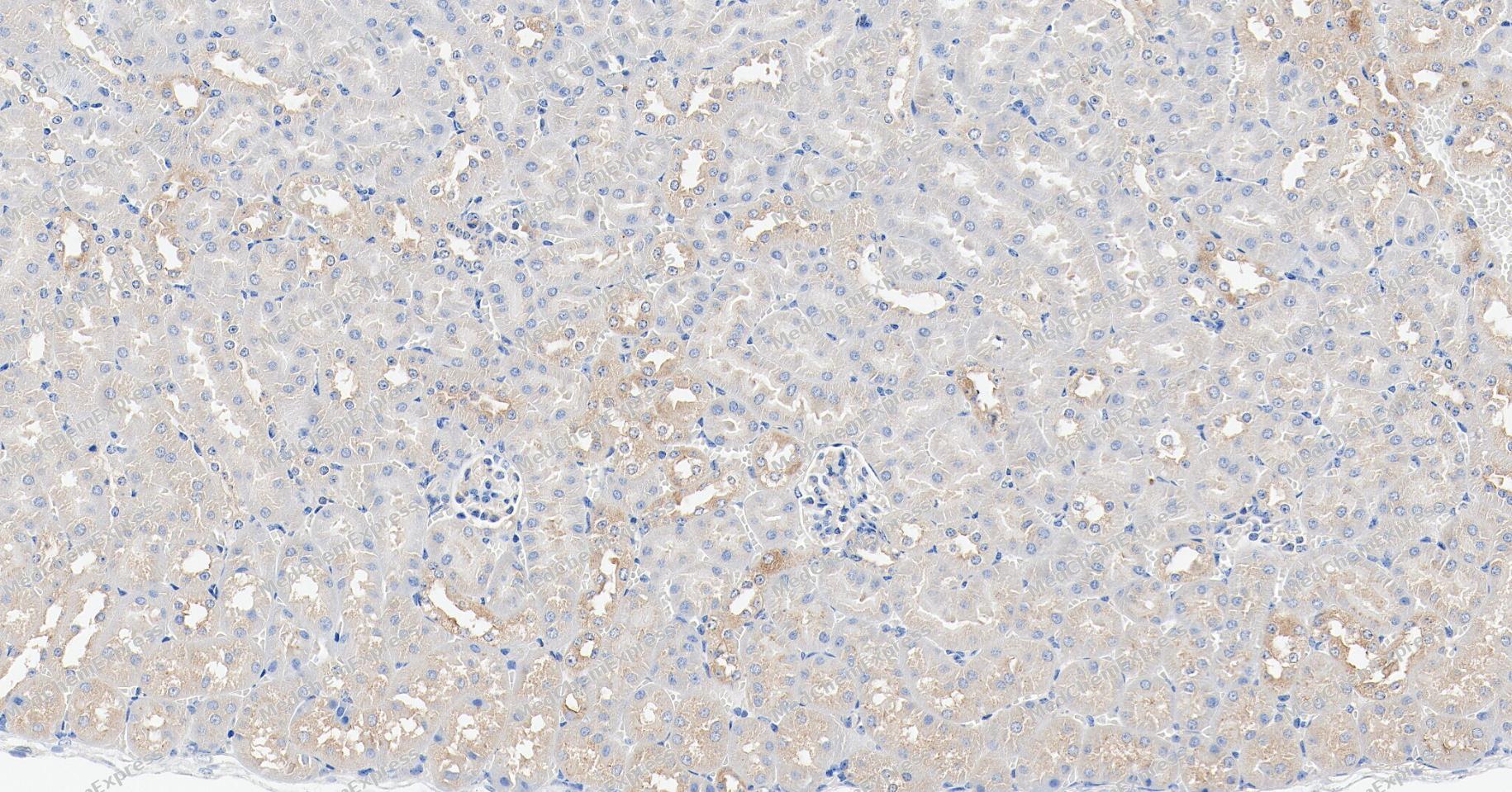 Immunohistochemical analysis of paraffin-embedded mouse kidney tissue using PI3 Kinase p85 alpha Antibody (YA5432). The section was pre-treated using heat mediated antigen retrieval with Tris/EDTA buffer (pH 9.0) for 20 minutes. The tissues were probed with the primary antibody (HY-P85740, 1/100) overnight at 4℃. The detection was performed using an HRP conjugated compact polymer system. DAB was used as the chromogen. Tissues were counterstained with hematoxylin and mounted with neutral balsam.
 -
 Immunocytochemistry analysis of Hela cells labeling PI3 Kinase p85 alpha with PI3 Kinase p85 alpha Antibody (HY-P85740) at 1/100 dilution. Cells were fixed in 4% paraformaldehyde for 15 minutes at room temperature, permeabilized with 0.1% Triton X-100 in PBS for 15 minutes at room temperature, then blocked with quick block buffer for 10 minutes at room temperature. Cells were then incubated with PI3 Kinase p85 alpha Antibody (HY-P85740) at 1/100 dilution in quick block buffer overnight at 4 ℃.AF488-conjugated Goat Anti-Mouse IgG H&L (HY-P8005, Green) was used as the secondary antibody at 1/1,000 dilution. PBS instead of the primary antibody was used as the secondary antibody only control. The Nuclear counterstain was DAPI (Blue).
 -
 Immunocytochemistry analysis of NIH-3T3 cells labeling PI3 Kinase p85 alpha with PI3 Kinase p85 alpha Antibody (HY-P85740) at 1/100 dilution. Cells were fixed in 4% paraformaldehyde for 15 minutes at room temperature, permeabilized with 0.1% Triton X-100 in PBS for 15 minutes at room temperature, then blocked with quick block buffer for 10 minutes at room temperature. Cells were then incubated with PI3 Kinase p85 alpha Antibody (HY-P85740) at 1/100 dilution in quick block buffer overnight at 4 ℃.AF488-conjugated Goat Anti-Mouse IgG H&L (HY-P8005, Green) was used as the secondary antibody at 1/1,000 dilution. PBS instead of the primary antibody was used as the secondary antibody only control. The Nuclear counterstain was DAPI (Blue).


Background
-
Function
PI3 Kinase p85 alpha binds to activated (phosphorylated) protein-Tyr kinases, through its SH2 domain, and acts as an adapter, mediating the association of the p110 catalytic unit to the plasma membrane. Necessary for the insulin-stimulated increase in glucose uptake and glycogen synthesis in insulin-sensitive tissues. Plays an important role in signaling in response to FGFR1, FGFR2, FGFR3, FGFR4, KITLG/SCF, KIT, PDGFRA and PDGFRB. Likewise, plays a role in ITGB2 signaling. Modulates the cellular response to ER stress by promoting nuclear translocation of XBP1 isoform 2 in a ER stress- and/or insulin-dependent manner during metabolic overloading in the liver and hence plays a role in glucose tolerance improvement[1][2][3][4].
-
Expression
Tissue_specificity:Isomer 2 is expressed in skeletal muscle and brain tissue, and at lower levels in the kidneys and cardiac muscle. Both isomer 2 and isomer 4 are present in skeletal muscle (protein level) . -
Isoforms & Post-Translational Modification
P27986 has 5 isomers: P27986-1: 83598 Da (predicted); P27986-2: 53486 Da (predicted); P27986-3: 49965 Da (predicted); P27986-4: 84474 Da (predicted); P27986-5: 42838 Da (predicted).
Polyubiquitinated in T-cells by CBLB; which does not promote proteasomal degradation but impairs association with CD28 and CD3Z upon T-cell activation;In adipose tissue, polyubiquitinated by the BCR(KBTBD2) E3 ubiquitin ligase complex; recognized by KBTBD2 through the SH2 domains, undergoes 'Lys-48'-linked polyubiquitination leading to its degradation;Phosphorylated. Tyrosine phosphorylated in response to signaling by FGFR1, FGFR2, FGFR3 and FGFR4. Phosphorylated by CSF1R. Phosphorylated by ERBB4. Phosphorylated on tyrosine residues by TEK/TIE2. Dephosphorylated by PTPRJ. Phosphorylated by PIK3CA at Ser-608; phosphorylation is stimulated by insulin and PDGF. The relevance of phosphorylation by PIK3CA is however unclear (By similarity). Phosphorylated in response to KIT and KITLG/SCF. Phosphorylated by FGR -
Subunit
Heterodimer of a regulatory subunit PIK3R1 and a p110 catalytic subunit (PIK3CA, PIK3CB or PIK3CD). Interacts (via SH2 domains) with CCDC88A/GIV (tyrosine-phosphorylated form); the interaction enables recruitment of PIK3R1 to the EGFR receptor, enhancing PI3K activity and cell migration (PubMed:21954290). Interacts (via SH2 domain) with CSF1R (tyrosine phosphorylated). Interacts with PIK3R2; the interaction is dissociated in an insulin-dependent manner (By similarity). Interacts with XBP1 isoform 2; the interaction is direct and induces translocation of XBP1 isoform 2 into the nucleus in a ER stress- and/or insulin-dependent but PI3K-independent manner (PubMed:20348923). Interacts with FER. Interacts (via SH2 domain) with TEK/TIE2 (tyrosine phosphorylated). Interacts with PTK2/FAK1 (By similarity). Interacts with phosphorylated TOM1L1. Interacts with phosphorylated LIME1 upon TCR and/or BCR activation. Interacts with SOCS7. Interacts with RUFY3. Interacts (via SH2 domain) with CSF1R (tyrosine phosphorylated). Interacts with LYN (via SH3 domain); this enhances enzyme activity (By similarity). Interacts with phosphorylated LAT, LAX1 and TRAT1 upon TCR activation. Interacts with CBLB. The SH2 domains interact with the YTHM motif of phosphorylated INSR in vitro. Also interacts with tyrosine-phosphorylated IGF1R in vitro. Interacts with CD28 and CD3Z upon T-cell activation. Interacts with IRS1, IRS2 and phosphorylated IRS4, as well as with NISCH and HCST (PubMed:8628286, PubMed:19109239). Interacts with FASLG, KIT and BCR. Interacts with AXL, FGFR1, FGFR2, FGFR3 and FGFR4 (phosphorylated). Interacts with FGR and HCK. Interacts with PDGFRA (tyrosine phosphorylated) and PDGFRB (tyrosine phosphorylated). Interacts with ERBB4 (phosphorylated). Interacts with NTRK1 (phosphorylated upon ligand-binding). Interacts with FAM83B; activates the PI3K/AKT signaling cascade (PubMed:23676467). Interacts with APPL1 and APPL2 (By similarity). Interacts with SRC (PubMed:28903391). Interacts with ALOX5; this interaction bridges ALOX5 with CD40 after CD40 ligation in B cells and leads to the production of reactive oxygen species (ROS) (PubMed:21200133). Interacts with TYK2 (PubMed:10995743). Interacts with nephrin NPHN1; the interaction is reduced by high glucose levels (PubMed:28955049). Interacts with CASP8 (phosphorylated on Tyr-380) (PubMed:27109099). Interacts with CD28 (PubMed:7568038). Interacts with ICOS (PubMed:30523347)
-
SwissProt ID
-
Synonyms
PIK3R1; GRB1; Phosphatidylinositol 3-kinase regulatory subunit alpha; PI3-kinase regulatory subunit alpha; PI3K regulatory subunit alpha; PtdIns-3-kinase regulatory subunit alpha; Phosphatidylinositol 3-kinase 85 kDa regulatory subunit alpha; PI3-kinase subunit p85-alpha; PtdIns-3-kinase regulatory subunit p85-alpha


Documentation
-
Data Sheet (234 KB)
-
SDS (252 KB)
- English - EN (252 KB)
- Français - FR (252 KB)
- Deutsch - DE (252 KB)
- Norwegian - NO (252 KB)
- Español - ES (252 KB)
- Swedish - SV (252 KB)
- Italian - IT (252 KB)
- Korean - KR (252 KB)
- Portuguese - PT (252 KB)
-
User Guide for Antibodies (1077 KB)

 Powered by Bioz
